. 2000 Feb;74(4):1674-85.
doi: 10.1128/jvi.74.4.1674-1685.2000.

Identification of a novel cleavage activity of the first papain-like proteinase domain encoded by open reading frame 1a of the coronavirus Avian infectious bronchitis virus and characterization of the cleavage products

Affiliations

Identification of a novel cleavage activity of the first papain-like proteinase domain encoded by open reading frame 1a of the coronavirus Avian infectious bronchitis virus and characterization of the cleavage products

K P Lim et al. J Virol. 2000 Feb.

Abstract

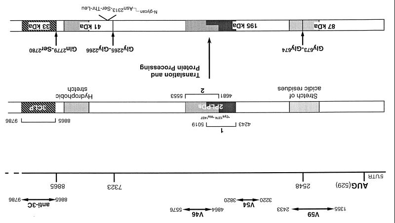
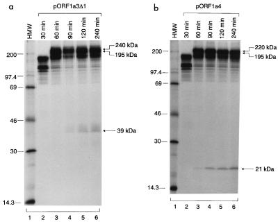
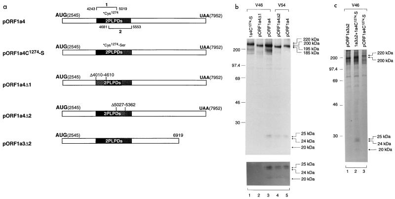
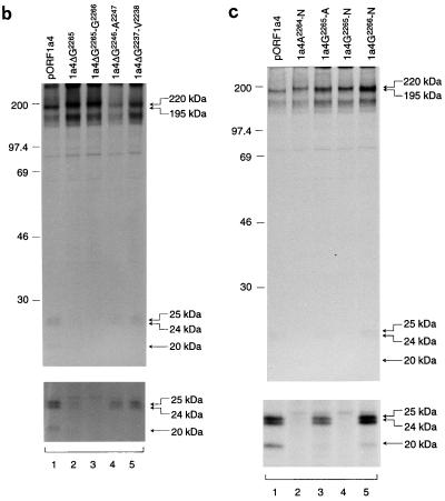
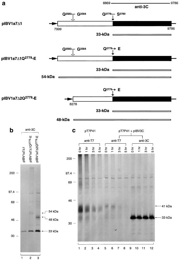
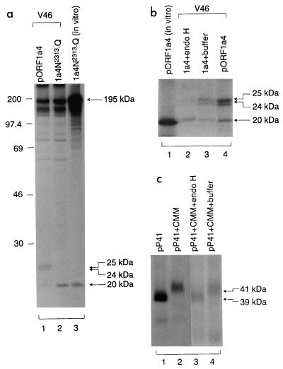
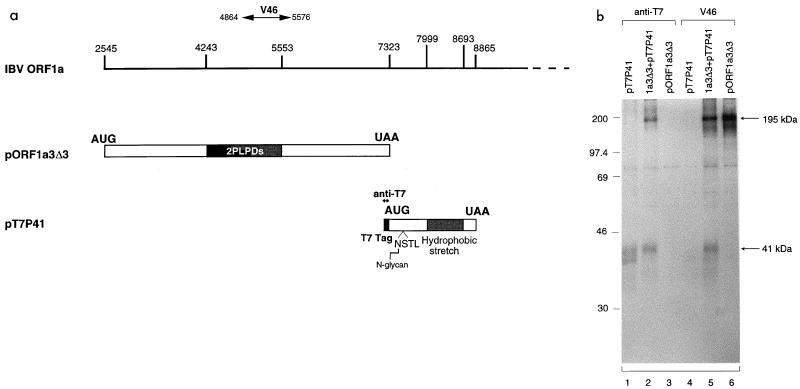
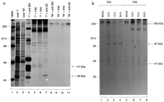
The coronavirus Avian infectious bronchitis virus (IBV) employs polyprotein processing as a strategy to express its gene products. Previously we identified the first cleavage event as proteolysis at the Gly(673)-Gly(674) dipeptide bond mediated by the first papain-like proteinase domain (PLPD-1) to release an 87-kDa mature protein. In this report, we demonstrate a novel cleavage activity of PLPD-1. Expression, deletion, and mutagenesis studies showed that the product encoded between nucleotides 2548 and 8865 was further cleaved by PLPD-1 at the Gly(2265)-Gly(2266) dipeptide bond to release an N-terminal 195-kDa and a C-terminal 41-kDa cleavage product. Characterization of the cleavage activity revealed that the proteinase is active on this scissile bond when expressed in vitro in rabbit reticulocyte lysates and can act on the same substrate in trans when expressed in intact cells. Both the N- and C-terminal cleavage products were detected in virus-infected cells and were found to be physically associated. Glycosidase digestion and site-directed mutagenesis studies of the 41-kDa protein demonstrated that it is modified by N-linked glycosylation at the Asn(2313) residue encoded by nucleotides 7465 to 7467. By using a region-specific antiserum raised against the IBV sequence encoded by nucleotides 8865 to 9786, we also demonstrated that a 33-kDa protein, representing the 3C-like proteinase (3CLP), was specifically immunoprecipitated from the virus-infected cells. Site-directed mutagenesis and expression studies showed that a previously predicted cleavage site (Q(2583)-G(2584)) located within the 41-kDa protein-encoding region was not utilized by 3CLP, supporting the conclusion that the 41-kDa protein is a mature viral product.


Figures

FIG. 1
FIG. 1
Diagram of the structure of the 5′-terminal 9-kb region of ORF 1a, illustrating the location of the 87-kDa protein, the 195-kDa protein with the two putative overlapping PLPDs, the 41-kDa glycoprotein, and 3CLP. The Gly673↓Gly674, Gly2265↓Gly2266, and Gln2779↓Ser2780 scissile bonds, the Asn2313 N-linked glycosylation site, and the catalytic residues of PLPD-1 (Cys1274 and His1437) are indicated. Also shown are the IBV sequence recognized by antisera V59, V54, V46, and anti-3C and the stretches of acidic and hydrophobic residues encoded by ORF1a.
FIG. 2
FIG. 2
(a) Diagram showing the IBV sequence contained in plasmids pORF1a3, pORF1a3Δ1, pORF1a4, and pORF1a3(M). (b) Analysis of transiently expressed ORF 1a products from cells transfected with pORF1a3(M), pORF1a3, pORF1a3Δ1, and pORF1a4. Plasmid DNAs were transiently expressed in Cos-7 cells, using the vaccinia virus-T7 expression system. Cells were labeled with [35S]methionine, lysates were prepared, and polypeptides were immunoprecipitated with anti-3C and V46. Gel electrophoresis of the polypeptides was performed on an SDS–12.5% polyacrylamide gel, and polypeptides were detected by fluorography. Numbers indicate molecular masses in kilodaltons.
FIG. 3
FIG. 3
Time course analysis of polypeptides synthesized in rabbit reticulocyte lysates from pORF1a3Δ1 (a) and pORF1a4 (b). A 10-fold excess of unlabeled methionine was added to the in vitro translation reaction mixture after incubation at 30°C for 90 min, and aliquots were taken from the reaction mixture after incubation for 30, 60, 90, 120, and 240 min. [35S]methionine-labeled translation products were separated on an SDS–15% polyacrylamide gel and detected by fluorography. HMW, high-molecular-mass markers (numbers indicate kilodaltons).
FIG. 4
FIG. 4
(a) Diagram showing the IBV sequences present in plasmids pORF1a4, pORF1a4C1274-S, pORF1a4Δ1, pORF1a4Δ2, and pORF1a3Δ2. (b) Analysis of transiently expressed ORF 1a products from transfection of pORF1a4, pORF1a4C1274-S, pORF1a4Δ1, and pORF1a4Δ2. Plasmid DNAs were transiently expressed in Cos-7 cells, using the vaccinia virus-T7 expression system. Cells were labeled with [35S]methionine, lysates were prepared, and polypeptides were immunoprecipitated with antisera V46 and V54. Gel electrophoresis of the polypeptides was performed on an SDS–12.5% polyacrylamide gel, and polypeptides were detected by fluorography. The upper panel was prepared from a gel exposed for 1 day, and the lower panel was prepared from the same gel exposed for 5 days. Numbers indicate molecular masses in kilodaltons. (c) trans-cleavage assay of PLPD-1 by coexpression of pORF1a3Δ2 and pORF1a4C1274-S. Cos-7 cells were transfected with either a single plasmid or two plasmids together and labeled with [35S]methionine. Lysates were prepared, and polypeptides were immunoprecipitated with antiserum V46. Gel electrophoresis of the polypeptides was performed on an SDS–12.5% polyacrylamide gel, and polypeptides were detected by fluorography. Numbers indicate molecular masses in kilodaltons.
FIG. 5
FIG. 5
(a) Effects of substrate deletions and mutations on the second cleavage activity mediated by PLPD-1. The amino acid sequence flanking the Gly2265-Gly2266 dipeptide bond and the deletions and mutations introduced are outlined. (b) Effects of deletions of the Gly2265, Gly2265-Gly2266, Gly2246-Ala2247, and Gly2237-Val2238 residues on cleavage activity. Plasmid DNAs were transiently expressed in Cos-7 cells, using the vaccinia virus-T7 expression system. [35S]methionine-labeled cell lysates were prepared, and polypeptides were immunoprecipitated with antiserum V46. Gel electrophoresis of the polypeptides was performed on an SDS–12.5% polyacrylamide gel, and polypeptides were detected by fluorography. The upper panel was prepared from a gel exposed for 1 day, and the lower panel was prepared from the same gel exposed for 5 days. Numbers indicate molecular masses in kilodaltons. (c) Effects of mutations of the A2264-N, G2265-A, G2265-N, and G2266-N residues on cleavage activity. The mutants were expressed and analyzed as described for panel b. The upper panel was prepared from a gel exposed for 1 day, and the lower panel was prepared from the same gel exposed for 5 days.
FIG. 5
FIG. 5
(a) Effects of substrate deletions and mutations on the second cleavage activity mediated by PLPD-1. The amino acid sequence flanking the Gly2265-Gly2266 dipeptide bond and the deletions and mutations introduced are outlined. (b) Effects of deletions of the Gly2265, Gly2265-Gly2266, Gly2246-Ala2247, and Gly2237-Val2238 residues on cleavage activity. Plasmid DNAs were transiently expressed in Cos-7 cells, using the vaccinia virus-T7 expression system. [35S]methionine-labeled cell lysates were prepared, and polypeptides were immunoprecipitated with antiserum V46. Gel electrophoresis of the polypeptides was performed on an SDS–12.5% polyacrylamide gel, and polypeptides were detected by fluorography. The upper panel was prepared from a gel exposed for 1 day, and the lower panel was prepared from the same gel exposed for 5 days. Numbers indicate molecular masses in kilodaltons. (c) Effects of mutations of the A2264-N, G2265-A, G2265-N, and G2266-N residues on cleavage activity. The mutants were expressed and analyzed as described for panel b. The upper panel was prepared from a gel exposed for 1 day, and the lower panel was prepared from the same gel exposed for 5 days.
FIG. 6
FIG. 6
(a) Diagram showing the IBV sequence contained in plasmids pIBV1a7Δ1, pIBV1a7Δ1Q2779-E, and pIBV1a7Δ2Q2779-E. (b) Analysis of transiently expressed ORF 1a products from cells transfected with pIBV1a7Δ1, pIBV1a7Δ1Q2779-E, and pIBV1a7Δ2Q2779-E. Plasmid DNAs were transiently expressed in Cos-7 cells, using the vaccinia virus-T7 expression system. Cells were labeled with [35S]methionine, lysates were prepared, and polypeptides were immunoprecipitated with anti-3C. Gel electrophoresis of the polypeptides was performed on an SDS–12.5% polyacrylamide gel, and polypeptides were detected by fluorography. Numbers indicate molecular masses in kilodaltons. (c) Time course analysis of the 41-kDa protein by coexpression of pT7P41 and pIBV3C. Cos-7 cells were transfected with either pT7P41 or pT7P41 and pIBV3C together and labeled with [35S]methionine for 4 h. The cells were washed three times with GMEM and were incubated in GMEM before they were harvested 0, 1, 3, and 5 h later. Lysates were prepared, and polypeptides were immunoprecipitated with antisera anti-T7 and anti-3C. Gel electrophoresis of the polypeptides was performed on an SDS–12.5% polyacrylamide gels, and polypeptides were detected by fluorography. Numbers indicate molecular masses in kilodaltons.
FIG. 7
FIG. 7
(a) Mutational analysis of the putative N-linked glycosylation site of the 41-kDa protein. Plasmids pORF1a4 and pORF1a4N2313-Q were transiently expressed in Cos-7 cells, using the vaccinia virus-T7 expression system. Cells were labeled with [35S]methionine, lysates were prepared, and polypeptides were immunoprecipitated with antiserum V46. Gel electrophoresis of the in vivo-synthesized polypeptides (lanes 1 and 2) together with the in vitro-synthesized products (lane 3) was performed on an SDS–12.5% polyacrylamide gel. Polypeptides were detected by fluorography. Numbers indicate molecular masses in kilodaltons. (b) Endo H treatment of the C-terminal cleavage products from expression of pORF1a4. Plasmid DNA was transfected into Cos-7 cells, using the vaccinia virus-T7 expression system. The [35S]methionine-labeled cell lysates were prepared, and polypeptides were immunoprecipitated with antiserum V46. The immunoprecipitated viral proteins were analyzed either by gel electrophoresis directly (lane 4) or after incubation with endo H (40 U/mg; lane 2). Incubation of the immunoprecipitated viral proteins with endo H buffer alone (lane 3) and the 20-kDa cleavage product obtained from in vitro expression of pORF1a4 (lane 1) were included as controls. The [35S]methionine-labeled polypeptides were subjected to gel electrophoresis on an SDS–12.5% polyacrylamide gel, and polypeptides were detected by fluorography. (c) Endo H treatment of the in vitro-synthesized 41-kDa protein. Plasmid pP41 was expressed in vitro, using the cell-free transcription-coupled translation system, in the presence or absence of 5 μl of canine pancreatic microsomal membranes. The [35S]methionine-labeled polypeptides were analyzed either by gel electrophoresis directly or after incubation with endo H (40 U/mg) or buffer only.
FIG. 8
FIG. 8
(a) Diagram showing the IBV sequences present in plasmids pORF1a3Δ3 and pT7P41. (b) Analysis of transiently expressed proteins from transfection of pORF1a3Δ3 and pT7P41. Plasmid DNAs were transiently expressed in Cos-7 cells, using the vaccinia virus-T7 expression system. Cells were labeled with [35S]methionine, lysates were prepared, and polypeptides were immunoprecipitated with anti-T7 or V46 antiserum. Gel electrophoresis of the polypeptides was performed on an SDS–12.5% polyacrylamide gel, and polypeptides were detected by fluorography. Numbers indicate molecular masses in kilodaltons.
FIG. 9
FIG. 9
(a) Detection of polypeptides encoded by ORF1a in IBV-infected Vero cells. IBV-infected (“I”) and mock-infected (“M”) Vero cell monolayers in 35-mm-diameter dishes were labeled with [35S]methionine for 4 h at 6 h p.i. Cell lysates were prepared, and polypeptides were either analyzed directly or immunoprecipitated with antisera anti-IBV, V46, V54, and anti-3C. Gel electrophoresis of the polypeptides was performed on an SDS–12.5% polyacrylamide gel, and polypeptides were detected by fluorography. Numbers indicate molecular masses in kilodaltons. (b) Time course analysis of three ORF 1a-specific polypeptides in IBV-infected Vero cells. Cells were infected with IBV at a multiplicity of infection of approximately 2. After incubation in complete GMEM for 5.5 h, the cells were incubated in methionine-free medium for 0.5 h and were labeled with [35S]methionine (100 μCi/ml) for 4 h. The cells were then washed three times with GMEM and incubated in GMEM until harvested at 10, 12, and 24 h p.i. Lysates were prepared, and polypeptides were immunoprecipitated with antisera V59 and V46. Gel electrophoresis of the polypeptides was performed on an SDS–12.5% polyacrylamide gel, and polypeptides were detected by fluorography. Numbers indicate molecular masses in kilodaltons.

References

    1. Bonilla P J, Hughes S A, Weiss S R. Characterization of a second cleavage site and demonstration of activity in trans by the papain-like proteinase of the murine coronavirus mouse hepatitis virus strain A59. J Virol. 1997;71:900–909. - PMC - PubMed
    1. Boursnell M E G, Brown T D K, Foulds I J, Green P F, Tomley F M, Binns M M. Completion of the sequence of the genome of the coronavirus avian infectious bronchitis virus. J Gen Virol. 1987;68:57–77. - PubMed
    1. Brierley I, Boursnell M E G, Binns M M, Bilimoria B, Blok V C, Brown T D K, Inglis S C. An efficient ribosomal frame-shifting signal in the polymerase-encoding region of the coronavirus IBV. EMBO J. 1987;6:3779–3785. - PMC - PubMed
    1. Brierley I, Digard P, Inglis S C. Characterization of an efficient coronavirus ribosomal frameshifting signal: requirement for a RNA pseudoknot. Cell. 1989;57:537–547. - PMC - PubMed
    1. Cormack B P, Valdivia R H, Falkow S. FACS-optimized mutant of the green fluorescent protein (GFP) Gene. 1996;173:33–38. - PubMed

Publication types

MeSH terms

LinkOut - more resources