. 2000 Feb;74(4):1686-93.
doi: 10.1128/jvi.74.4.1686-1693.2000.

The central proline of an internal viral fusion peptide serves two important roles

Affiliations

The central proline of an internal viral fusion peptide serves two important roles

S E Delos et al. J Virol. 2000 Feb.

Abstract

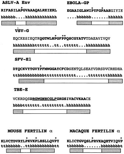
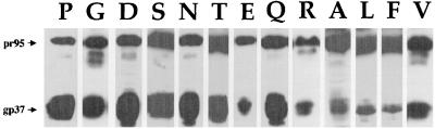
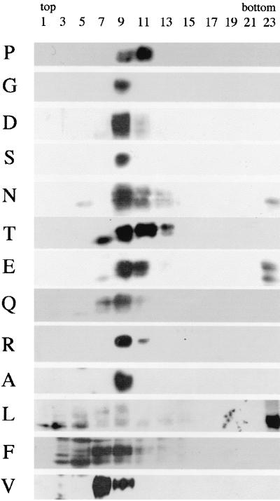
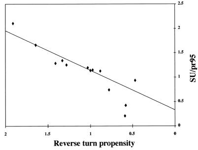
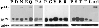
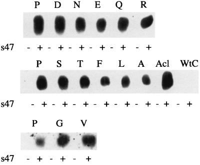
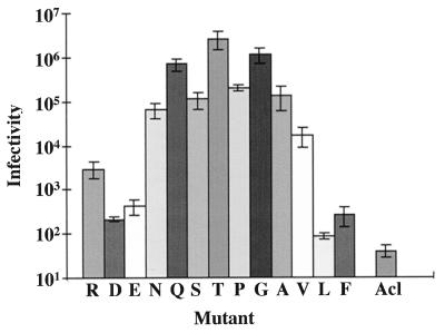
The fusion peptide of the avian sarcoma/leukosis virus (ASLV) envelope protein (Env) is internal, near the N terminus of its transmembrane (TM) subunit. As for most internal viral fusion peptides, there is a proline near the center of this sequence. Robson-Garnier structure predictions of the ASLV fusion peptide and immediate surrounding sequences indicate a region of order (beta-sheet), a tight reverse turn containing the proline, and a second region of order (alpha-helix). Similar motifs (order, turn or loop, order) are predicted for other internal fusion peptides. In this study, we made and analyzed 12 Env proteins with substitutions for the central proline of the fusion peptide. Env proteins were expressed in 293T cells and in murine leukemia virus pseudotyped virions. We found the following. (i) All mutant Envs form trimers, but when the bulky hydrophobic residues phenylalanine or leucine are substituted for proline, trimerization is weakened. (ii) Surprisingly, the proline is required for maximal processing of the Env precursor into its surface and TM subunits; the amount of processing correlates linearly with the propensity of the substituted residue to be found in a reverse turn. (iii) Nonetheless, proteolytically processed forms of all Envs are preferentially incorporated into pseudotyped virions. (iv) All Envs bind receptor with affinity greater than or equal to wild-type affinity. (v) Residues that support high infectivity cluster with proline at intermediate hydrophobicity. Infectivity is not supported by mutant Envs in which charged residues are substituted for proline, nor is it supported by the trimerization-defective phenylalanine and leucine mutants. Our findings suggest that the central proline in the ASLV fusion peptide is important for the formation of the native (metastable) Env structure as well as for membrane interactions that lead to fusion.


Figures

FIG. 1
FIG. 1
Predicted structures for internal fusion peptide regions. Protein sequences were subjected to structure prediction using the Robson-Garnier algorithm (21) in the Genetics Computer Group program package as described in Materials and Methods. b, β-sheet; ., random coil; h, α-helix; t, reverse turn. The segments of order (α-helix or β-sheet) are indicated by gray boxes; segments of random coil or reverse turn are indicated by white boxes; prolines are starred. The crystal structure of TBE-E reveals extension of the ordered sequences within the fusion peptide region into the predicted turn segment (43). The residues that actually form the turn are underlined. Fusion peptides, as described in the literature, are in boldface (ASLV EnvA [30], Ebola virus G2 [Ebola-GP] [19, 32, 44], VSV-G [14, 50, 55], SVF-E1 [33], TBE-E [43], mouse fertilin α [51]; macaque fertilin α [40].)
FIG. 2
FIG. 2
Expression of mutant EnvAs. 293T cells were transfected with pCB6-EnvA DNA, induced, harvested, and lysed as described in Materials and Methods. Samples were resolved by SDS-PAGE and processed for Western blot analysis with the anti-Ngp37 antibody. Both the uncleaved pr95 (upper band) and cleaved gp37 (lower band) are indicated.
FIG. 3
FIG. 3
Trimerization of mutant EnvAs. Cell lysates were prepared as described for Fig. 2 and subjected to sucrose density centrifugation as described in Materials and Methods. Fractions of 500 μl were collected and processed for Western blot analysis with the anti-Ngp37 antibody. The gp37 band is shown.
FIG. 4
FIG. 4
Processing of mutant EnvAs into SU (gp85) and TM (gp37) subunits. Cells expressing wild-type and mutant EnvAs were radiolabeled with Tran35S-label, lysed, subjected to immunoprecipitation using the anti-EnvA tail antibody, and treated with N-glycosidase F. Proteins were resolved by SDS-PAGE, visualized using a PhosphorImager, and quantified using ImageQuant. The data were subjected to linear regression analysis and fit best to the linear equation y = 0.81× + 0.33 with r2 = 0.84. The data (triangles) represent the ratio of gp85 to unprocessed pr95. The solid line represents the linear least squares fit of the data. The two points significantly below the line are for the poorly trimerized F and L mutants.
FIG. 5
FIG. 5
Incorporation of mutant EnvAs into MLV-pseudotyped virions. EnvA-MLV pseudotyped virions were prepared and concentrated as described in Materials and Methods. Virions were diluted into SDS sample buffer, resolved by SDS-PAGE, transferred to nitrocellulose, and probed with the anti-Ngp37 antibody. Parallel blots were probed with an antibody recognizing MLV capsid (gp30).
FIG. 6
FIG. 6
Coimmunoprecipitation of s47 with mutant EnvAs. Lysates of EnvA-expressing cells were prepared as described for Fig. 2 and incubated with biotinylated s47 for 30 min at 4°C. The mixtures were then immunoprecipitated with the anti-EnvA tail antibody (anti-EnvC tail antibody for EnvC, the Env protein of ASLV, Prague C, used as a negative control), resolved by SDS-PAGE, transferred to nitrocellulose, and probed with horseradish peroxidase-conjugated streptavidin.
FIG. 7
FIG. 7
Infectivity of mutant EnvAs. EnvA-MLV pseudotyped virions encoding β-galactosidase were prepared as for Fig. 4. Serial dilutions of pseudotyped viral supernatants were added to Tva-expressing NIH 3T3(PG950) cells, incubated at 37°C for 48 h, and assayed for β-galactosidase as described in Materials and Methods. Data from at least three independent experiments were averaged and are presented as a function of increasing hydrophobicity of the mutant residue. Ranking of relative hydrophobicity is described in the footnote to Table 2. Error bars represent standard error of the mean.

References

    1. Baker K A, Dutch R E, Lamb R A, Jardetzky T S. Structural basis for paramyxovirus-mediated membrane fusion. Mol Cell. 1999;3:309–319. - PubMed
    1. Balliet J W, Berson J, D'Cruz C M, Huang J, Crane J, Gilbert J M, Bates P. Production and characterization of a soluble, active form of Tva, the subgroup A avian sarcoma and leukosis virus receptor. J Virol. 1999;73:3054–3061. - PMC - PubMed
    1. Bigler D, Chen M, Waters S, White J M. A model for sperm-egg binding and fusion based on ADAMs and integrins. Trends Cell Biol. 1997;7:220–225. - PubMed
    1. Blobel C P, Wolfsberg T G, Turck C W, Myles D G, Primakoff P, White J M. A potential fusion peptide and an integrin ligand domain in a protein active in sperm-egg fusion. Nature. 1992;356:248–252. - PubMed
    1. Bullough P A, Hughson F M, Skehel J J, Wiley D C. Structure of influenza haemagglutinin at the pH of membrane fusion. Nature. 1994;371:37–43. - PubMed

Publication types

MeSH terms