. 2000 Feb;74(4):1718-26.
doi: 10.1128/jvi.74.4.1718-1726.2000.

Multiple integrations of human foamy virus in persistently infected human erythroleukemia cells

Affiliations

Multiple integrations of human foamy virus in persistently infected human erythroleukemia cells

C D Meiering et al. J Virol. 2000 Feb.

Abstract

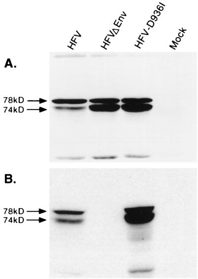
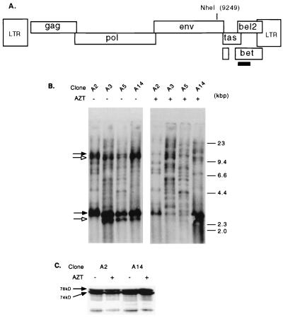
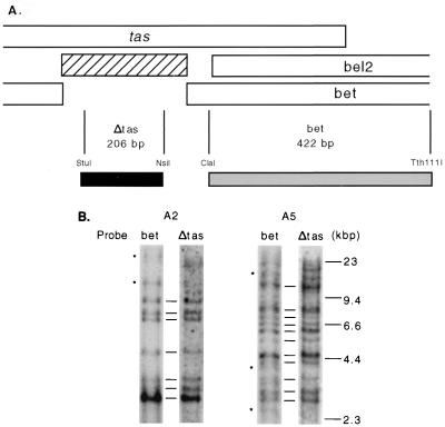
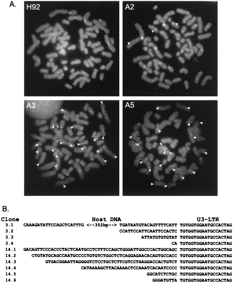
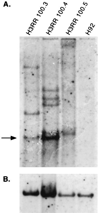
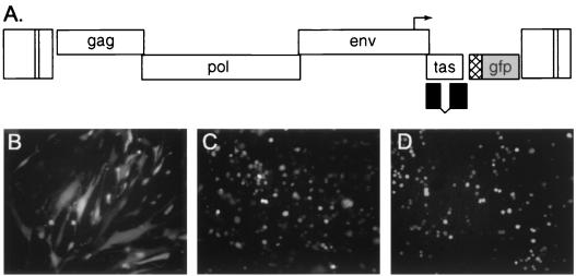
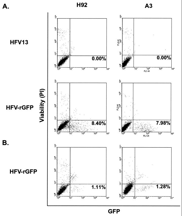
Foamy viruses are complex retroviruses whose replication strategy resembles that of conventional retroviruses. However, foamy virus replication also resembles that of hepadnaviruses in many respects. Because hepadnaviruses replicate in an integrase-independent manner, we were interested in investigating the characteristics of human foamy virus (HFV) integration. We have shown that HFV requires a functional integrase protein for infectivity. Our analyses have revealed that in single-cell clones derived from HFV-infected erythroleukemia-derived cells (H92), there were up to 20 proviral copies per host cell genome as determined by Southern blot and fluorescent in situ hybridization analysis. Use of specific probes has also shown that a majority of the proviruses contain the complete tas gene, which encodes the viral transactivator, and are not derived from Deltatas cDNAs, which have been shown to arise rapidly in infected cells. To demonstrate that the multiple proviral sequences are due to integration instead of recombination, we have sequenced the junctions between the proviral sequences and the host genome and found that the proviruses have authentic long terminal repeat ends and that each integration is at a different chromosomal site. A virus lacking the Gag nuclear localization signal accumulates fewer proviruses, suggesting that nuclear translocation is important for high proviral load. Since persistently infected H92 clones are not resistant to superinfection, the relative importance of an intracellular versus extracellular mechanism in proviral acquisition has yet to be determined.


Figures

FIG. 1
FIG. 1
Analysis of wt HFV, envelope deletion mutant (HFVΔEnv), or IN active site mutant (pHFV-D936I) virus expression in transfected FAB cells. (A) Whole cell lysates subjected to Western blotting and probed with anti-Gag antiserum. Uncleaved 78-kDa Gag protein is indicated by the solid arrow, and cleaved 74-kDa Gag protein is marked by the open arrow. (B) Cell-free virus supernatants as tested in panel A.
FIG. 2
FIG. 2
Proviral copy number in persistently infected H92 cells. (A) Schematic of the HFV genome showing the location of the unique NheI restriction site. The [32P]dCTP-labeled probe is indicated by a solid rectangle. (B) Southern blot of persistently infected H92 clones either untreated or treated with AZT. Upper arrows indicate uncut unintegrated viral DNA, while lower arrows indicate NheI-digested, unintegrated viral DNA. Solid arrows indicate undigested (upper band) or NheI-digested (lower band) full-length unintegrated viral DNA. Open arrows indicate undigested (upper band) or NheI-digested (lower band) unintegrated HFVΔtas DNA. (C) Western blot analysis using Gag antiserum on culture supernatants from clones A2 and A14 grown with or without AZT.
FIG. 3
FIG. 3
Identification of HFVΔtas proviruses in persistently infected H92 clones A2 and A5 treated with AZT. (A) The 301-bp deletion lacking in HFVΔtas is indicated by a hatched rectangle. The gray rectangle indicates the 422-bp bet probe, which detects both HFV and HFVΔtas; the black rectangle indicates the Δtas probe, which detects only wt HFV provirus. (B) Southern blot of genomic DNA from clones A2 and A5, using probes described for panel A. The asterisks indicate possible HFVΔtas integrations.
FIG. 4
FIG. 4
Analysis of multiple integrations in persistently infected H92 cells. (A) FISH analysis of uninfected H92 cells and persistently infected H92 clones A2, A3, and A5. Arrows indicate locations where the FITC-labeled HFV DNA probe is clearly visible on both sister chromatids. (B) Sequence of PCR-amplified DNAs at the junction between the HFV provirus 5′ LTR and host cell DNA in HFV-infected clones A3 (3.x) and A14 (14.x).
FIG. 5
FIG. 5
Proviral copy number in H92 cells persistently infected with H3RR virus. (A) Southern blot analysis of H3RR proviral integrations in clones 100.3, 100.4, and 100.5 and in uninfected H92 cells. Arrow indicates unintegrated, NheI-digested viral DNA. (B) The same blot stripped and reprobed with human c-myc, control cDNA.
FIG. 6
FIG. 6
Schematic diagram and expression of pHFV-rGFP. (A) Diagram of pHFV-rGFP indicating expression of GFP from the RSV promoter (cross-hatched box) as well as a truncated Bet protein (black boxes). Tas is expressed from the HFV internal promoter (arrow). (B) GFP expression at 48 h post infection in BHK cells after direct infection with cell-free HFV-rGFP at a multiplicity of infection of 1:5. (C) GFP expression in H92 after infection with HFV-rGFP by coculture with infected HEL cells; see Materials and Methods. (D) As for panel C except using clone A3.
FIG. 7
FIG. 7
FACS analysis of HFV-rGFP-superinfected A3 cells. Vertical axes show cell viability as measured by PI uptake, while horizontal axes show intensity of GFP fluorescence. The percentages of cells which are viable and GFP positive are indicated in the lower right quadrant. (A) H92 cells and A3 cells infected with either HFV13 or HFV-rGFP via coculture; see Materials and Methods. (B) Infection of H92 and A3 cells with cell-free HFV-rGFP.

Similar articles

Cited by

References

    1. Achong B G, Mansell P W A, Epstein M A, Clifford P. An unusual virus in cultures from a human nasopharyngeal carcinoma. J Natl Cancer Inst. 1971;46:299–307. - PubMed
    1. Ausubel F M, Brent R, Kingston R E, Moore D D, Seidman J G, Smith J A, Struhl K, editors. Current protocols in molecular biology. Vol. 1. New York, N.Y: John Wiley & Sons, Inc.; 1998.
    1. Baldwin D N, Linial M L. The roles of Pol and Env in the assembly pathway of human foamy virus. J Virol. 1998;72:3658–3665. - PMC - PubMed
    1. Birren B W, Tachi-iri Y, Kim U J, Nguyen M, Shizuya H, Korenberg J R, Simon M I. A human chromosome 22 fosmid resource: mapping and analysis of 96 clones. Genomics. 1996;34:97–106. - PubMed
    1. Bock C T, Schwinn S, Schroder C H, Velhagen I, Zentgraf H. Localization of hepatitis B virus core protein and viral DNA at the nuclear membrane. Virus Genes. 1996;12:53–63. - PubMed

Publication types

LinkOut - more resources