. 2000 Feb;74(4):1736-41.
doi: 10.1128/jvi.74.4.1736-1741.2000.

Hepatitis C virus core protein interacts with 14-3-3 protein and activates the kinase Raf-1

Affiliations

Hepatitis C virus core protein interacts with 14-3-3 protein and activates the kinase Raf-1

H Aoki et al. J Virol. 2000 Feb.

Abstract

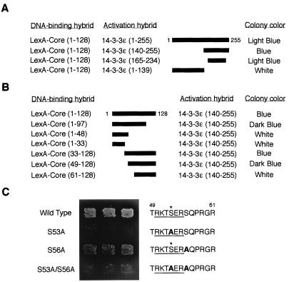
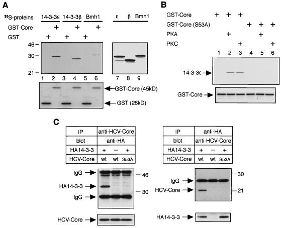
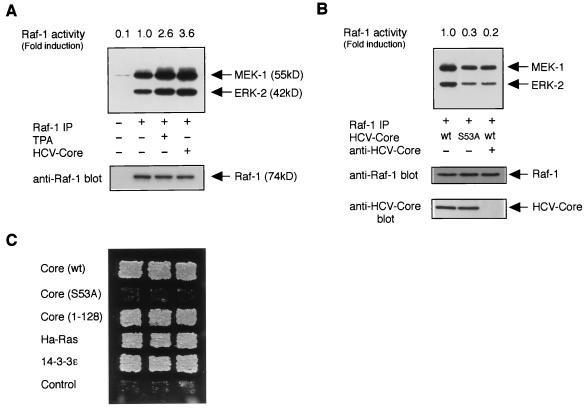
Persistent hepatitis C virus (HCV) infection is a major cause of chronic liver dysfunction in humans and is epidemiologically closely associated with the development of human hepatocellular carcinoma. Among HCV components, core protein has been reported to be implicated in cell growth regulation both in vitro and in vivo, although mechanisms explaining those effects are still unclear. In the present study, we identified that members of the 14-3-3 protein family associate with HCV core protein. 14-3-3 protein bound to HCV core protein in a phosphoserine-dependent manner. Introduction of HCV core protein caused a substantial increase in Raf-1 kinase activity in HepG2 cells and in a yeast genetic assay. Furthermore, the HCV core-14-3-3 interaction was essential for Raf-1 kinase activation by HCV core protein. These results suggest that HCV core protein may represent a novel type of Raf-1 kinase-activating protein through its interaction with 14-3-3 protein and may contribute to hepatocyte growth regulation.


Figures

FIG. 1
FIG. 1
Interaction of HCV core protein with 14-3-3 protein in the yeast two-hybrid system. (A) Determination of binding domain within 14-3-3 protein. Various activation hybrid mutants were expressed in the yeast strain EGY48 with LexA-Core protein (amino acids 1 to 128). A schematic of these 14-3-3 deletion mutants is shown with thick bars. Protein interaction was monitored by activation of the lacZ reporter gene (determined by colony color; blue colony represents a positive interaction) on an SC plate containing 2% galactose and 80 mg of X-Gal per liter with 3 days of incubation at 30°C. (B) Determination of binding domain within HCV core protein. LexA fusion proteins containing the indicated sequences of HCV core protein were expressed with an activation domain hybrid of 14-3-3 protein, 14-3-3ɛ (amino acids 140 to 255), in the yeast strain EGY48. Interactions were monitored as described for panel A. (C) HCV core protein interacts with 14-3-3 protein in a phosphoserine-dependent manner in yeast. LexA fusion proteins containing wild-type and S53A, S56A, and S53A/S56A mutants of HCV core protein (amino acids 1 to 128) were expressed with 14-3-3ɛ, an activation hybrid of 14-3-3 protein (amino acids 140 to 255), in the yeast strain EGY48, and the phosphoserine-dependent interaction was monitored by activation of leucine reporter gene and by colony color on a leucine-deficient SC plate containing galactose and X-Gal for 3 days. The putative 14-3-3 binding motifs (underlined) within HCV core proteins are indicated on the right. Substituted alanine residues are indicated by boldface type. Asterisks indicate phosphorylation sites for PKA and PKC.
FIG. 2
FIG. 2
Interaction of HCV core protein with 14-3-3 protein in vitro and in mammalian cells. (A) HCV core protein interacts with several 14-3-3 protein isoforms in vitro. Three isoforms of 35S-labeled 14-3-3 proteins were subjected to a GST pull-down experiment using budding yeast-produced GST-Core protein (top panel, lanes 2, 4, and 6) and GST alone (top panel, lanes 1, 3, and 5). Positions of molecular weight standards (in kilodaltons) are shown at the left. Input of 35S-labeled 14-3-3 proteins (14-3-3ɛ, 14-3-3β, and Bmh1) are shown in lanes 7, 8, and 9, respectively. Coomassie blue-stained GST and GST-Core from the same gel are aligned to show protein content (bottom panel, lanes 1 to 6). (B) Phosphorylation of serine-53 by PKA or PKC is essential for HCV core–14-3-3 interaction in vitro. Bacterially produced GST-Core proteins (wild type and the S53A mutant) were treated with or without recombinant PKA or recombinant PKC and subjected to GST pull-down experiments using 35S-labeled 14-3-3ɛ protein. (C) HCV core protein interacts with 14-3-3 protein in mammalian cells. HepΔNCTH cells expressing wild-type (wt) HCV core protein and HepΔNCTH cells expressing the S53A mutant of HCV core protein (S53A) were transiently transfected with pCAHA-14-3-3ɛ [HA14-3-3(+)] or pCAHA empty vector [HA14-3-3(−)]. After 48 h, cell lysate was subjected to immunoprecipitation (IP) and Western blot (blot) with the antibodies indicated above the upper panels. The presence of HCV core protein and HA14-3-3 protein in extracts was verified by reprobing the same membranes with antibodies for immunoprecipitation (bottom lanes). Positions of molecular mass standards (in kilodaltons) are shown at the right.
FIG. 3
FIG. 3
HCV core protein activates the kinase Raf-1. (A) An in vitro-coupled kinase assay for Raf-1 was performed with extracts from HepG2 (untreated or treated with TPA) or HepΔNCTH [HCV core (+)] cells. Phosphorylated MEK-1 and ERK-2 are indicated (top panel, arrows). The levels of immunoprecipitated Raf-1 are shown (bottom panel, arrows). Relative Raf-1 activities (fold induction; the 32P incorporation into ERK-2) shown are the means for duplicate determination and are representative of three experiments. (B) Extracts from HepΔNCTH cells expressing wild-type HCV core protein (wt) and HepΔNCTH (S53A) cells expressing the S53A mutant of HCV core protein (S53A) were subjected to an in vitro-coupled kinase assay. (Top panel) In the right lane, HCV core protein in cell extract was preabsorbed by using anti-HCV core antibody. (Bottom panel) The presence of HCV core protein in precleared lysates was verified by Western blot analysis. (C) Mammalian Raf-1 activation assay in yeast. Yeast strain SY1984RP cells were transformed with plasmids expressing wild-type [Core (wt)] or S53A mutant [Core (S53A)] of full-length HCV core protein, truncated HCV core protein (Core 1-128), Ha-Ras, 14-3-3ɛ, and pVT102-L empty vector (Control). Activation of Raf-1 was monitored by growth on a histidine-deficient SC plate with 3 days of incubation at 30°C.

References

    1. Aitken A. 14-3-3 proteins on the MAP. Trends Cell Biol. 1996;6:341–347. - PubMed
    1. Alter H J, Purcell R H, Shih J W, Melpolder J C, Houghton M, Choo Q-L, Kuo G. Detection of antibody to hepatitis C virus in prospectively followed transfusion recipients with acute and chronic non-A, non-B hepatitis. N Engl J Med. 1989;321:1494–1500. - PubMed
    1. Barba G, Harper F, Harada T, Kohara M, Goulinet S, Matsuura Y, Eder G, Schaff Z, Chapman M J, Miyamura T, Brechot C. Hepatitis C protein shows a cytoplasmic localization and associates to cellular lipid storage droplets. Proc Natl Acad Sci USA. 1997;94:1200–1205. - PMC - PubMed
    1. Chang J, Yang S H, Cho Y G, Hwang S B, Hahn Y S, Sung Y C. Hepatitis C virus core from two different genotypes has an oncogenic potential but is not sufficient for transforming primary rat embryo fibroblasts in cooperation with the H-ras oncogene. J Virol. 1998;72:3060–3065. - PMC - PubMed
    1. Chen C-M, You L-R, Hwang L-H, Lee Y-H W. Direct interaction of hepatitis C virus core protein with the cellular lymphotoxin-β receptor modulates the signal pathway of the lymphotoxin-β receptor. J Virol. 1997;71:9417–9426. - PMC - PubMed

Publication types

MeSH terms