. 2000 Feb;74(4):1781-6.
doi: 10.1128/jvi.74.4.1781-1786.2000.

Role of the influenza virus M1 protein in nuclear export of viral ribonucleoproteins

Affiliations

Role of the influenza virus M1 protein in nuclear export of viral ribonucleoproteins

M Bui et al. J Virol. 2000 Feb.

Abstract

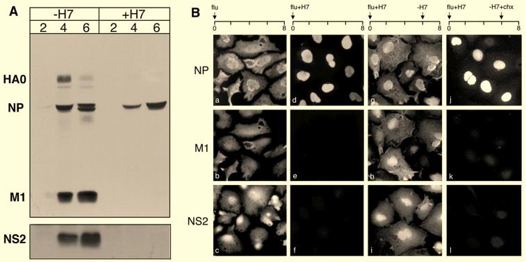
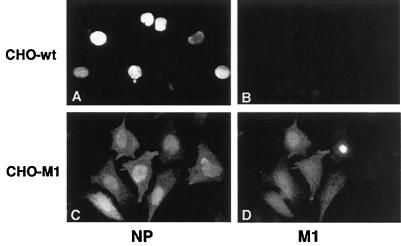
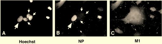
The protein kinase inhibitor H7 blocks influenza virus replication, inhibits production of the matrix protein (M1), and leads to a retention of the viral ribonucleoproteins (vRNPs) in the nucleus at late times of infection (K. Martin and A. Helenius, Cell 67:117-130, 1991). We show here that production of assembled vRNPs occurs normally in H7-treated cells, and we have used H7 as a biochemical tool to trap vRNPs in the nucleus. When H7 was removed from the cells, vRNP export was specifically induced in a CHO cell line stably expressing recombinant M1. Similarly, fusion of cells expressing recombinant M1 from a Semliki Forest virus vector allowed nuclear export of vRNPs. However, export was not rescued when H7 was present in the cells, implying an additional role for phosphorylation in this process. The viral NS2 protein was undetectable in these systems. We conclude that influenza virus M1 is required to induce vRNP nuclear export but that cellular phosphorylation is an additional factor.


Figures

FIG. 1
FIG. 1
Extraction of vRNPs from infected cell nuclei. The figure shows SDS-PAGE analysis of influenza virus-infected cells treated with PBS (lane 1), PBS–0.1% Triton X-100 (lane 2), Triton X-100–EcoRI (lane 3), or Triton X-100–EcoRI–1 M NaCl (lane 4). Cells were infected with influenza virus for 8 h, lysed, and separated into an insoluble (pellet) and a soluble (supernatant) fraction. vRNPs were detected by Western blotting using anti-NP antibodies.
FIG. 2
FIG. 2
Gradient isolation of vRNPs from H7-treated and untreated cells. Cells were infected with influenza virus for 8 h, in the presence or absence of H7. Cells were lysed in PBS–Triton X-100–EcoRI–1 M NaCl and separated into an insoluble (pellet) and a soluble (supernatant) fraction. Soluble material was loaded onto a discontinuous glycerol gradient and centrifuged to isolate vRNPs. Gradient fractions were analyzed by SDS-PAGE, and vRNPs were detected by Western blotting using anti-NP antibodies. Fractions 1 to 3 and 9 to 16 are shown.
FIG. 3
FIG. 3
Effect of H7 on viral protein synthesis. (A) SDS-PAGE analysis of influenza virus-infected cells in the presence (+H7) or absence (−H7) of H7 at 2, 4, and 6 h p.i.. HA0, NP, and M1 were detected by Western blotting using the anti-influenza virus antibody IBO. NS2 was detected by immunoprecipitation with anti-NS2 sera. (B) Immunofluorescence microscopy of influenza virus-infected cells using anti-NP, anti-M1, and anti-NS2 antibodies. The panels showing anti-NP and anti-M1 staining represent the same field, whereas panels showing anti-NS2 were a separate sample processed in parallel. Cells were analyzed at 8 h p.i. and were infected in the absence of H7 (a to c), in the presence of H7 (+H7) (d to f), with H7 washed out (−H7) at 6 h p.i. (g to i), and with H7 washed out at 6 h and cycloheximide (−H7+chx) added (j to l).
FIG. 4
FIG. 4
Stable expression of M1 promotes vRNP export. wt CHO (A and B) and CHO-M1 (C and D) cells were infected with influenza virus in the presence of H7. At 6 h p.i., H7 was washed out by transferring cells to medium lacking H7 but containing cycloheximide. Cells were analyzed at 8 h p.i. by immunofluorescence microscopy with a monoclonal anti-NP antibody (A and C) or a polyclonal anti-M1 antibody (B and D). The presynthesized M1 in CHO-M1 cells was sufficient to include vRNP export.
FIG. 5
FIG. 5
M1 provided by fusion of SFV-M1-infected cells promotes vRNP export. A cell fusion approach was used to study the effect of H7 and M1 expression on the nuclear export of vRNPs. HeLa cells (large arrows) were infected with influenza virus in the presence of H7, and L929 cells (small arrows) were fused at 6 h p.i. L929 cells were either uninfected (A to C; no SFV-M1) or infected with SFV-M1 (D to F; +SFV-M1). After fusion, cells were transferred to medium containing cycloheximide. Cells were analyzed by fluorescence microscopy using the DNA stain Hoechst 33258 (A and D), with anti-NP antibodies (B and E), and with anti-M1 antibodies (C and F). In the absence of M1, no shuttling of vRNPs occurred, but vRNP export occurred rapidly upon fusion of M1-expressing cells.
FIG. 6
FIG. 6
M1 cannot promote vRNP nuclear export in the presence of H7. HeLa cells (large arrows) were infected with influenza virus in the presence of H7, and L929 cells (small arrows) were fused at 6 h p.i. L929 cells were infected with SFV-M1. After fusion, cells were transferred to medium containing cycloheximide, in the presence of H7. Cells were analyzed by fluorescence microscopy using the DNA stain Hoechst 33258 (A), with anti-NP antibodies (B), and with anti-M1 antibodies (C). In the presence of H7, no export of vRNPs occurred, despite high levels of M1.

References

    1. Bogerd H P, Fridell R A, Madore S, Cullen B R. Identification of a novel cellular cofactor for the Rev/Rex class of retroviral regulatory proteins. Cell. 1995;82:485–494. - PubMed
    1. Bui M, Whittaker G, Helenius A. Effect of M1 protein and low pH on nuclear transport of influenza virus ribonucleoproteins. J Virol. 1996;70:8391–8401. - PMC - PubMed
    1. Bukrinskaya A G, Vorkunova G K, Vorkunova N K. Cytoplasmic and nuclear input virus RNPs in influenza virus-infected cells. J Gen Virol. 1979;45:557–567. - PubMed
    1. Davey J, Dimmock N J, Colman A. Identification of the sequence responsible for the nuclear accumulation of the influenza virus nucleoprotein in Xenopus oocytes. Cell. 1985;40:667–675. - PubMed
    1. Deppert W, Schirmbeck R. The nuclear matrix and virus function. Int Rev Cytol. 1995;162A:485–537. - PubMed

Publication types

MeSH terms

LinkOut - more resources