. 2000 Feb;74(4):1827-39.
doi: 10.1128/jvi.74.4.1827-1839.2000.

Anterograde transport of herpes simplex virus type 1 in cultured, dissociated human and rat dorsal root ganglion neurons

Affiliations

Anterograde transport of herpes simplex virus type 1 in cultured, dissociated human and rat dorsal root ganglion neurons

M Miranda-Saksena et al. J Virol. 2000 Feb.

Abstract

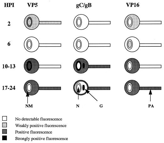
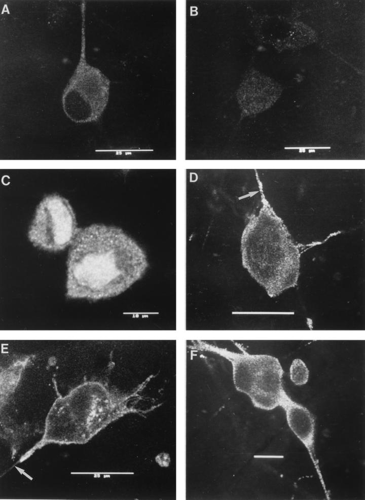
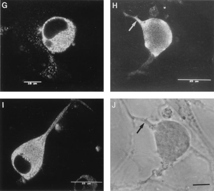
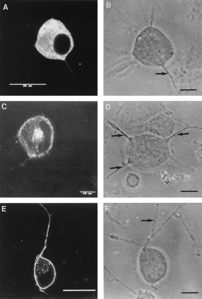
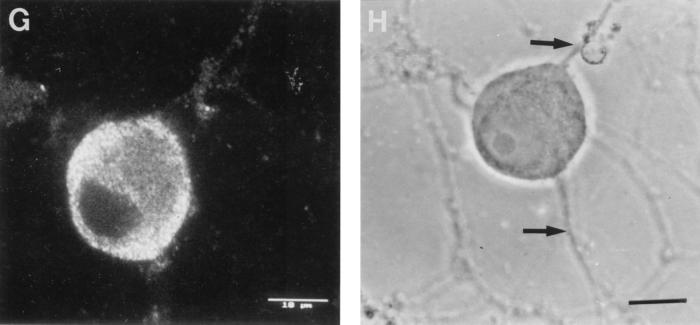
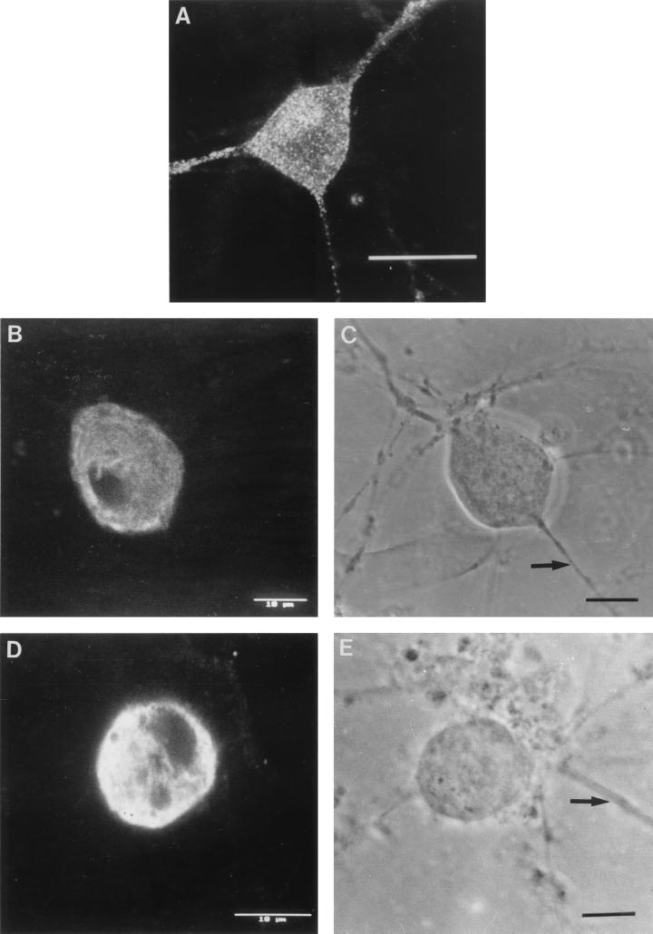
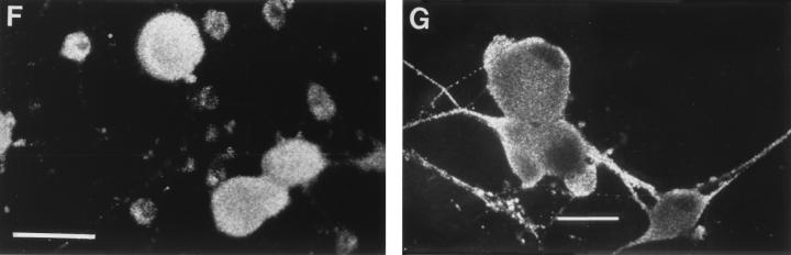
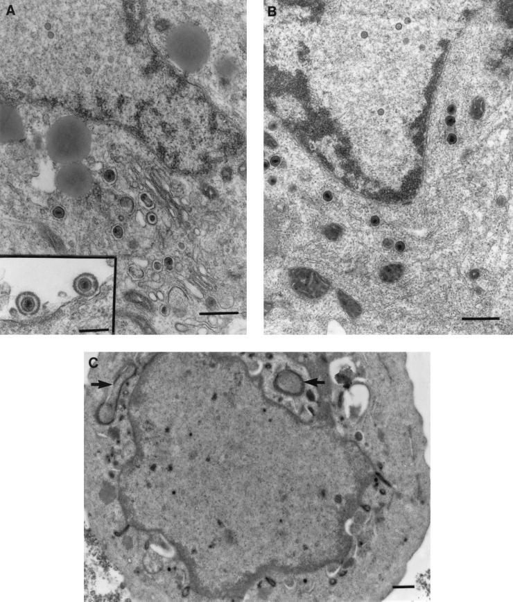
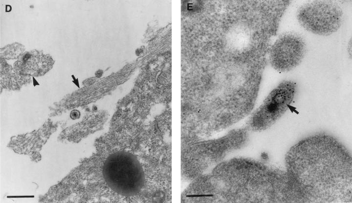
The mechanism of anterograde transport of herpes simplex virus was studied in cultured dissociated human and rat dorsal root ganglion neurons. The neurons were infected with HSV-1 to examine the distribution of capsid (VP5), tegument (VP16), and glycoproteins (gC and gB) at 2, 6, 10, 13, 17, and 24 h postinfection (p.i.) with or without nocodazole (a microtubule depolymerizer) or brefeldin A (a Golgi inhibitor). Retrogradely transported VP5 was detected in the cytoplasm of the cell body up to the nuclear membrane at 2 h p.i. It was first detected de novo in the nucleus and cytoplasm at 10 h p.i., the axon hillock at 13 h p.i., and the axon at 15 to 17 h p.i. gC and gB were first detected de novo in the cytoplasm and the axon hillock at 10 h p.i. and then in the axon at 13 h p.i., which was always earlier than the detection of VP5. De novo-synthesized VP16 was first detected in the cytoplasm at 10 to 13 h p.i. and in the axon at 16 to 17 h p.i. Nocodazole inhibited the transport of all antigens, VP5, VP16, and gC or gB. The kinetics of inhibition of VP5 and gC could be dissociated. Brefeldin A inhibited the transport of gC or gB and VP16 but not VP5 into axons. Transmission immunoelectron microscopy confirmed that there were unenveloped nucleocapsids in the axon with or without brefeldin A. These findings demonstrate that glycoproteins and capsids, associated with tegument proteins, are transported by different pathways with slightly differing kinetics from the nucleus to the axon. Furthermore, axonal anterograde transport of the nucleocapsid can proceed despite the loss of most VP16.


Figures

FIG. 1
FIG. 1
Diagrams of HSV-1 infection of cultured dissociated human fetal DRG neurons, showing the kinetics of the appearance, distribution, and axonal transport of capsid (VP5), glycoprotein (gC and gB), and tegument (VP16) antigens in different cellular compartments over the first 24 h p.i. (HPI). Gray and black shading indicates increasing degrees of positive staining for viral antigen. N, nucleus; NM, nuclear membrane; G, Golgi; PA, primary axon.
FIG. 2
FIG. 2
Confocal photomicrographs of the kinetics of appearance and distribution of capsid (VP5), glycoprotein (gC), and tegument (VP16) antigens in human neurons at selected times p.i. (A) VP5 at 2 h p.i., showing retrograde transport within an axon to the cell body. (B) VP5 at 6 h p.i., showing no detectable antigen. Photographic sensitivity was enhanced on this image to allow visualization of the cell. Under the photographic conditions used for preparation of the rest of this panel, no cell staining would normally be seen. (C) VP5 at 13 h p.i. showing intense nuclear staining and antigen diffusely distributed in the cytoplasm, extending into the axon hillock. (D) VP5 at 17 h p.i. showing cytoplasmic and axonal distribution. The arrow indicates the principal axon. Bar, 25 μm. (E) gC at 13 h p.i. showing cytoplasmic and axonal distribution. Distal regions of the stained axon (arrow) are out of the confocal plane. (F) gC at 17 h p.i. showing disappearance of antigen from the Golgi and cytoplasm. Bar, 10 μm. (G) VP16 at 13 h p.i. showing that this protein was present only in the cytoplasm. (H and J) VP16 at 15 h p.i. showing distribution in only the proximal axon. Note the antigen front (arrow). Under bright-field microscopy, the axon continued distally (for 120 μm) beyond this front (arrow) (J). Bar in panel J, 10 μm. (I) VP16 at 17 h p.i. showing that this protein is present in both the cytoplasm and the full length of the axon.
FIG. 2
FIG. 2
Confocal photomicrographs of the kinetics of appearance and distribution of capsid (VP5), glycoprotein (gC), and tegument (VP16) antigens in human neurons at selected times p.i. (A) VP5 at 2 h p.i., showing retrograde transport within an axon to the cell body. (B) VP5 at 6 h p.i., showing no detectable antigen. Photographic sensitivity was enhanced on this image to allow visualization of the cell. Under the photographic conditions used for preparation of the rest of this panel, no cell staining would normally be seen. (C) VP5 at 13 h p.i. showing intense nuclear staining and antigen diffusely distributed in the cytoplasm, extending into the axon hillock. (D) VP5 at 17 h p.i. showing cytoplasmic and axonal distribution. The arrow indicates the principal axon. Bar, 25 μm. (E) gC at 13 h p.i. showing cytoplasmic and axonal distribution. Distal regions of the stained axon (arrow) are out of the confocal plane. (F) gC at 17 h p.i. showing disappearance of antigen from the Golgi and cytoplasm. Bar, 10 μm. (G) VP16 at 13 h p.i. showing that this protein was present only in the cytoplasm. (H and J) VP16 at 15 h p.i. showing distribution in only the proximal axon. Note the antigen front (arrow). Under bright-field microscopy, the axon continued distally (for 120 μm) beyond this front (arrow) (J). Bar in panel J, 10 μm. (I) VP16 at 17 h p.i. showing that this protein is present in both the cytoplasm and the full length of the axon.
FIG. 3
FIG. 3
Effects of nocodazole on the anterograde transport of capsid (VP5), glycoprotein (gC), and tegument (VP16) antigens into the axons of dissociated human DRG neurons. (A) VP5 after incubation with nocodazole at 14 to 16 h p.i. demonstrating inhibition of initiation of axonal transport by nocodazole. Note the lack of axonal staining. (B) Corresponding bright-field micrograph showing the presence of the axons. The arrow labels the axon belonging to the neuron. Other axons are derived from neurons out of frame. Bar, 10 μm. (C) gC after incubation with nocodazole at 10 to 13 h p.i., also showing inhibition of transport. (D) Corresponding bright-field micrograph showing the presence of the axons. Arrows label axons belonging to the neuron. Bar, 10 μm. (E) gC after incubation with nocodazole at 14 to 16 h p.i., showing the presence of antigen in the axon prior to incubation with the inhibitor. Bar, 25 μm. (F) Corresponding bright-field micrograph showing the axon (arrow) belonging to the neuron. Bar, 10 μm. (G) VP16 after incubation with nocodazole at 14 to 17 h p.i. showing inhibition of anterograde axonal transport. (H) Corresponding bright-field micrograph showing the presence of the axon. Arrows indicate axons belonging to the neuron. Bar, 10 μm.
FIG. 3
FIG. 3
Effects of nocodazole on the anterograde transport of capsid (VP5), glycoprotein (gC), and tegument (VP16) antigens into the axons of dissociated human DRG neurons. (A) VP5 after incubation with nocodazole at 14 to 16 h p.i. demonstrating inhibition of initiation of axonal transport by nocodazole. Note the lack of axonal staining. (B) Corresponding bright-field micrograph showing the presence of the axons. The arrow labels the axon belonging to the neuron. Other axons are derived from neurons out of frame. Bar, 10 μm. (C) gC after incubation with nocodazole at 10 to 13 h p.i., also showing inhibition of transport. (D) Corresponding bright-field micrograph showing the presence of the axons. Arrows label axons belonging to the neuron. Bar, 10 μm. (E) gC after incubation with nocodazole at 14 to 16 h p.i., showing the presence of antigen in the axon prior to incubation with the inhibitor. Bar, 25 μm. (F) Corresponding bright-field micrograph showing the axon (arrow) belonging to the neuron. Bar, 10 μm. (G) VP16 after incubation with nocodazole at 14 to 17 h p.i. showing inhibition of anterograde axonal transport. (H) Corresponding bright-field micrograph showing the presence of the axon. Arrows indicate axons belonging to the neuron. Bar, 10 μm.
FIG. 4
FIG. 4
Effects of BFA on anterograde transport of capsid (VP5), glycoprotein (gC), and tegument (VP16) antigens into axons of human DRG neurons at 24 h p.i. (A) VP5, showing no inhibition of transport into (multiple) axons. Bar, 25 μm. (B) gC, showing complete BFA inhibition of axonal transport. (D) VP16, showing BFA inhibition of transport into the axon in this neuron. (C and E) Bright-field micrographs corresponding to panels B and D, respectively, showing the presence of the axon (arrows). Bars, 10 μm. (F) Low-magnification confocal micrograph showing complete BFA inhibition of axonal transport of gC. Bar, 25 μm. (G) Low-magnification confocal micrograph showing axonal transport of gC in untreated control for panel F. Bar, 25 μm.
FIG. 4
FIG. 4
Effects of BFA on anterograde transport of capsid (VP5), glycoprotein (gC), and tegument (VP16) antigens into axons of human DRG neurons at 24 h p.i. (A) VP5, showing no inhibition of transport into (multiple) axons. Bar, 25 μm. (B) gC, showing complete BFA inhibition of axonal transport. (D) VP16, showing BFA inhibition of transport into the axon in this neuron. (C and E) Bright-field micrographs corresponding to panels B and D, respectively, showing the presence of the axon (arrows). Bars, 10 μm. (F) Low-magnification confocal micrograph showing complete BFA inhibition of axonal transport of gC. Bar, 25 μm. (G) Low-magnification confocal micrograph showing axonal transport of gC in untreated control for panel F. Bar, 25 μm.
FIG. 5
FIG. 5
Transmission electron micrographs with or without immunolabelling of human DRG neurons treated with BFA showing its effects on egress and transport of HSV-1 virions into axons. (A) Control untreated but HSV-infected neurons at 24 h p.i. showing unenveloped nucleocapsids in the nucleus and enveloped virions in the cytoplasm (bar, 500 nm) and in the extracellular space (inset; bar, 200 nm). (B) TEM of enveloped nucleocapsids concentrated in the perinuclear zone of the cytoplasm of the cell body of a BFA-treated HSV-infected neuron. Bar, 500 nm. (C) TIEM of a BFA-treated, HSV-infected neuron showing the cell body with convoluted nuclear membranes and long tubulovesicular structures (arrows). Bar, 500 nm. (D) TEM of axonal processes showing microtubules in longitudinal (arrow) and transverse (arrowhead) section in close relationship to the cell body of an untreated, HSV-infected neuron. Bar, 500 nm. (E) TIEM showing an unenveloped nucleocapsid (arrow) within the axonal process of cultured human DRG neurons immunolabelled for VP5. Bar, 200 nm.
FIG. 5
FIG. 5
Transmission electron micrographs with or without immunolabelling of human DRG neurons treated with BFA showing its effects on egress and transport of HSV-1 virions into axons. (A) Control untreated but HSV-infected neurons at 24 h p.i. showing unenveloped nucleocapsids in the nucleus and enveloped virions in the cytoplasm (bar, 500 nm) and in the extracellular space (inset; bar, 200 nm). (B) TEM of enveloped nucleocapsids concentrated in the perinuclear zone of the cytoplasm of the cell body of a BFA-treated HSV-infected neuron. Bar, 500 nm. (C) TIEM of a BFA-treated, HSV-infected neuron showing the cell body with convoluted nuclear membranes and long tubulovesicular structures (arrows). Bar, 500 nm. (D) TEM of axonal processes showing microtubules in longitudinal (arrow) and transverse (arrowhead) section in close relationship to the cell body of an untreated, HSV-infected neuron. Bar, 500 nm. (E) TIEM showing an unenveloped nucleocapsid (arrow) within the axonal process of cultured human DRG neurons immunolabelled for VP5. Bar, 200 nm.

Similar articles

Cited by

References

    1. Avitabile E, DiGaeta S, Torrisi M R, Ward P L, Roizman B, Campadelli-Fiume G. Redistribution of microtubules and Golgi apparatus in herpes simplex virus-infected cells and their role in viral exocytosis. J Virol. 1995;69:7472–7482. - PMC - PubMed
    1. Browne H, Bell S, Minson T, Wilson D W. An endoplasmic reticulum-retained herpes simplex virus glycoprotein H is absent from secreted virions: evidence for re-envelopment during egress. J Virol. 1996;70:4311–4316. - PMC - PubMed
    1. Campadelli-Fiume G, Farabegoli F, Di Gaeta S, Roizman B. Origin of unenveloped capsids in the cytoplasm of cells infected with herpes simplex virus 1. J Virol. 1991;65:1589–1595. - PMC - PubMed
    1. Chatterjee S, Sarkar S. Studies on endoplasmic reticulum-Golgi complex cycling pathway in herpes simplex virus-infected and brefeldin A-treated human fibroblast cells. Virology. 1992;191:327–337. - PubMed
    1. Cheung P, Banfield B W, Tufaro F. Brefeldin A arrests the maturation and egress of herpes simplex virus particles during infection. J Virol. 1991;65:1893–1904. - PMC - PubMed

Publication types

MeSH terms

Substances

LinkOut - more resources