

doi: 10.1128/jvi.74.4.2005-2010.2000.
Varicella-zoster virus proteins in skin lesions: implications for a novel role of ORF29p in chickenpox
Affiliations
- PMID: 10644373
- PMCID: PMC111678
- DOI: 10.1128/jvi.74.4.2005-2010.2000
Item in Clipboard
Varicella-zoster virus proteins in skin lesions: implications for a novel role of ORF29p in chickenpox
J Virol.
2000 Feb.
Abstract
Skin biopsy samples from varicella-zoster virus (VZV)-infected patients examined by immunohistochemistry demonstrated VZV replication in nonepithelial cell types. ORF29p, a nonstructural nuclear protein, was found in nerves of two of six patients with chickenpox. In tissue culture, ORF29p was secreted by VZV-infected fibroblasts. Extracellular ORF29p can be taken up through endocytosis by human neurons, implying a novel role for this protein in pathogenesis.
Figures
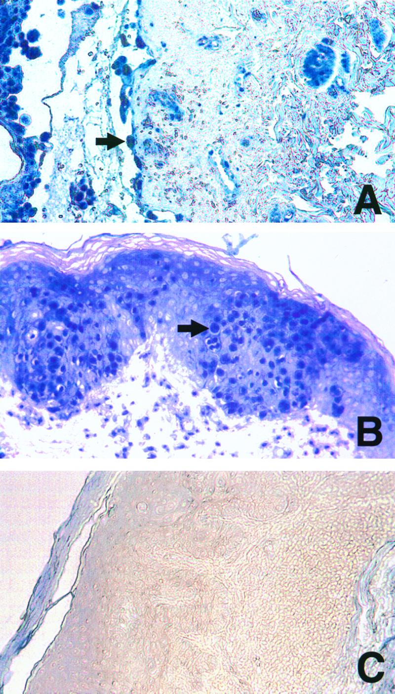
Immunohistochemical detection of ORF29p in skin biopsy samples. Chickenpox (A), zoster (B), and Grover's disease (C) skin lesions were analyzed for ORF29p as previously described (18), with the following exceptions. All washes were performed in Tris-buffered saline, and the signal was developed for 10 min in AP substrate (Vector Laboratories, Inc., Burlingame, Calif.), according to the manufacturer's recommendations, in the presence of levamisole to inhibit endogenous alkaline phosphatase activity. Arrows indicate positive epithelial cells. Magnification, ×100.
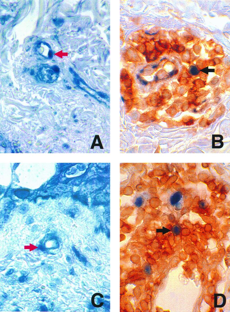
Immunohistochemical detection of ORF29p and CD43 in skin biopsy samples. Skin biopsy samples from a patient with chickenpox (A and B) or a patient with zoster (C and D) were probed for the presence of ORF29p (A and C) or ORF29p and CD43 (B and D) as described in the legend to Fig. 1. Red arrows indicate endothelial cells containing ORF29p. Black arrows indicate cells expressing CD43 that contain ORF29p. Magnification, ×600.
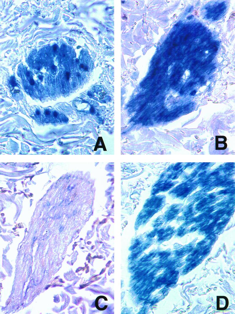
Immunohistochemical detection of ORF29p and gC in skin biopsy samples. Sections of nerves in the dermis underlying chickenpox (A and B) or zoster (C and D) lesions underwent immunohistochemistry for ORF29p (A and C) or gC (B and D) as described in the legend to Fig. 1. Magnification, ×400.
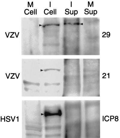
Western blot analyses of VZV and HSV-1 proteins. ORF29p, ORF21p, and ICP8 were detected in mock-infected cell extracts (M Cell) and supernatants (M Sup) or cell extracts (I Cell) and supernatants (I Sup) infected with the viruses denoted on the left. The proteins were immunoprecipitated and detected using the antibodies denoted on the right. Arrowheads denote the proteins of interest.
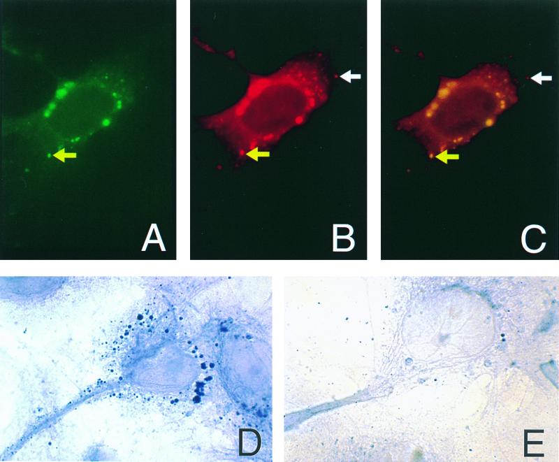
Immunohistochemical detection of ORF29p in hNTs. hNTs treated with VZV-infected cell supernatants and LysoTracker were analyzed by immunohistochemistry for the presence of ORF29p. Yellow arrows indicate ORF29p (A), LysoTracker (B), and colocalization of ORF29p and LysoTracker in the merged image (C). White arrows indicate an endocytic vesicle that does not contain ORF29p (B and C). ORF 29p is restricted to cyptoplasmic vesicles in the treated hNTs (D). Untreated hNTs do not contain ORF29p (E).
References
-
- Annunziato P, Lungu O, Gershon A, Silvers D, LaRussa P, Silverstein S. In situ hybridization detection of varicella zoster virus in paraffin-embedded skin biopsy samples. Clin Diagn Virol. 1996;7:69–76. - PubMed
-
- Arvin A. Varicella-zoster virus. In: Fields B N, Knipe D M, Howley P M, editors. Fields virology. 3rd ed. Vol. 2. Philadelphia, Pa: Lippincott-Raven Publishers; 1996. pp. 2547–2585.
-
- Asano Y, Itakura N, Hiroishi Y, Hirose S, Nagai T, Ozaki T, Yazaki T, Yamanishi Y, Takahashi M. Viremia is present in incubation period in nonimmunocompromised children with varicella. J Pediatr. 1985;106:69–71. - PubMed
-
- Assouline J G, Levin M J, Major E O, Forghani B, Straus S, Ostrove J M. Varicella-zoster virus infection of human astrocytes, Schwann cells, and neurons. Virology. 1990;179:834–843. - PubMed
-
- Cohen J, Straus S. Varicella-zoster virus and its replication. In: Fields B N, Knipe D M, Howley P M, editors. Fields virology. 3rd ed. Vol. 2. Philadelphia, Pa: Lippincott-Raven Publishers; 1996. pp. 2525–2546.
Publication types
MeSH terms
Substances
Grants and funding
LinkOut - more resources
Full Text Sources
Other Literature Sources
