. 2000 Feb;74(4):2029-37.
doi: 10.1128/jvi.74.4.2029-2037.2000.

Characterization of gammaherpesvirus 68 gene 50 transcription

Affiliations

Characterization of gammaherpesvirus 68 gene 50 transcription

S Liu et al. J Virol. 2000 Feb.

Abstract

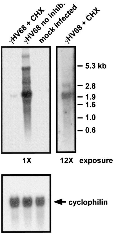
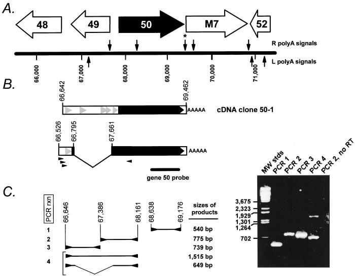
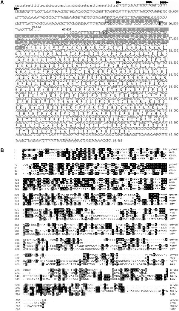
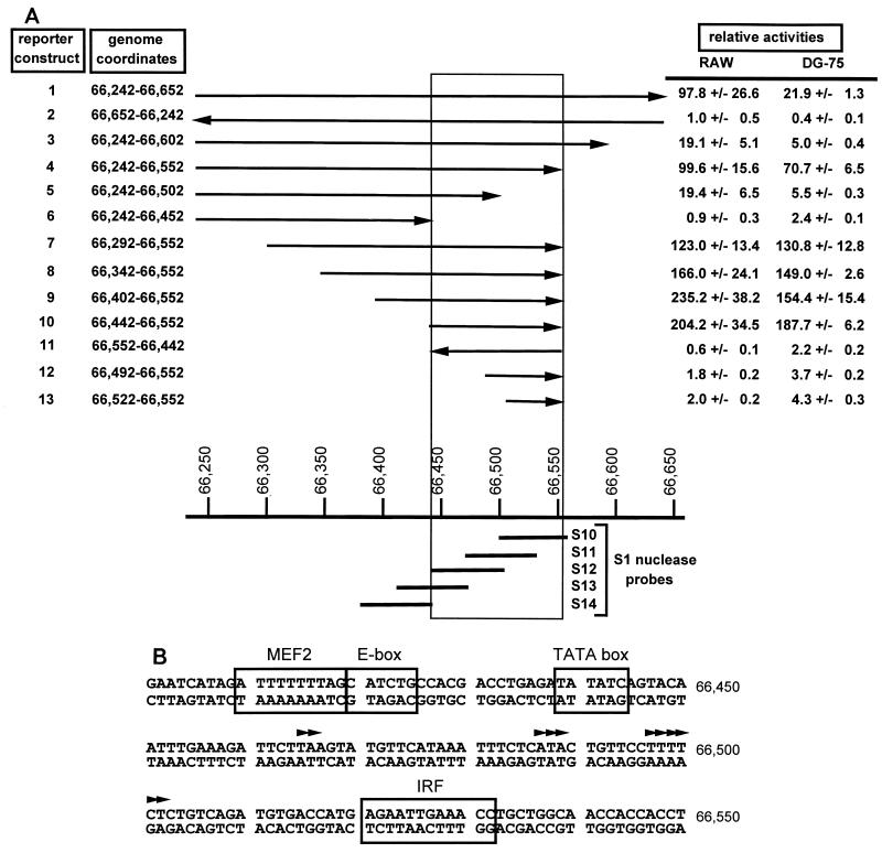
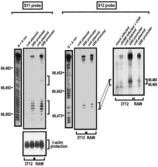
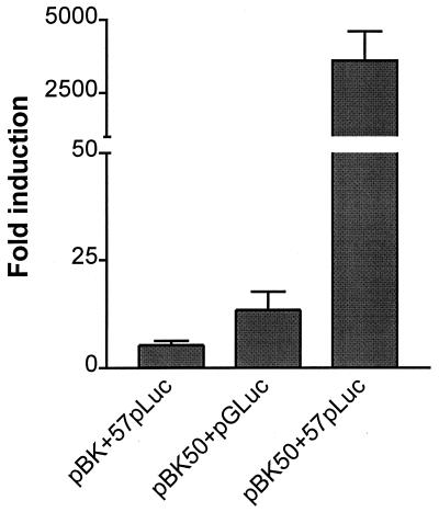
Gene 50 is the only immediate-early gene that appears to be conserved among the characterized gammaherpesviruses. It has recently been demonstrated for the human viruses Epstein-Barr virus (EBV) and Kaposi's sarcoma-associated herpesvirus (KSHV) that ectopic expression of the gene 50-encoded product in some latently infected cell lines can lead to the induction of virus replication, indicating that gene 50 is likely to play a pivotal role in regulating gammaherpesvirus reactivation. Here we demonstrate that the murine gammaherpesvirus 68 (gammaHV68) gene 50 is an immediate-early gene and that transcription of gammaHV68 gene 50 leads to the production of both spliced and unspliced forms of the gene 50 transcript. Splicing of the transcript near the 5' end serves to extend the gene 50 open reading frame, as has been observed for the gene 50 transcripts encoded by KSHV and herpesvirus saimiri (Whitehouse et al., J. Virol. 71:2550-2554, 1997; Lukac et al., Virology 252:304-312, 1998; Sun et al., Proc. Natl. Acad. Sci. USA 95:10866-10871, 1998). Reverse transcription-PCR analyses, coupled with S1 nuclease protection assays, provided evidence that gene 50 transcripts initiate at several sites within the region from bp 66468 to 66502 in the gammaHV68 genome. Functional characterization of the region upstream of the putative gene 50 transcription initiation site demonstrated orientation-dependent promoter activity and identified a 110-bp region (bp 66442 to 66552) encoding the putative gene 50 promoter. Finally, we demonstrate that the gammaHV68 gene 50 can transactivate the gammaHV68 gene 57 promoter, a known early gene target of the gene 50-encoded transactivator in other gammaherpesviruses. These studies show that the gammaHV68 gene 50 shares several important molecular similarities with the gene 50 homologs in other gammaherpesviruses and thus provides an impetus for future studies analyzing the role of the gammaHV68 gene 50-encoded protein in acute virus replication and reactivation from latency in vivo.


Figures

FIG. 1
FIG. 1
Northern blot analysis of gene 50-encoded transcripts. Ten micrograms of polyadenylated RNA prepared from NIH 3T12 fibroblasts infected with γHV68 at a multiplicity of infection of 7 for 8 h was loaded in each lane. Cells were either (i) mock infected, (ii) infected in the absence of any inhibitors, or (iii) infected in the presence of the protein synthesis inhibitors cycloheximide (final concentration, 40 μM) and anisomycin (final concentration, 10 μM) (γHV68 + CHX) in a volume of 2 ml of Dulbecco's modified Eagle medium containing 10% fetal calf serum for 1 h. After a 1-h incubation, an additional 15 ml of medium was added (with or without the indicated inhibitors), and the flasks were incubated at 37°C under a 5% CO2 atmosphere until harvesting. Infected cells were harvested 8 h postinfection, and total RNA was prepared as previously described (3). RNA blotting was carried out by fractionating RNA on 1.2% agarose gels containing 6.6% formaldehyde, 40 mM MOPS [3-(N-morpholino)propanesulfonic acid] (pH 7.0), 10 mM sodium acetate, and 1 mM EDTA, followed by capillary blotting in 20× SSC (1× SSC is 0.15 M NaCl and 0.015 M sodium citrate, pH 7.0) onto Hybond nylon membranes (Amersham Corp.). The RNA was covalently cross-linked to the nylon membrane by exposure to shortwave UV light, followed by hybridization with a 32P-labeled gene 50 probe overnight and washed under standard conditions (29). The upper left panel shows a 1-day exposure of the blot, while the upper right panel shows a 12-day exposure of the lane containing RNA isolated from cells infected in the presence of cycloheximide and anisomycin. The migration of molecular weight standards is shown to the right of the blots. The blot was stripped and rehybridized with a 32P-labeled rat cyclophilin probe (6) to assess RNA loading (lower panel). All probes were radiolabeled by the Megaprime DNA labeling system (Amersham, Arlington Heights, Ill.) in accordance with the manufacturer's protocol. The gene 50 fragment was generated by PCR with a sense primer which extended from bp 68660 to 66682 in the viral genome and an antisense primer which extended from bp 69176 to 69154 in the viral genome (see Fig. 2B for the location of probe relative to gene 50).
FIG. 2
FIG. 2
(A) Schematic illustration of the gene 50 region of the γHV68 genome (39). The locations of the putative viral genes adjacent to gene 50 are indicated, as are the locations of consensus poly(A) signals, which are indicated above (for those associated with the R strand of the viral genome) and below (for those associated with the L strand of the viral genome) the map of the viral genome. The asterisk denotes the poly(A) signal which was utilized in the transcript from which the cDNA clone 50-1 was generated. (B) Schematic structure of the cDNA clone 50-1, which is unspliced and contains viral sequences from bp 66642 to 69462. Below the schematic of the cDNA clone 50-1 is the deduced structure of the ∼2.0-kb spliced gene 50 transcript, based on RT-PCR analysis (see discussion in text). The small arrowheads below the spliced transcript denote PCR primers used to amplify the spliced form of the gene 50 cDNA. Also shown is the position of the gene 50 probe used in the Northern blot shown in Fig. 1. The solid black arrow indicates the gene 50 open reading frame, which is extended by the splice to the upstream exon, as illustrated in Fig. 3. The grey arrowheads indicate the presence of small (encoding >25 amino acids) open reading frames upstream of gene 50. (C) RT-PCR analysis of gene 50 transcripts. The structures and sizes of the RT-PCR products obtained employing the indicated PCR primers (arrowheads) are shown to the left of the ethidium bromide-stained agarose gel. The viral genomic coordinates (39) of the PCR primers employed for each reaction were as follows: PCR 1, upstream primer bp 68638 to 68660 and downstream primer bp 69176 to 69154; PCR 2, upstream primer bp 67386 to 67407 and downstream primer bp 68161 to 68141; PCR 3, upstream primer bp 66646 to 66667 and downstream primer bp 67385 to 67364; and PCR 4, upstream primer bp 66646 to 66667 and downstream primer bp 68161 to 68141. Also shown is an ethidium bromide-stained agarose gel of the PCR products generated with the primers indicated. A negative control RT-PCR, to detect the presence of contaminating viral genomic DNA in the RNA preparation, is shown (primers for PCR 2 were used to amplify DNA present in a cDNA synthesis reaction lacking reverse transcriptase [PCR 2, no RT]). All the PCR products visible by ethidium bromide staining were cloned and sequenced.
FIG. 3
FIG. 3
(A) Nucleotide and deduced protein sequences of gene 50 and its product (39). The deduced protein sequence is above the nucleotide sequence. The genome coordinates are to the right of the nucleotide sequence. The arrowheads above the 5′ untranslated sequence indicate candidate transcription initiation sites, determined by S1 nuclease protection analyses (see Fig. 5 and discussion in text). The nucleotide sequence shown in lowercase letters denotes the genomic sequence, based on nuclease protection analyses, upstream of the site of transcription initiation. The polyadenylation signal utilized in cDNA clone 50-1, located at bp 69434 in the viral genome, is boxed, as are the splice acceptor and donor sites. The spliced form of the gene 50 transcripts extends the open reading frame to encode an additional 94 amino acids (extended amino-terminal sequence is in shaded box above the nucleotide sequence). (B) Alignment of the predicted gene 50-encoded proteins of γHV68, HVS, KSHV, and EBV. The alignment was performed by Clustal analysis using MegAlign (DNASTAR) and is presented without further editing after the initial alignment. The first in-frame methionine encoded within the second exon of the spliced form of the gene 50 transcript is denoted by a shaded circle for the γHV68, HVS, and KSHV gene 50-encoded proteins.
FIG. 4
FIG. 4
(A) Identification of the gene 50 promoter. The structures of γHV68 genomic fragments used to map the gene 50 promoter are shown, along with the genomic map coordinates. All viral genomic fragments were cloned upstream of the luciferase reporter gene in the pGL2 Basic vector (Promega, Madison, Wis.). The arrows pointing right indicate the genomic fragments that were cloned in the sense orientation, with the luciferase gene downstream of the putative gene 50 promoter, while the arrows pointing left indicate the genomic fragments that were cloned in the opposite orientation. The indicated reporter constructs (2 μg) were transfected into both the murine macrophage cell line RAW (RAW 264.7) and the human EBV-negative Burkitt's lymphoma B cell line DG-75. RAW cells were transfected with the lipid-based transfection reagent SuperFect according to the manufacturer's protocol (Qiagen, Santa Clara, Calif.). DG-75 cells were transfected with DEAE-dextran, as previously described (28). Cells lysates were prepared 48 h posttransfection and assayed for luciferase activity (7). The data were compiled from three independent experiments for each cell line, and the standard error of the mean is shown. Also shown are the positions of the single-stranded probes used in the S1 nuclease protection analyses (see text for discussion and Fig. 5). The boxed region denotes those sequences which, based on this analysis, are required for gene 50 promoter activity. Relative luciferase activity is the fold increase (mean ± standard error of the mean) over that observed with the parent pGL2 Basic reporter construct (Promega), which was assigned a relative activity of 1.0. (B) Computer analysis of the minimal gene 50 promoter for the presence of transcription factor binding sites. The gene 50 promoter sequenced was analyzed by MatInspector Professional software (23). IRF, interferon response factor; MEF2, myocyte enhancer binding factor 2; E box, binding site for the E family of bHLH transcription factors.
FIG. 5
FIG. 5
Mapping of the 5′ ends of the gene 50 transcripts by S1 nuclease protection. Analysis of transcription initiation from the putative gene 50 promoter in transiently transfected NIH 3T12 or RAW cells. The bp 66242 to 66652 gene 50 promoter-driven luciferase reporter construct, or the parent pGL2 Basic reporter construct (Promega), was transfected into NIH 3T12 and RAW cells by the lipid-based transfection reagent SuperFect (Qiagen) as described in the legend to Fig. 4. RNA was prepared (3) from cells harvested 48 h posttransfection, and 40 μg of total RNA was hybridized with 4 ng of the indicated single-stranded oligonucleotide S1 nuclease probes, which had been 32P labeled at the 5′ end with T4 polynucleotide kinase (Boehringer Mannheim, Indianapolis, Ind.) (24, 46). The sequences of the probes used were as follows: S11, 665315′-GTTTCAATTCTCATGGTCACATCTGACAGAGAAAAGGAACAGTATGAGAAATTTATGAAC-3′66472; S12, 665015′-GAAAAGGAACAGTATGAGAAATTTATGAACATACTTAAGAATCTTTCAAATTGTACTGAT-3′66442 (see Fig. 4 for locations of the S11 and S12 probes). Following hybridization overnight at 42°C, the reaction mixture was digested with 300 U of S1 nuclease (Promega) as previously described (24, 46). The protected fragments were recovered and fractionated on a 10% denaturing acrylamide gel. Chemical cleavages of the 32P-labeled oligonucleotide probes (G + A rxn) were used as size markers in the indicated lanes. The right panel shows an analysis of transcription initiation from the gene 50 promoter in γHV68-infected NIH 3T12 cells. NIH 3T12 cells were either mock infected or infected at a multiplicity of infection of 7 in the presence of cycloheximide (final concentration, 40 μM) and anisomycin (final concentration, 10 μM) (+ CHX). Total cellular RNA was prepared (3) from cells harvested 8 h postinfection, followed by isolation of polyadenylated RNA with the PolyA Spin mRNA Isolation kit (New England Biolabs, Beverly, Mass.). Ten micrograms of poly(A) RNA isolated from mock-infected or γHV68-infected cells was hybridized with 32P-labeled S12 probe, followed by digestion with S1 nuclease as described above. In parallel, total RNA isolated from NIH 3T12 and RAW cells transfected with the bp 66242 to 66652 open reading frame 50 luciferase reporter construct was also hybridized with the S12 probe, as described above. Protected fragments were fractionated on a denaturing 10% acrylamide gel.
FIG. 6
FIG. 6
Transcriptional activation of the γHV68 gene 57 promoter by the γHV68 gene 50-encoded protein. A 565-bp fragment, spanning from bp 75218 to 75782 in the γHV68 genome and containing the gene 57 promoter (based on previous transcript mapping [20]), was cloned into pGL2 Basic (Promega). The 50-1 cDNA clone (see Fig. 2) was cloned into the NheI and KpnI sites of pBK-CMV (Stratagene). NIH 3T12 cells were cotransfected with 1 μg of either pGL2-Basic (pGLuc) or pGL2 Basic containing the gene 57 promoter (57pLuc) and 1 μg of either pBK-CMV (pBK) or pBK-CMV containing gene 50 (pBK50) with the lipid-based transfection reagent Superfect according to the manufacturer's protocol (Qiagen). Cell lysates were prepared 48 h posttransfection and assayed for luciferase activity (7). The data were compiled from four independent experiments, and the standard error of the mean is indicated.

Similar articles

Cited by

References

    1. Borras A M, Strominger J L, Speck S H. Characterization of the ZI domains in the Epstein-Barr virus BZLF1 gene promoter: role in phorbol ester induction. J Virol. 1996;70:3894–3901. - PMC - PubMed
    1. Buisson M, Manet E, Trescol-Biemont M C, Gruffat H, Durand B, Sergeant A. The Epstein-Barr virus (EBV) early protein EB2 is a posttranscriptional activator expressed under the control of EBV transcription factors EB1 and R. J Virol. 1989;63:5276–5284. - PMC - PubMed
    1. Chomczynski P, Sacchi N. Single-step method of RNA isolation by acid guanidinium thiocyanate-phenol-chloroform extraction. Anal Biochem. 1987;162:156–159. - PubMed
    1. Countryman J, Jenson H, Seibl R, Wolf H, Miller G. Polymorphic proteins encoded within BZLF1 of defective and standard Epstein-Barr viruses disrupt latency. J Virol. 1987;61:3672–3679. - PMC - PubMed
    1. Daibata M, Speck S H, Mulder C, Sairenji T. Regulation of the BZLF1 promoter of Epstein-Barr virus by second messengers in anti-immunoglobulin-treated B cells. Virology. 1994;198:446–454. - PubMed

Publication types

MeSH terms

LinkOut - more resources