

A preliminary gene map for the Van der Woude syndrome critical region derived from 900 kb of genomic sequence at 1q32-q41
- PMID: 10645953
- PMCID: PMC310500
A preliminary gene map for the Van der Woude syndrome critical region derived from 900 kb of genomic sequence at 1q32-q41
Abstract
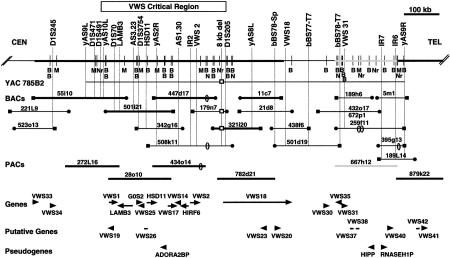
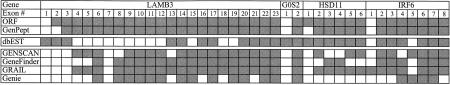
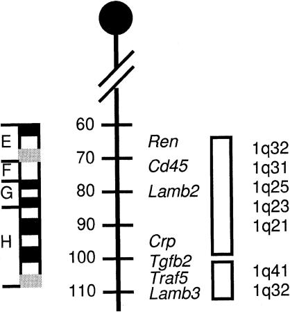
Van der Woude syndrome (VWS) is a common form of syndromic cleft lip and palate and accounts for approximately 2% of all cleft lip and palate cases. Distinguishing characteristics include cleft lip with or without cleft palate, isolated cleft palate, bilateral lip pits, hypodontia, normal intelligence, and an autosomal-dominant mode of transmission with a high degree of penetrance. Previously, the VWS locus was mapped to a 1.6-cM region in 1q32-q41 between D1S491 and D1S205, and a 4.4-Mb contig of YAC clones of this region was constructed. In the current investigation, gene-based and anonymous STSs were developed from the existing physical map and were then used to construct a contig of sequence-ready bacterial clones across the entire VWS critical region. All STSs and BAC clones were shared with the Sanger Centre, which developed a contig of PAC clones over the same region. A subset of 11 clones from both contigs was selected for high-throughput sequence analysis across the approximately 1.1-Mb region; all but two of these clones have been sequenced completely. Over 900 kb of genomic sequence, including the 350-kb VWS critical region, were analyzed and revealed novel polymorphisms, including an 8-kb deletion/insertion, and revealed 4 known genes, 11 novel genes, 9 putative genes, and 3 psuedogenes. The positional candidates LAMB3, G0S2, HIRF6, and HSD11 were excluded as the VWS gene by mutation analysis. A preliminary gene map for the VWS critical region is as follows: [see text] 41-TEL. The data provided here will help lead to the identification of the VWS gene, and this study provides a model for how laboratories that have a regional interest in the human genome can contribute to the sequencing efforts of the entire human genome.
Figures
References
-
- Aberdam D, Galliano MF, Mattei M-G, Pisani-Spadafora A, Ortonne JP, Meneguzzi G. Assignment of mouse nicein genes to chromosomes 1 and 18. Mamm Genome. 1994;5:229–233. - PubMed
-
- Adachi M, Sekiya M, Isobe M, Kumura Y, Ogita Z, Hinoda Y, Imai K, Yachi A. Molecular cloning and chromosomal mapping of a human protein-tyrosine phosphatase LC-PTP. Biochem Biophys Res Commun. 1992;186:1607–1615. - PubMed
-
- Altschul SF, Gish W, Miller W, Myers EW, Lipman DJ. Basic local alignment search tool. J Mol Biol. 1990;215:403–410. - PubMed
-
- Becker W, Joost HG. Structural and functional characteristics of Dyrk, a novel subfamily of protein kinases with dual specificity. Prog Nucleic Acid Res Mol Biol. 1999;62:1–17. - PubMed
Publication types
MeSH terms
Substances
Associated data
- Actions
- Actions
- Actions
- Actions
- Actions
- Actions
- Actions
- Actions
- Actions
- Actions
Grants and funding
LinkOut - more resources
Full Text Sources
Other Literature Sources
Medical
Molecular Biology Databases
Miscellaneous