. 2000 Feb;74(2):574-81.
doi: 10.1046/j.1471-4159.2000.740574.x.

Mu-opioid agonist inhibition of kappa-opioid receptor-stimulated extracellular signal-regulated kinase phosphorylation is dynamin-dependent in C6 glioma cells

Affiliations

Mu-opioid agonist inhibition of kappa-opioid receptor-stimulated extracellular signal-regulated kinase phosphorylation is dynamin-dependent in C6 glioma cells

L M Bohn et al. J Neurochem. 2000 Feb.

Abstract

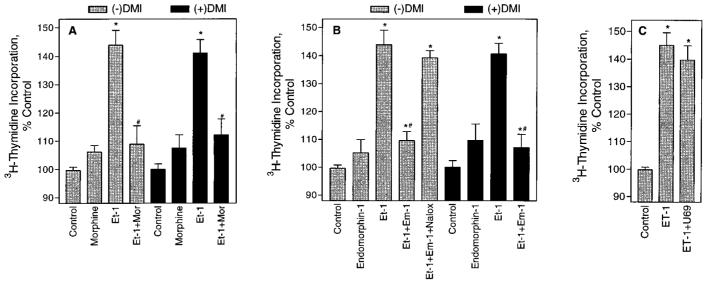
In previous studies we found that mu-opioids, acting via mu-opioid receptors, inhibit endothelin-stimulated C6 glioma cell growth. In the preceding article we show that the kappa-selective opioid agonist U69,593 acts as a mitogen with a potency similar to that of endothelin in the same astrocytic model system. Here we report that C6 cell treatment with mu-opioid agonists for 1 h results in the inhibition of kappa-opioid mitogenic signaling. The mu-selective agonist endomorphin-1 attenuates kappa-opioid-stimulated DNA synthesis, phosphoinositide turnover, and extracellular signal-regulated kinase phosphorylation. To investigate the role of receptor endocytosis in signaling, we have examined the effects of dynamin-1 and its GTPase-defective, dominant suppressor mutant (K44A) on opioid modulation of extracellular signal-regulated kinase phosphorylation in C6 cells. Overexpression of dynamin K44A in C6 cells does not affect kappa-opioid phosphorylation of extracellular signal-regulated kinase. However, it does block the inhibitory action on kappa-opioid signaling mediated by the kappa-opioid receptor. Our results are consistent with a growing body of evidence of the opposing actions of mu- and kappa-opioids and provide new insight into the role of opioid receptor trafficking in signaling.


Figures

FIG. 1
FIG. 1
μ-Opioids selectively inhibit endothelin-stimulated DNA synthesis. C6 cells were kept in “low MEM” for 48 h before treatment. For the final 20 h of this 48-h period, 5 μM DMI was included for some assays. Cells were treated with opioids for 1 h, and then endothelin-1 (Et-1) was added (in the presence of opioids). After 30 min, [3H]thymidine (0.5 μCi/ml) was included in the medium, and [3H]thymidine incorporation was determined after 23 h by scintillation counting. Data have been normalized to represent percent incorporation over unstimulated controls. A: Morphine (1 μM) inhibits ET-1 (30 nM) stimulated [3H]thymidine incorporation. *Significantly greater than control, #significantly less than ET-1, p < 0.01. Data are mean ± SEM (bars) values of three to six experiments performed in triplicate. B: Endomorphin-1 (Em-1; 1 μM) inhibits ET-1 (30 nM) stimulated [3H]thymidine incorporation. These effects are reversed by the opioid antagonist naloxone (Nalox; 1 μM, 1 h before Em-1). *Significantly greater than control, #significantly less than ET-1, p < 0.01. Data are mean ± SEM (bars) values of four to nine experiments performed in triplicate. C: The κ-OR-selective opioid U69,593 (U69; 1 μM) does not significantly affect ET-1-stimulated DNA synthesis. *Significantly greater than control, p < 0.01. Data are mean ± SEM (bars) values of five experiments performed in triplicate. Basal [3H]thymidine incorporation yielded an average of 15,970 ± 2,259 dpm per well for unstimulated controls and 8,982 ± 1,100 dpm per well for unstimulated, DMI-treated controls.
FIG. 2
FIG. 2
Endomorphins inhibit κ-opioid-stimulated DNA synthesis. C6 cells were kept in “low MEM” for 48 h before treatment. In this same medium, endomorphin-1 (Em-1) or -2 (Em-2; 10 nM) was added 1 h before U69,593 (U69; 1 μM) in assays where both agonists are present. After 30 min, [3H]thymidine (0.5 μCi/ml) was added to the medium, and [3H]thymidine incorporation was assayed after 23 h by scintillation counting. CTAP (1 μM) was added 1 h before addition of either Em-1 or -2. Data are average ± SEM (bars) values of three experiments performed in triplicate. *Significantly greater than control, #significantly less than U69, φsignificantly greater than U69 + Em-1 and U69 + Em-2, p < 0.01. Basal [3H]thymidine incorporation yielded an average of 14,370 ± 1,985 dpm per well.
FIG. 3
FIG. 3
Endomorphin-1 (Em-1) inhibits κ-opioid-stimulated PI turnover. C6 cells were kept in “low MEM” for 48 h and then labeled with 1.5 μCi of myo-[3H]inositol/ml per well in the same medium overnight. Medium was changed, and cells were incubated in “low MEM” plus 5 mM LiCl for 1 h before drug treatment. Em-1 (10 nM) was added 1 h before stimulation with U69,593 (U69; 1 μM). After a 10-min U69 treatment, cells were collected, and extracted 3H-IPx fractions were eluted from anion exchange columns and assayed by scintillation counting. *Significantly greater than control, p < 0.01; #significantly less than U69, p < 0.01; φsignificantly greater than U69 + Em-1, p < 0.05. Data are mean ± SEM (bars) values from three to seven experiments performed in triplicate. Basal 3H-IPx accumulation was measured as 35,920 ± 3,916 dpm per well.
FIG. 4
FIG. 4
Endomorphin-1 (Em-1) inhibits κ-opioid phosphorylation of ERK1/2. C6 cells were kept in “low MEM” for 48 h before addition of opioids. Em-1 (10 nM) was added 1 h before a 10-min stimulation with U69,593 (U69; 10 nM). CTAP (1 μM) was added 1 h before Em-1 where indicated. In some cases, cells were treated for 10 min or 1 h with Em-1 alone. Data are mean ± SEM (bars) values of three experiments. *Significantly greater than control, p < 0.001; #significantly less than U69, p < 0.001; φsignificantly greater than U69 + Em-1, p < 0.01. Also shown is a representative membrane, blotted first with anti-phospho(P)ERK1/2 (top) and then stripped and reblotted with anti-ERK1 (bottom).
FIG. 5
FIG. 5
Time dependence for endomorphin-1 (Em-1) inhibition of U69,593 (U69)-stimulated ERK1/2 phosphorylation. C6 cells were kept in “low MEM” for 48 h before addition of opioids. Em-1 (10 nM) was added at the times indicated before a 10-min stimulation with U69 (10 nM). Data are mean ± SEM (bars) values of three or four experiments. *Significantly greater than control, p < 0.005; #significantly less than U69 ( p < 0.01) and U69 + Em-1 [0 ( p < 0.01), 10, and 30 min ( p < 0.05)]. Also shown is a representative membrane, blotted first with antiphospho(P)ERK1/2 (top) and then stripped and reblotted with anti-ERK1 (bottom).
FIG. 6
FIG. 6
Dynamin K44A mutant has no effect on κ-OR-mediated phosphorylation of ERK1/2, yet blocks the inhibitory effects of endomorphin-1 (Em-1). C6 cells were transiently transfected with 1 μg of dynamin (Dyn; left panels) or dynamin K44A (K44A; right panels) cDNA and then maintained in “low MEM” for 48 h before treatment. Em-1 (10 nM) was added 1 h before the 10-min U69,593 (U69; 10 nM) stimulation. Data are mean ± SEM (bars) values of four to seven experiments. *Significantly greater than control, #significantly less than U69, p < 0.001. Also shown are representative membranes, blotted first with anti-phospho(P)ERK1/2 (top) and then stripped and reblotted with anti-ERK1 (bottom).

References

    1. Ahn S, Maudsley S, Luttrel LM, Lefkowitz RJ, Daaka Y. Src-mediated tyrosine phosphorylation of dynamin is required for β2-adrenergic receptor internalization and mitogen-activated protein kinase signaling. J. Biol. Chem. 1999;274:1185–1188. - PubMed
    1. Albouz S, Tocque B, Hauw JJ, Boutry JM, LeSaux F, Bourdoin R, Baumann N. Tricyclic antidepressant desipramine induces stereospecific opiate binding and lipid modification in rat glioma C6 cells. Life Sci. 1982;31:2549–2554. - PubMed
    1. Barg J, Belcheva M, Bem WT, Lambourne B, McLachlan JA, Tolman KC, Johnson FE, Coscia CJ. Desipramine modulation of sigma and opioid peptide receptor expression in glial cells. Peptides. 1991;12:845–849. - PubMed
    1. Barg J, Belcheva MM, Coscia CJ. Evidence for the implication of phosphoinositol signal transduction in μ-opioid inhibition of DNA synthesis. J. Neurochem. 1992;59:1145–1152. - PMC - PubMed
    1. Barg J, Belcheva MM, Rowiński J, Coscia CJ. κ-Opioid agonist modulation of [3H]thymidine incorporation into DNA: evidence for the involvement of pertussis toxin-sensitive G protein-coupled phosphoinositide turnover. J. Neurochem. 1993a;60:1505–1511. - PMC - PubMed

Publication types

MeSH terms