

Cloning, expression, and purification of a thermostable nonhomodimeric restriction enzyme, BslI
- PMID: 10648519
- PMCID: PMC94369
- DOI: 10.1128/JB.182.4.949-955.2000
Cloning, expression, and purification of a thermostable nonhomodimeric restriction enzyme, BslI
Abstract
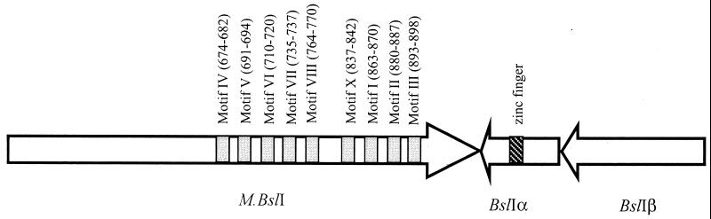
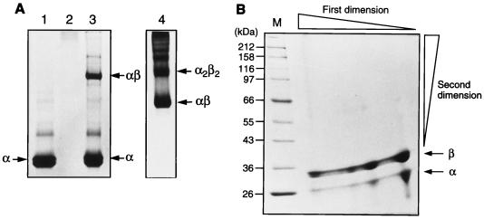
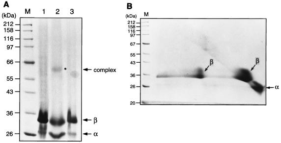
BslI is a thermostable type II restriction endonuclease with interrupted recognition sequence CCNNNNN/NNGG (/, cleavage position). The BslI restriction-modification system from Bacillus species was cloned and expressed in Escherichia coli. The system is encoded by three genes: the 2,739-bp BslI methylase gene (bslIM), the bslIRalpha gene, and the bslIRbeta gene. The alpha and beta subunits of BslI can be expressed independently in E. coli in the absence of BslI methylase (M.BslI) protection. BslI endonuclease activity can be reconstituted in vitro by mixing the two subunits together. Gel filtration chromatography and native polyacrylamide gel electrophoresis indicated that BslI forms heterodimers (alphabeta), heterotetramers (alpha(2)beta(2)), and possibly oligomers in solution. Two beta subunits can be cross-linked by a chemical cross-linking agent, indicating formation of heterotetramer BslI complex (alpha(2)beta(2)). In DNA mobility shift assays, neither subunit alone can bind DNA. DNA mobility shift activity was detected after mixing the two subunits together. Because of the symmetric recognition sequence of the BslI endonuclease, we propose that its active form is alpha(2)beta(2). M.BslI contains nine conserved motifs of N-4 cytosine DNA methylases within the beta group of aminomethyltransferase. Synthetic duplex deoxyoligonucleotides containing cytosine hemimethylated or fully methylated at N-4 in BslI sites in the first or second cytosine are resistant to BslI digestion. C-5 methylation of the second cytosine on both strands within the recognition sequence also renders the site refractory to BslI digestion. Two putative zinc fingers are found in the alpha subunit of BslI endonuclease.
Figures
Similar articles
-
Glucocorticoid receptor-like Zn(Cys)4 motifs in BslI restriction endonuclease.J Mol Biol. 2003 Nov 28;334(3):595-603. doi: 10.1016/j.jmb.2003.09.043. J Mol Biol. 2003. PMID: 14623197
-
Cloning and nucleotide sequences of the BanI restriction-modification genes in Bacillus aneurinolyticus.J Biochem. 1990 Apr;107(4):645-9. doi: 10.1093/oxfordjournals.jbchem.a123101. J Biochem. 1990. PMID: 2358438
-
Cloning of the BssHII restriction-modification system in Escherichia coli : BssHII methyltransferase contains circularly permuted cytosine-5 methyltransferase motifs.Nucleic Acids Res. 1997 Oct 15;25(20):3991-4. doi: 10.1093/nar/25.20.3991. Nucleic Acids Res. 1997. PMID: 9321648 Free PMC article.
-
Characterization of an extremely thermostable restriction enzyme, PspGI, from a Pyrococcus strain and cloning of the PspGI restriction-modification system in Escherichia coli.Appl Environ Microbiol. 1998 Oct;64(10):3669-73. doi: 10.1128/AEM.64.10.3669-3673.1998. Appl Environ Microbiol. 1998. PMID: 9758783 Free PMC article.
-
The Pvu II restriction-modification system: cloning, characterization and use in revealing an E. coli barrier to certain methylases or methylated DNAs.Gene Amplif Anal. 1987;5:227-45. Gene Amplif Anal. 1987. PMID: 3333367 Review. No abstract available.
Cited by
-
Restriction endonucleases: classification, properties, and applications.Mol Biotechnol. 2003 Mar;23(3):225-43. doi: 10.1385/mb:23:3:225. Mol Biotechnol. 2003. PMID: 12665693 Review.
-
Pullulanase and Starch Synthase III Are Associated with Formation of Vitreous Endosperm in Quality Protein Maize.PLoS One. 2015 Jun 26;10(6):e0130856. doi: 10.1371/journal.pone.0130856. eCollection 2015. PLoS One. 2015. PMID: 26115014 Free PMC article.
-
Analysis of the zinc finger domain of TnpA, a DNA targeting protein encoded by mobilizable transposon Tn4555.Plasmid. 2007 Jul;58(1):23-30. doi: 10.1016/j.plasmid.2006.11.005. Epub 2007 Jan 3. Plasmid. 2007. PMID: 17204325 Free PMC article.
-
Structure and function of type II restriction endonucleases.Nucleic Acids Res. 2001 Sep 15;29(18):3705-27. doi: 10.1093/nar/29.18.3705. Nucleic Acids Res. 2001. PMID: 11557805 Free PMC article. Review.
-
A genetic dissection of the LlaJI restriction cassette reveals insights on a novel bacteriophage resistance system.BMC Microbiol. 2006 Apr 28;6:40. doi: 10.1186/1471-2180-6-40. BMC Microbiol. 2006. PMID: 16646963 Free PMC article.
References
-
- Athanasiadis A, Blassi M, Kotsifaki D, Tucker P A, Wilson K S, Kokkinidis M. Crystal structure of PvuII endonuclease reveals extensive structural homologies to EcoRV. Nat Struct Biol. 1994;1:469–475. - PubMed
-
- Bozic D, Grazulis S, Siksnys V, Huber R. Crystal structure of Citrobacter freundii restriction endonuclease Cfr10I at 2.15 Å resolution. J Mol Biol. 1996;255:176–186. - PubMed
-
- Chong S, Mersha F B, Comb D G, Scott M E, Landry D, Vence L M, Perler F B, Benner J, Kucera R B, Hirvonen C A. Single-column purification of free recombinant proteins using a self-cleavable affinity tag derived from a protein splicing element. Gene. 1997;192:271–281. - PubMed
-
- Dreyer S D, Zhou L, Machado M A, Horton W A, Zabel B, Winterpacht A, Lee B. Cloning, characterization and chromosomal assignment of the human ortholog of murine Zfp-37, a candidate gene for Nager syndrome. Mamm Genome. 1998;9:458–462. - PubMed
MeSH terms
Substances
Associated data
- Actions
LinkOut - more resources
Full Text Sources
Other Literature Sources
Molecular Biology Databases
Miscellaneous
