

Visualization of phospholipid domains in Escherichia coli by using the cardiolipin-specific fluorescent dye 10-N-nonyl acridine orange
- PMID: 10648548
- PMCID: PMC94398
- DOI: 10.1128/JB.182.4.1172-1175.2000
Visualization of phospholipid domains in Escherichia coli by using the cardiolipin-specific fluorescent dye 10-N-nonyl acridine orange
Abstract
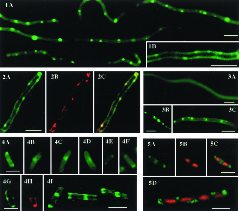
Cardiolipin (CL)-specific fluorescent dye 10-N-nonyl-acridine orange (NAO) was used to visualize CL distribution in Escherichia coli cells of different phospholipid compositions. In a filamentous mutant containing only anionic phospholipids, green fluorescent spots were observed along the filaments at approximately regular intervals. Three-dimensional image reconstruction obtained by optical sectioning and a deconvolution algorithm revealed NAO-binding domains in the plane of the cell membrane. Substantial red fluorescence emission of bound NAO supported labeling of CL-containing domains. These structures were not found in mutants deficient in CL biosynthesis. The domains were also observed mostly in the septal region and on the poles in cells of normal size with wild-type phospholipid composition.
Figures
References
-
- Bergelson L. Special issue on domain organization in biological membranes. Introductory remarks. Mol Membr Biol. 1995;12:3.
-
- Brasitus T A, Tall A R, Schachter D. Thermotropic transitions in rat intestinal plasma membranes studied by differential scanning calorimetry and fluorescence polarization. Biochemistry. 1980;19:1256–1261. - PubMed
-
- Chang S-C, Heacock P N, Mileykovskaya E, Voelker D R, Dowhan W. Isolation and characterization of the gene (CLS1) encoding cardiolipin synthase in Saccharomyces cerevisiae. J Biol Chem. 1998;273:14933–14941. - PubMed
-
- Christensen H, Garton N J, Horobin R W, Minnikin D E, Barer M R. Lipid domains of mycobacteria studied with fluorescent molecular probes. Mol Microbiol. 1999;31:1561–1572. - PubMed
-
- DeChavigny A, Heacock P N, Dowhan W. Sequence and inactivation of the pss gene of Escherichia coli. Phosphatidylethanolamine may not be essential for cell viability. J Biol Chem. 1991;266:5323–5332. - PubMed
Publication types
MeSH terms
Substances
Grants and funding
LinkOut - more resources
Full Text Sources
