. 2000 Jan 24;148(2):239-47.
doi: 10.1083/jcb.148.2.239.

Nuclear eukaryotic initiation factor 4E (eIF4E) colocalizes with splicing factors in speckles

Affiliations

Nuclear eukaryotic initiation factor 4E (eIF4E) colocalizes with splicing factors in speckles

J Dostie et al. J Cell Biol. .

Abstract

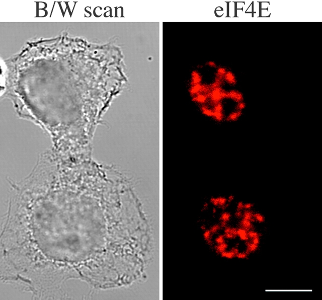
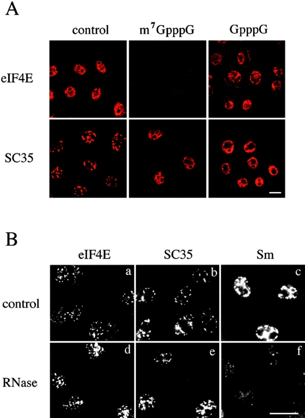
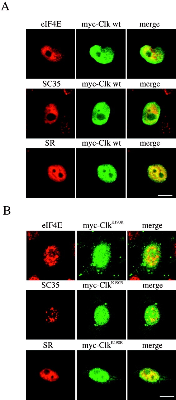
The eukaryotic initiation factor 4E (eIF4E) plays a pivotal role in the control of protein synthesis. eIF4E binds to the mRNA 5' cap structure, m(7)GpppN (where N is any nucleotide) and promotes ribosome binding to the mRNA. It was previously shown that a fraction of eIF4E localizes to the nucleus (Lejbkowicz, F., C. Goyer, A. Darveau, S. Neron, R. Lemieux, and N. Sonenberg. 1992. Proc. Natl. Acad. Sci. USA. 89:9612-9616). Here, we show that the nuclear eIF4E is present throughout the nucleoplasm, but is concentrated in speckled regions. Double label immunofluorescence confocal microscopy shows that eIF4E colocalizes with Sm and U1snRNP. We also demonstrate that eIF4E is specifically released from the speckles by the cap analogue m(7)GpppG in a cell permeabilization assay. However, eIF4E is not released from the speckles by RNase A treatment, suggesting that retention of eIF4E in the speckles is not RNA-mediated. 5,6-dichloro-1-beta-d-ribofuranosylbenzimidazole (DRB) treatment of cells causes the condensation of eIF4E nuclear speckles. In addition, overexpression of the dual specificity kinase, Clk/Sty, but not of the catalytically inactive form, results in the dispersion of eIF4E nuclear speckles.


Figures

Figure 1
Figure 1
Nuclear eIF4E localizes to speckles. The localization of endogenous eIF4E was determined by indirect immunofluorescence with a monoclonal anti-mouse eIF4E antibody, 10C6, and Texas red-conjugated secondary antibody. HeLa cells were fixed and processed for immunofluorescence as described in Materials and Methods. Images were taken from a 63× objective of a Zeiss LSM 410 microscope and analyzed with a Kontron IBAS confocal imaging system. Bar, 10 μm.
Figure 2
Figure 2
eIF4E colocalizes with Sm and U1snRNP antigens to speckled regions in the nucleus. A, a–c, SC35 colocalizes with Sm. The localization of endogenous SC35 was determined by indirect immunofluorescence with a monoclonal anti-mouse SC35 antibody and Texas red-conjugated anti-mouse IgG (a and c), and that of Sm with a human anti-Sm sera and fluorescein-conjugated anti-human IgG (b and c). d–f, SC35 colocalizes with U1snRNP. Same as in a–c, except that a human anti-U1snRNP sera was used in e and f. Bar, 5 μm. B, a–c, eIF4E colocalizes with Sm. The localization of eIF4E was determined by indirect immunofluorescence with the monoclonal anti-mouse eIF4E antibody 10C6 and Texas red-conjugated anti-mouse IgG (a and c), and that of Sm with a human anti-Sm sera and fluorescein-conjugated anti-human IgG (b and c). d–f, Same as in a–c, except that a human anti-U1snRNP sera was used in e and f. Bar, 10 μm. In c and f (in both A and B), the colocalization appears yellow. CV-1 monkey kidney cells were prepared as described in Materials and Methods. Images were taken from a 63× objective of a BioRad MRC-600 confocal imaging system mounted on a Nikon Diaphot-TMD microscope as previously described ( Lejbkowicz et al. 1992).
Figure 3
Figure 3
Effect of transcription inhibition on the localization of eIF4E. HeLa cells were incubated with buffer alone (a, e, and i), cycloheximide (b, f, and j), DRB (c, g, and k), or in the presence of cycloheximide and DRB (d, h, and l) for 3 h. Cells were fixed and the localization of eIF4E, SC35, and Sm was determined by indirect immunofluorescence with the anti-eIF4E mAb 10C6 (top), anti-mouse SC35 mAb (middle), and with a human anti-Sm antisera (bottom). Primary antibodies were detected with Texas red-conjugated secondary antibodies and analyzed with a Kontron IBAS confocal imaging system. Images were taken from a 63× objective of a Zeiss LSM 410 microscope. Bar, 10 μm.
Figure 4
Figure 4
Nuclear eIF4E is released from the speckles by the monomethylated cap structure, but not by RNase treatment. A, HeLa cells were permeabilized with digitonin and incubated with buffer alone (left), m7GpppG (middle), or GpppG (right) for 25 min. The cells were rinsed and fixed as described in Materials and Methods. The localization of eIF4E (top) and SC35 (bottom) was determined by indirect immunofluorescence with the anti-eIF4E mAb 10C6 and monoclonal anti-SC35, respectively. Primary antibodies were detected with Texas red-conjugated mouse secondary antibodies and analyzed with a Kontron IBAS confocal imaging system. Images were taken from a 63× objective of a Zeiss LSM 410 microscope. Bar, 10 μm. B, CV-1 cells were fixed with methanol and incubated with PBS (a–c) or 100 μg/ml RNase A. The localization of eIF4E (a and d), SC35 (b and e), and Sm (c and f) was determined by indirect immunofluorescence with the anti-eIF4E mAb 10C6, monoclonal anti-mouse SC35, and human anti-Sm sera, respectively. Primary antibodies were detected with Texas red-conjugated mouse secondary antibodies. Images were taken from a 63× objective of a BioRad MRC-600 confocal imaging system mounted on a Nikon Diaphot-TMD microscope as previously described ( Lejbkowicz et al. 1992). Bar, 10 μm.
Figure 5
Figure 5
The nuclear localization of eIF4E is regulated by Clk/Sty. HeLa cells were transiently transfected with myc-Clk/Sty wild-type (A) or myc-Clk/StyK190R (B) for 24 h. The localization of endogenous eIF4E and SC35, or SR splicing factors was determined with mAbs 10C6, anti-SC35, and anti-SR 104, respectively, and revealed with Texas red-conjugated secondary antibody. The distribution of transiently transfected Clk/Sty was determined with a rabbit anti-myc antibody and fluorescein-conjugated secondary antibody. Images were taken from a 63× objective of a Zeiss LSM 410 microscope and analyzed with a Kontron IBAS confocal imaging system. Bar, 10 μm.

References

    1. Adam S.A., Marr R.S., Gerace L. Nuclear protein import in permeabilized mammalian cells requires soluble cytoplasmic factors. J. Cell Biol. 1990;111:807–816 . - PMC - PubMed
    1. Beyer A.L., Osheim Y.N. Splice site selection, rate of splicing, and alternative splicing on nascent transcripts. Genes Dev. 1988;2:754–765 . - PubMed
    1. Bregman D.B., Du L., van der Zee S., Warren S.L. Transcription-dependent redistribution of the large subunit of RNA polymerase II to discrete nuclear domains. J. Cell Biol. 1995;129:287–298 . - PMC - PubMed
    1. Bringmann P., Rinke J., Appel B., Reuter R., Luhrmann R. Purification of snRNPs U1, U2, U4, U5 and U6 with 2,2,7-trimethylguanosine-specific antibody and definition of their constituent proteins reacting with anti-Sm and anti-(U1)RNP antisera. EMBO (Eur. Mol. Biol. Organ.) J. 1983;2:1129–1135 . - PMC - PubMed
    1. Caceres J.F., Screaton G.R., Krainer A.R. A specific subset of SR proteins shuttles continuously between the nucleus and the cytoplasm. Genes Dev. 1998;12:55–66 . - PMC - PubMed

Publication types

MeSH terms