Comparative Study
. 2000 Jan 24;148(2):375-84.
doi: 10.1083/jcb.148.2.375.

Caenorhabditis elegans UNC-45 is a component of muscle thick filaments and colocalizes with myosin heavy chain B, but not myosin heavy chain A

Affiliations
Comparative Study

Caenorhabditis elegans UNC-45 is a component of muscle thick filaments and colocalizes with myosin heavy chain B, but not myosin heavy chain A

W Ao et al. J Cell Biol. .

Abstract

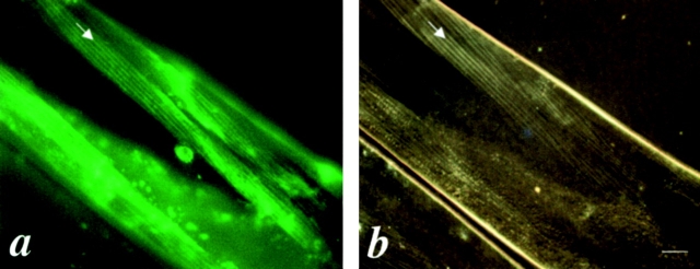
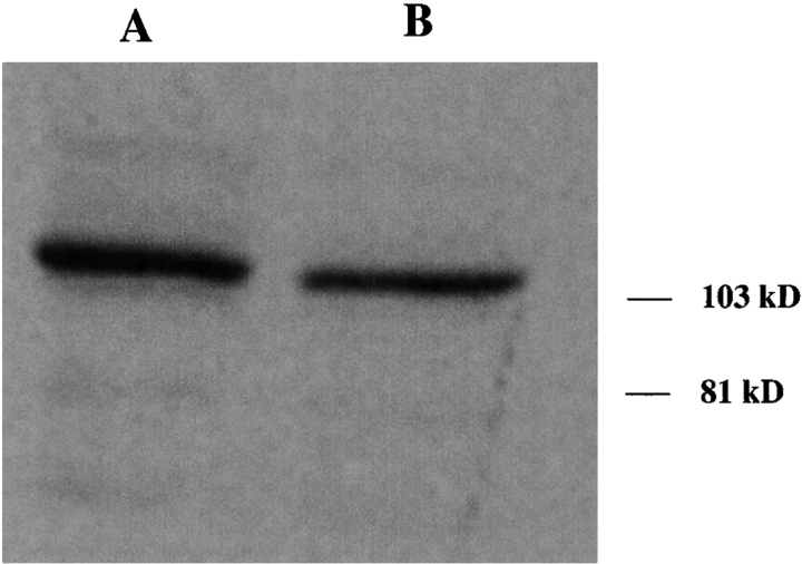
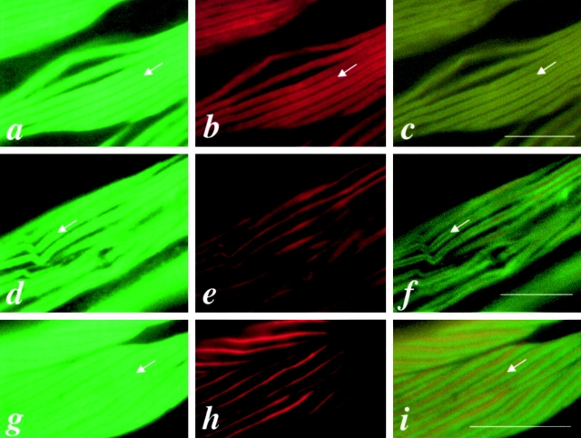
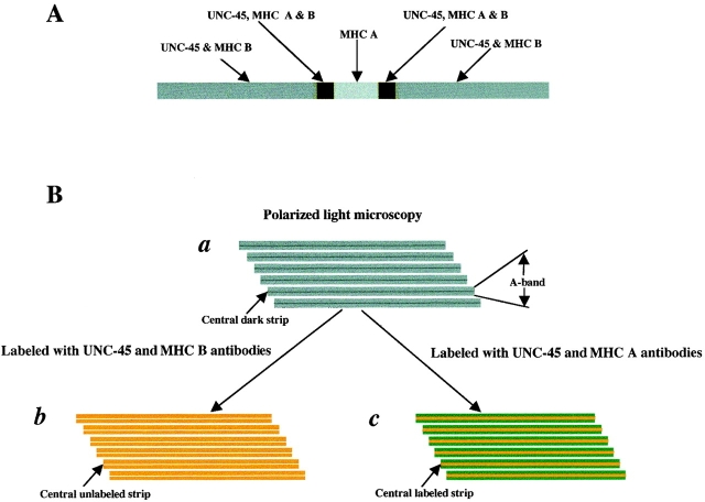
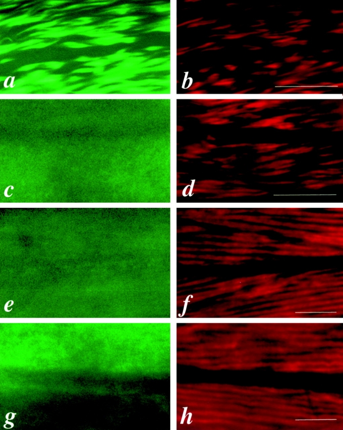
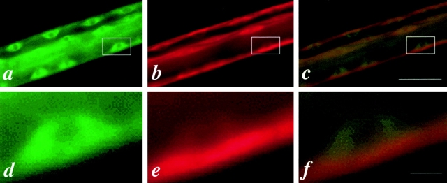
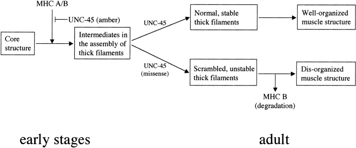
In the nematode Caenorhabditis elegans, animals mutant in the gene encoding the protein product of the unc-45 gene (UNC-45) have disorganized muscle thick filaments in body wall muscles. Although UNC-45 contains tetratricopeptide repeats (TPR) as well as limited similarity to fungal proteins, no biochemical role has yet been found. UNC-45 reporters are expressed exclusively in muscle cells, and a functional reporter fusion is localized in the body wall muscles in a pattern identical to thick filament A-bands. UNC-45 colocalizes with myosin heavy chain (MHC) B in wild-type worms as well as in temperature-sensitive (ts) unc-45 mutants, but not in a mutant in which MHC B is absent. Surprisingly, UNC-45 localization is also not seen in MHC B mutants, in which the level of MHC A is increased, resulting in near-normal muscle thick filament structure. Thus, filament assembly can be independent of UNC-45. UNC-45 shows a localization pattern identical to and dependent on MHC B and a function that appears to be MHC B-dependent. We propose that UNC-45 is a peripheral component of muscle thick filaments due to its localization with MHC B. The role of UNC-45 in thick filament assembly seems restricted to a cofactor for assembly or stabilization of MHC B.


Figures

Figure 1
Figure 1
Comparison of the functional UNC-45::GFP protein distribution and the muscle filament pattern under polarized light microscopy in the body wall muscles of the rescued unc-45 (r450). a is the GFP signal associated with the UNC-45::GFP fusion protein. b is the same field showing the muscle filament pattern as visualized by polarized light microscopy. The arrows indicate the thick filaments containing A-bands. Bar, 10 μm.
Figure 2
Figure 2
The 7N5 antiserum generated against an NH2-terminal fragment of UNC-45 reacts specifically with a polypeptide of about 105 K (MWr) on Western blots of wild-type worms. The polypeptide detected is consistent with the predicted size of UNC-45 (∼107 kD). The same amount of protein extract was used for each lane. The 7N5 antiserum was used in dilutions of 1:5,000 and 1:10,000, respectively, in lanes A and B. The preimmune serum used at 1:5,000 dilution did not show any specific reaction on Western blots (data not shown).
Figure 3
Figure 3
UNC-45 colocalizes with MHC B in the thick filaments of body wall muscles of wild-type adult worms. a, b, and c are the same field of body wall muscle labeled with anti–UNC-45 (7N5), anti–MHC B (DM 5-8), or double-labeled with 7N5 and DM 5-8, respectively. d, e, and f are the same field of body wall muscle labeled with 7N5, anti–MHC A (DM 5-6), or double-labeled with 7N5 and DM 5-6, respectively. g, h, and i are the same field of body wall muscle labeled with DM 5-8, DM 5-6, or double-labeled with DM 5-8 and DM 5-6, respectively. Note that DM 5-8 reacts with MHC B, which is localized on the two polar regions of the A-bands, and DM 5-6 reacts with MHC A, which is localized in the middle of the A-bands. c shows the overlapped pattern of UNC-45 and MHC B at the two polar regions of the thick filaments. f and i show a similar pattern with a central labeled strip when double-labeled with UNC-45 and DM 5-6, or DM 5-8 and DM 5-6, respectively. The arrows indicate the central unlabeled strip for UNC-45 (a and d) and for MHC B (b and g), the central unlabeled strip for double-staining (c) as shown in Fig. 4 B, panel b, and the central labeled strip for double-staining (f and i) in Fig. 4 B, panel c. Bars, 10 μm.
Figure 4
Figure 4
Schematic diagram of muscle thick filaments. (A) Thick filament, depicting the localization of UNC-45, MHC A, and B. (B, panel a) Schematic view of thick filaments as visualized by polarized light microscopy, showing A-band and the central dark strip in the A-band that corresponds to the region containing only the thick filaments but not thin filaments. (B, panel b) Schematic view of thick filaments when labeled with both UNC-45 (green) and MHC B (red) antibodies as shown in Fig. 3 c. The color appears as light yellow, since the UNC-45 and MHC B are completely overlapped. The central unlabeled strip corresponds to the region containing only MHC A, but not MHC B or UNC-45. (B, panel c) Schematic view of thick filaments when labeled with both UNC-45 (green) and MHC A (red) antibodies as shown in Fig. 3 f. The central labeled strip corresponds to the whole region containing MHC A and appears as dark yellow due to the overlapping parts of MHC A and UNC-45 and the central part containing only MHC A.
Figure 6
Figure 6
Motility assay for RW2329 (unc-54 [e190]; sup-3 [e1407st90st92]) and RW2665 (unc-54 [e190]; unc-45 [m94]; sup-3 [e1407st90st92]) mutants. a, b, c, and d are plates showing the traces of worms after crawling for 1 h at 25°C for RW2665 (unc-54 [e190]; unc-45 [m94]; sup-3 [e1407st90st92]), RW2329 (unc-54 [e190]; sup-3 [e1407st90st92]), N2, and unc-45 (m94), respectively. The dots in the centers of the plates are the start points and the others are the end points. N2 and unc-45 (m94) were used as controls. Five young adults were assayed for each strain and all showed similar motility. There is no apparent difference for the motility between RW2329 and RW2665.
Figure 5
Figure 5
UNC-45 and MHC A staining in mutant worms. a and b are the same field of body wall muscle from unc-45 (r450) mutant animals grown at the restrictive temperature (22°C) labeled with 7N5 and DM 5-6, respectively. c and d are the same field of body wall muscle from unc-54 (e190) mutants labeled with 7N5 and DM 5-6, respectively. e and f are the same field of body wall muscle from unc-54 (e190); sup-3 (e1407st90st92) mutants labeled with 7N5 and DM 5-6, respectively. g and h are the same field of body muscle from unc-54 (e190); unc-45 (m94); sup-3 (e1407st90st92) mutants labeled with 7N5 and DM 5-6, respectively. Note that UNC-45 is localized to thick filaments in unc-45 (ts) mutants (a), but not in unc-54 (0) mutants (c) in which MHC B is absent and unc-54 (0); sup-3 mutants (e and g) in which MHC B is absent, but the level of MHC A is increased. Bars, 10 μm.
Figure 7
Figure 7
UNC-45 may be added into thick filaments after MHC isoforms. a, b, and c are the same part of a wild-type worm at the L1 larval stage labeled with 7N5, DM 5-8, or double-labeled with 7N5 and DM 5-8, respectively. d, e, and f are enlarged for the boxed areas of a, b, and c, respectively, showing one muscle cell. Note that UNC-45 is diffuse in the cytoplasm (d), whereas MHC B has been assembled into thick filaments near the cell membrane along the face of the muscle cell that is adjacent to the hypodermis (e). Bars, 10 μm.
Figure 8
Figure 8
A model for UNC-45 function. UNC-45 is required for the initiation of thick filament assembly in the early stages of development. It may function as a catalyst to modify MHC B (and/or MHC A) before assembly or directly participate in the assembly itself. In the case of UNC-45 amber mutations, UNC-45 cannot function properly and causes embryonic lethality. Later, UNC-45 is localized to thick filaments in the same patterns as MHC B. In the adult, UNC-45 normally functions as a stabilizer for MHC B. In animals carrying UNC-45 missense mutations in the CRO1/SHE4 domain at the restrictive temperature, MHC B turnover is increased, either due to inherent MHC B instability on the thick filament or an inability to properly assemble MHC B into the thick filament, followed by increased turnover of unassembled MHC B.

References

    1. Ardizzi J.P., Epstein H.F. Immunochemical localization of myosin heavy chain isoforms and paramyosin in developmentally and structurally diverse muscle cell types of the nematode C. elegans . J. Cell Biol. 1987;105:2763–2770 . - PMC - PubMed
    1. Ausubel, F.M., R. Brent, R.E. Kingston, D.D. Moore, J.A. Smith, J.G. Seidman, and K. Struh, editors. 1991. Current Protocols in Molecular Biology. John Wiley and Sons, Inc., New York. 1,600 pp.
    1. Barral J.M., Bauer C.C., Ortiz I., Epstein H.F. Unc-45 mutations in Caenorhabditis elegans implicates a CRO1/She4p-like domain in myosin assembly. J. Cell Biol. 1998;143:1215–1225 . - PMC - PubMed
    1. Bejsovec A., Anderson R.P. Myosin heavy chain mutations that disrupt C. elegans thick filament assembly. Genes Dev. 1988;2:1307–1317 . - PubMed
    1. Berteaux-Lecellier V., Zickler D., Debuchy R., Panvier-Adoutte A., Thompson-Coffe C., Picard M. A homologue of the yeast SHE4 gene is essential for the transition between the syncytial and cellular stages during sexual reproduction of the fungus Podospora anserina . EMBO (Eur. Mol. Biol. Organ.) J. 1998;17:1248–1258 . - PMC - PubMed

Publication types

MeSH terms