. 2000 Jan 24;148(2):385-97.
doi: 10.1083/jcb.148.2.385.

ErbB2 is necessary for induction of carcinoma cell invasion by ErbB family receptor tyrosine kinases

Affiliations

ErbB2 is necessary for induction of carcinoma cell invasion by ErbB family receptor tyrosine kinases

K S Spencer et al. J Cell Biol. .

Abstract

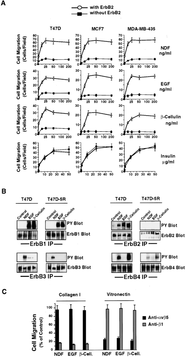
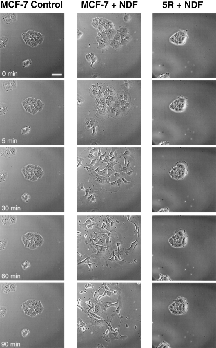
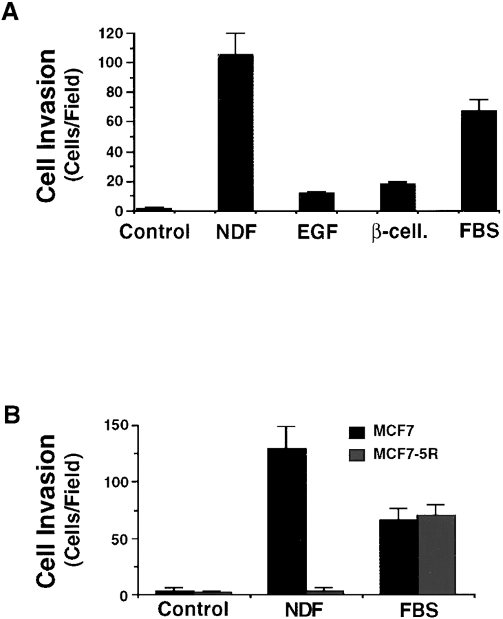
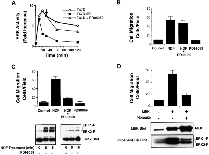
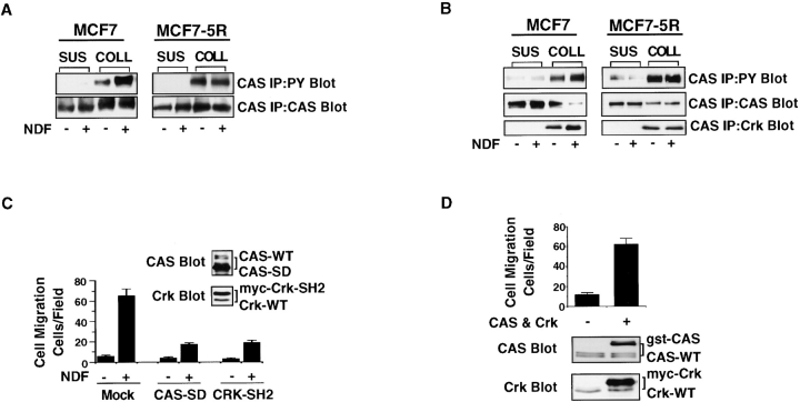
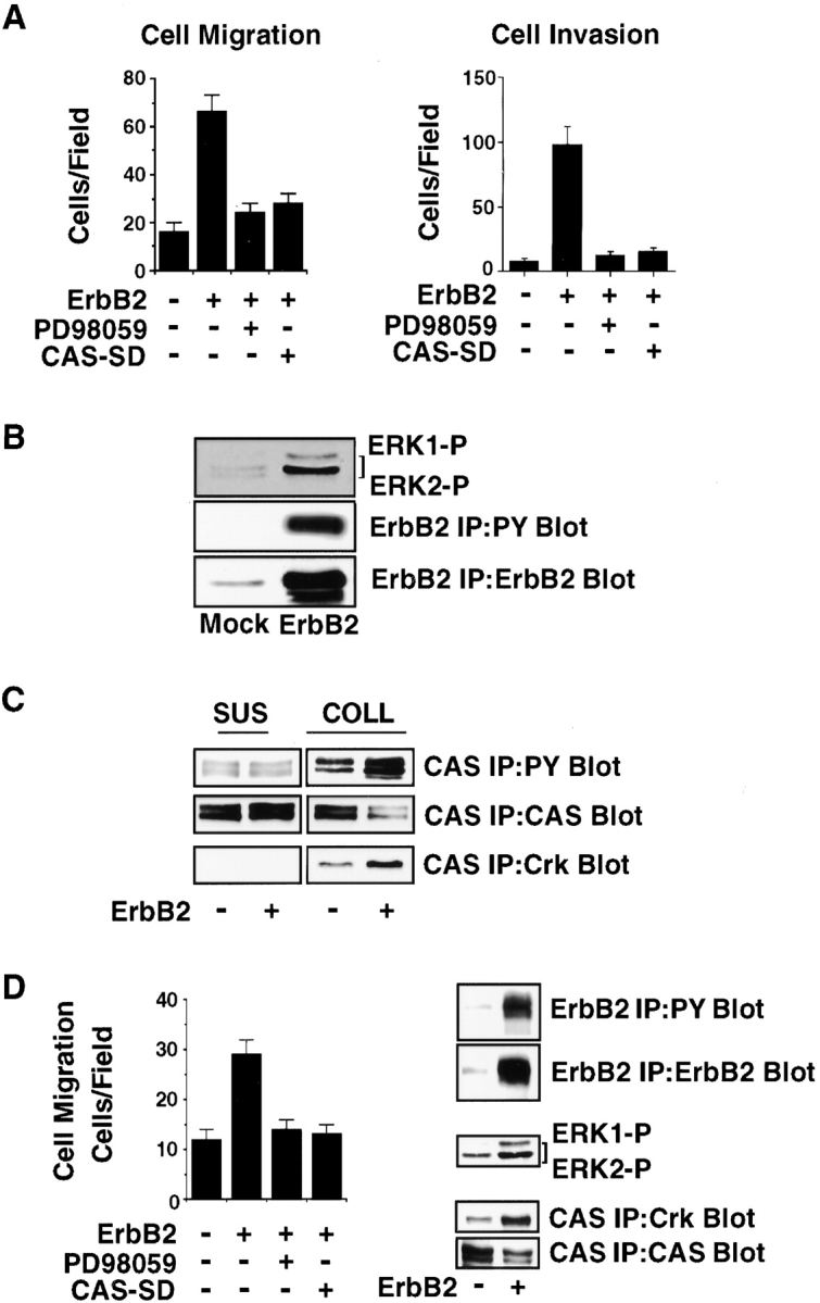
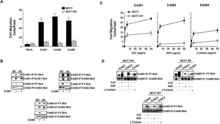
The epidermal growth factor (EGF) family of tyrosine kinase receptors (ErbB1, -2, -3, and -4) and their ligands are involved in cell differentiation, proliferation, migration, and carcinogenesis. However, it has proven difficult to link a given ErbB receptor to a specific biological process since most cells express multiple ErbB members that heterodimerize, leading to receptor cross-activation. In this study, we utilize carcinoma cells depleted of ErbB2, but not other ErbB receptor members, to specifically examine the role of ErbB2 in carcinoma cell migration and invasion. Cells stimulated with EGF-related peptides show increased invasion of the extracellular matrix, whereas cells devoid of functional ErbB2 receptors do not. ErbB2 facilitates cell invasion through extracellular regulated kinase (ERK) activation and coupling of the adaptor proteins, p130CAS and c-CrkII, which regulate the actin-myosin cytoskeleton of migratory cells. Overexpression of ErbB2 in cells devoid of other ErbB receptor members is sufficient to promote ERK activation and CAS/Crk coupling, leading to cell migration. Thus, ErbB2 serves as a critical component that couples ErbB receptor tyrosine kinases to the migration/invasion machinery of carcinoma cells.


Figures

Figure 1
Figure 1
Carcinoma cell migration induced by EGF family peptides requires functional ErbB2 receptors. A, Mammary adenocarcinoma cells lines (MCF7, T47D, and MDA-MB-435) with or without cell surface ErbB2 receptors were serum-starved and allowed to migrate on collagen-coated Transwell membranes for 2 h in the presence of various concentrations of NDF, EGF, BTC, or insulin as described in Materials and Methods. Each point represents the mean ± SEM of at least three independent experiments. The background migration in cells with or without ErbB2 receptors was the same and has been subtracted. B, ErbB family receptors immunoprecipitated from T47D cells with or without cell surface ErbB2 (5R) after exposure to 50 ng/ml of NDF, EGF, BTC, or buffer only (control) for 10 min. Immunoprecipitates (IP) were examined by immunoblotting for the presence of ErbB receptors, as well as changes in phosphotyrosine (PY) as described in Materials and Methods. C, T47D cells were allowed to migrate on collagen I or vitronectin-coated Transwell membranes for 2 h in the presence of 50 ng/ml NDF, EGF, BTC (β-cell.) with or without 25 μg/ml of function blocking antibodies to β1 (P4C10) or αvβ5 (P1F6) integrins. Data are expressed as percent of ligand-induced migration in the absence of antibodies and represents the mean ± SEM of at least three independent experiments.
Figure 2
Figure 2
Induction of carcinoma cell migration by NDF is associated with loss of cell–cell contacts, epithelial cell scattering, and random migration. Serum-starved MCF7 cells with or without cell surface ErbB2 receptors (5R) were cultured on collagen-coated coverslips before being stimulated with NDF (50 ng/ml) or buffer only (control) and examined by time-lapse phase microscopy for changes in cell morphology and migration at the indicated times. Bar, 50 μm.
Figure 3
Figure 3
Functional ErbB2 receptors are required for induction of carcinoma cell invasion of the ECM in response to EGF family peptides. A, MCF7 cells were serum-starved and allowed to invade Transwell membranes coated with the reconstituted basement membrane material Matrigel for 24 h in the presence or absence of 50 ng/ml of NDF, EGF, BTC (β-cell.), or 10% FBS as described in Materials and Methods. Each bar represents the mean ± SEM of at least three independent experiments. B, MCF7 cells with and without cell surface ErbB2 receptors (MCF7-5R) were serum-starved, then examined for their ability to invade the reconstituted basement membrane as described above in the presence or absence of 50 ng/ml of NDF or 10% FBS. Each bar represents the mean ± SEM of at least three independent experiments.
Figure 4
Figure 4
ErbB2-mediated cell migration requires ERK activity. A, Kinase activity of ERK2 immunoprecipitated from T47D cells with or without cell surface ErbB2 receptors (T47D-5R) after treatment with 50 ng/ml NDF for the indicated times as described in Materials and Methods. In some cases, PD98059 (50 μM) was added to T47D cells 30 min after exposure to NDF to block ERK activation by MEK. Arrow indicates time of exposure of cells to PD98059. B, T47D cells pretreated for 30 min with or without NDF (50 ng/ml) and then allowed to migrate on collagen-coated Transwell membranes for 2 h in the presence or absence of PD98059 (50 μM). Each bar represents the mean ± SEM of at least three independent experiments. C, T47D cells pretreated for 1 h with or without PD98059 (50 μM) and were then allowed to migrate on collagen-coated Transwell membranes in the presence or absence of NDF for 2 h. Each bar represents the mean ± SEM of at least three independent experiments. An aliquot of cells treated as for the migration experiment above was lysed in detergent then analyzed for changes in ERK activity by immunoblotting with antibodies to the phosphorylated activated form of ERK1 and ERK2. Immunoblots were stripped and reprobed with antibodies to ERK2 to confirm equal amounts of ERK protein were present in these lysates. D, MCF7-5R cells were transiently transfected with a β-gal reporter construct, together with either the empty vector or the vector containing mutationally activated MEK. Cells were allowed to migrate on collagen-coated Transwell membranes in the presence or absence of PD98059 for 3 h and were then stained with X-gal reagent to detect transfected cells. Transfection efficiency, as well as cell adhesion to collagen, was monitored to assure that equal numbers of cells were transfected and loaded into migration chambers as described in Materials and Methods. Each bar represents the mean ± SEM of at least three independent experiments. An aliquot of cells treated as for the migration experiment above was lysed in detergent and analyzed for MEK expression or changes in ERK activity by immunoblotting with antibodies to MEK or the phosphorylated activated forms of ERK1 and ERK2, respectively.
Figure 5
Figure 5
Coupling of the adaptor proteins CAS and Crk are necessary for ErbB2-mediated cell migration. A, CAS immunoprecipitated from MCF7 cells with and without cell surface ErbB2 receptors (MCF7-5R) and either held in suspension (SUS) or allowed to attach to collagen-coated (COLL) dishes for 15 min in the presence or absence of NDF (50 ng/ml), and then lysed in boiling 1% SDS to completely solubilize cellular proteins. The proteins were diluted in buffer and CAS immunoprecipitated and examined for tyrosine phosphorylation by immunoblotting with anti-phosphotyrosine antibodies as described in Materials and Methods. B, CAS immunoprecipitated from cells treated, as described in A, and examined for CAS tyrosine phosphorylation, as well as Crk binding by immunoblotting as described in Materials and Methods. 1% Triton X-100 was used to solubilize cellular proteins in these experiments in place of SDS. The milder lysis conditions allows for the retention CAS/Crk complexes. However, some of the CAS protein is not completely solubilized in adherent cells, most likely because it strongly associates with the detergent-insoluble cytoskeleton in cells attached to ECM proteins ( Polte and Hanks 1997). C, MCF7 cells were transiently transfected with a β-gal reporter construct, together with either the empty vector or the vector containing CAS with the Crk binding sites deleted (CAS-SD) or Crk with its CAS binding domain mutated (Crk-SH2). Cells were allowed to migrate on collagen-coated Transwell membranes in the presence or absence of NDF (50 ng/ml) for 3 h and were then stained with X-gal reagent to detect transfected cells. Transfection efficiency, as well as cell adhesion to collagen, was monitored to assure that equal numbers of cells were transfected and loaded into migration chambers as described in Materials and Methods. Each bar represents the mean ± SEM of at least three independent experiments. An aliquot of cells treated as for the migration experiment above was lysed in detergent then analyzed for CAS and Crk expression by immunoblotting with antibodies to CAS and Crk. Note that the Crk-SH2 is myc-tagged and shows reduced mobility, compared with the endogenous wild-type Crk (Crk-WT) protein as the result of the molecular tag. CAS-SD shows faster mobility, compared with endogenous CAS (CAS-WT) since its substrate domain has been truncated. D, MCF7-5R cells without cell surface ErbB2 receptors were transiently transfected with a β-gal reporter construct, together with either the empty vector or the vector containing CAS and Crk, and then allowed to migrate as described in C. An aliquot of cells treated as for the migration experiment above was lysed in detergent and then analyzed for CAS and Crk expression by immunoblotting with antibodies to CAS and Crk. Note that Crk and CAS are myc- and gst-tagged, respectively, and show reduced mobility, compared with endogenous wild-type (WT) forms of these proteins.
Figure 6
Figure 6
Overexpression of ErbB2 is sufficient to induce breast carcinoma cell migration and invasion, as well as ERK activation and CAS/Crk coupling. A, Serum-starved MCF7 cells were transiently transfected with the empty vector or the vector (pcDNA3) containing ErbB2 along with CAS-SD and the β-gal reporter gene. Cells were allowed to either migrate on collagen-coated Transwell membranes for 3 h or to invade the basement membrane-like material Matrigel for 24 h and then stained with X-gal to identify transfected migratory/invasive cells. Transfection efficiency, as well as cell adhesion to collagen and Matrigel, was monitored to assure that equal numbers of cells were transfected and loaded into the migration chamber as described in Materials and Methods. In some cases, cells were allowed to migrate or invade in the presence of PD98059 (50 μM). Each bar represents the mean ± SEM of at least three independent experiments. B, Serum-starved MCF7 cells transiently transfected with the empty vector or the vector (pcDNA3) containing ErbB2 were lysed and examined for ErbB2 expression and tyrosine phosphorylation, as well as ERK activation by immunoblotting as described in Materials and Methods. C, CAS immunoprecipitated from protein lysates of cells transfected as described in B were either held in suspension (SUS) or allowed to attach to collagen-coated (COLL) dishes for 15 min. The immunoprecipitates were examined for tyrosine phosphorylation of CAS, as well as Crk, binding by immunoblotting as described in Materials and Methods. D, Serum-starved CHO cells were transiently transfected with the empty vector or the vector (pcDNA3) containing ErbB2 or CAS-SD along with the β-gal reporter gene. Cells were allowed to migrate on fibronectin-coated Transwell membranes for 3 h in the presence or absence of PD98059 (50 μM) as described in A. An aliquot of cells treated as for the migration experiments above was lysed and examined for ErbB2 expression and tyrosine phosphorylation, as well as ERK activation, by immunoblotting as described in Materials and Methods. CAS was also immunoprecipitated from protein lysates of cells transfected with ErbB2 or the empty control vector after attachment to fibronectin-coated dishes for 15 min and examined for Crk binding by immunoblotting with Crk antibodies.
Figure 7
Figure 7
ErbB2 activation is necessary for cell migration induced by overexpression of ErbB family receptors. A, Serum-starved MCF7 cells with or without cell surface ErbB2 receptors (MCF7-5R) were transiently transfected with either the empty vector (Mock) or the vector (pcDNA3) containing either ErbB1, ErbB3, or ErbB4 along with the β-gal reporter gene. Cells were allowed to migrate on collagen-coated Transwell membranes for 3 h and then stained with X-gal to identify transfected migratory cells. Transfection efficiency, as well as cell adhesion to collagen, was monitored to assure that equal numbers of transfected cells were loaded into the migration chamber as described in Materials and Methods. B, Immunoprecipitation of ErbB1, -3, or -4 from cells treated as for the migration experiments above and examined for expression, as well as changes in tyrosine phosphorylation by immunoblotting as described in Materials and Methods. Note that endogenous ErbB2 was also immunoprecipitated from these cells and examined for changes in tyrosine phosphorylation. In these experiments, ErbB1, -3, and -4 were overexpressed above endogenous proteins by ∼56-, 30-, and 48-fold, respectively, as determined by immunoblotting with ErbB antibodies and ECL. MCF7 cells express moderate levels of endogenous ErbB3, -4, and a low level of ErbB1 receptors, which are not visible at the short exposure time (5 s) used to visualize the overexpressed proteins in these experiments. C, Cells transfected as described in A were allowed to migrate for 3 h in the presence of various concentrations of EGF, NDF, or BTC as described above. The basal migration of these cells in the absence of EGF-like peptides has been subtracted as background. D, Immunoprecipitation of ErbB1, -3, or -4 from cells treated as for the migration experiments in C. ErbB receptor (ErbBR) expression, as well as changes in tyrosine phosphorylation, were determined in cells exposed to the indicated ligand (50 ng/ml for 10 min) by immunoblotting as described in Materials and Methods. Note that endogenous ErbB2 was also immunoprecipitated from these cells and examined for changes in tyrosine phosphorylation. Each bar or data point represents the mean ± SEM of at least three independent experiments.

Similar articles

Cited by

References

    1. Alroy I., Yarden Y. The ErbB signaling network in embryogenesis and oncogenesissignal diversification through combinatorial ligand-receptor interactions. FEBS Lett. 1997;410:83–86 . - PubMed
    1. Bacus S.S., Gudkov A., Zelnick C.R., Chin D., Stern R., Stancovski L., Peles E., Ben-Baruch N., Farbstein H., Lupu R. Neu differentiation factor (Heregulin) induces expression of intracellular adhesion molecule 1implications for mammary tumors. Cancer Res. 1993;53:5251–5261 . - PubMed
    1. Beerli R.R., Hynes N.E. Epidermal growth factor-related peptides activate distinct subsets of ErbB receptors and differ in their biological activities. J. Biol. Chem. 1996;271:6071–6076 . - PubMed
    1. Beerli R.R., Wels W., Hynes N.E. Intracellular expression of single-chain antibodies reverts ErbB-2 transformation. J. Biol. Chem. 1994;269:23931–23936 . - PubMed
    1. Ben-Levy R.H., Paterson F., Marshall C.J., Yarden Y. A single autophosphorylation site confers oncogenicity to the Neu/ErbB-2 receptor and enables coupling to the MAP kinase pathway. EMBO (Eur. Mol. Biol. Organ.) J. 1994;13:3302–3311 . - PMC - PubMed

Publication types

MeSH terms