. 2000 Feb;20(4):1095-103.
doi: 10.1128/MCB.20.4.1095-1103.2000.

Kinetics of ribosomal pausing during programmed -1 translational frameshifting

Affiliations

Kinetics of ribosomal pausing during programmed -1 translational frameshifting

J D Lopinski et al. Mol Cell Biol. 2000 Feb.

Abstract

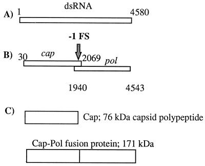
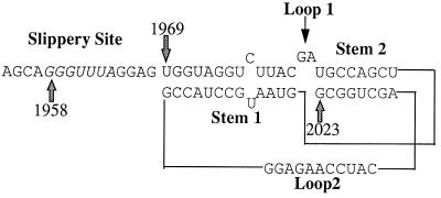
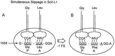
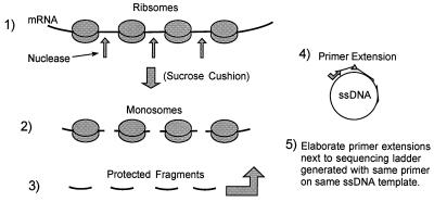
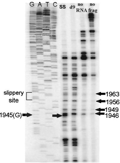
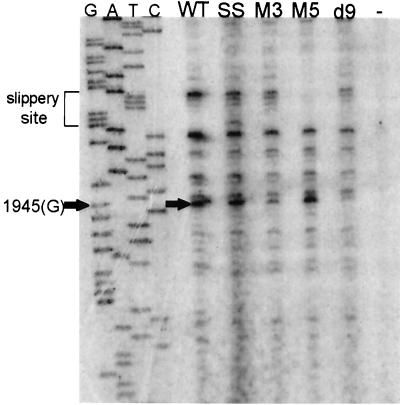
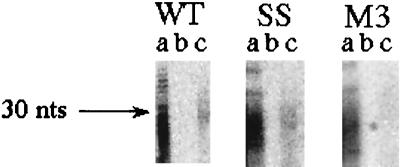
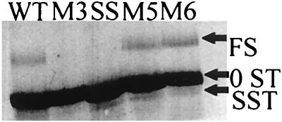
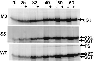
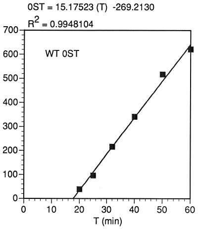
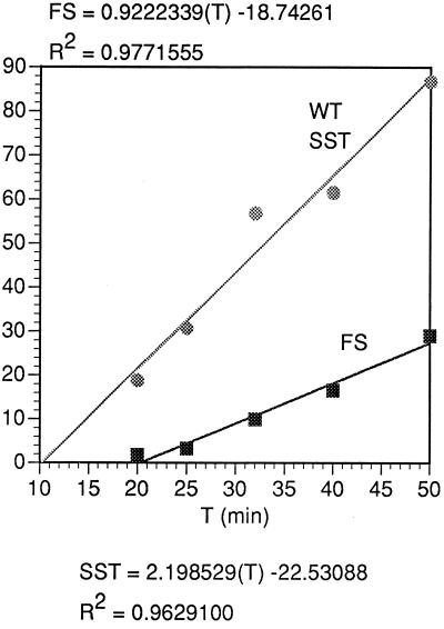
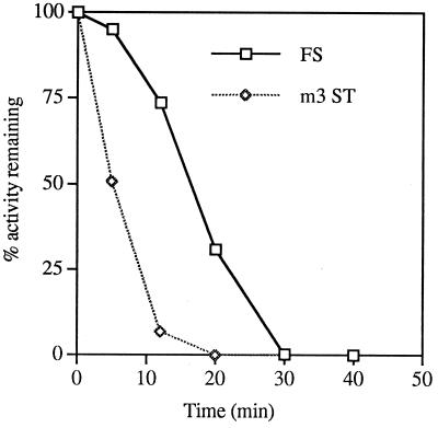
In the Saccharomyces cerevisiae double-stranded RNA virus, programmed -1 ribosomal frameshifting is responsible for translation of the second open reading frame of the essential viral RNA. A typical slippery site and downstream pseudoknot are necessary for this frameshifting event, and previous work has demonstrated that ribosomes pause over the slippery site. The translational intermediate associated with a ribosome paused at this position is detected, and, using in vitro translation and quantitative heelprinting, the rates of synthesis, the ribosomal pause time, the proportion of ribosomes paused at the slippery site, and the fraction of paused ribosomes that frameshift are estimated. About 10% of ribosomes pause at the slippery site in vitro, and some 60% of these continue in the -1 frame. Ribosomes that continue in the -1 frame pause about 10 times longer than it takes to complete a peptide bond in vitro. Altering the rate of translational initiation alters the rate of frameshifting in vivo. Our in vitro and in vivo experiments can best be interpreted to mean that there are three methods by which ribosomes pass the frameshift site, only one of which results in frameshifting.


Figures

FIG. 1
FIG. 1
(A) The 4.58-kb dsRNA genome of ScV-L-A. (B) The two ORFs, cap and pol, of the ScV-L-A plus strand, separated by the −1 frameshift within the overlapping region. (C) The ScV-L-A 76-kDa 0 frame protein product Cap and the 171-kDa fusion protein Cap-Pol.
FIG. 2
FIG. 2
Possible structure of the ScV-L-A pseudoknot. The slippery site is in italics starting at base 1958. The second stem is shown with two more base pairs than in some formulations (14).
FIG. 3
FIG. 3
Simultaneous slippage model (22) shown here for ScV-L-A. A tRNAGly-tRNALeu pair in the 0 frame (underlined) P and A site of the ribosome slips back one nucleotide to pair with the −1 frame (pol) Gly-Phe codons, leaving mispaired nucleotides in the wobble position of each −1 frame codon.
FIG. 4
FIG. 4
Heelprinting technique (42). In step 1, translating ribosomes are stopped on the template with cycloheximide and EDTA and unprotected mRNA is digested away, liberating monosomes. In step 2, the monosomes are collected through a sucrose cushion and the ribosomal proteins are stripped away, yielding protected RNA fragments. In step 4, the fragments are annealed to an ssDNA that has a region complementary to the message used in the translation. The heteroduplexes stop the extension of a primer at the 5′ ends of the RNA fragments that were protected from nuclease by the ribosome. In step 5, the primer extensions are elaborated next to a DNA sequencing ladder generated with the same primer on the same ssDNA template, thereby mapping the 5′ end of the protected RNA fragment.
FIG. 5
FIG. 5
Heelprinting results with micrococcal nuclease. Lanes represent the SS and d9 mutants, control with mRNA left out of translation, and control with no reticulocyte or RNA used in the heelprinting reaction (no frag).
FIG. 6
FIG. 6
WT and mutant heelprints and a heelprint without added RNA (−).
FIG. 7
FIG. 7
Quantitative protected fragment recovery. Protected RNA fragments from the WT, SS, and M3 constructs are shown after recovery from the hybridization procedure (see Materials and Methods) with no DNA oligonucleotide and no RNase (lanes a), no DNA with RNase (lanes b), or with DNA and with RNase (lanes c).
FIG. 8
FIG. 8
Standard translation of WT, SS, M3, M5, and M6 constructs. Positions of 0ST, SST, and FS products are indicated. These translations had no chase with nonradioactive methionine.
FIG. 9
FIG. 9
Pulse-chase in vitro translations. Reaction mixtures were incubated for 20 min and divided in half. One half was chased with cold methionine, and aliquots were taken from each half (with [+] or without [−] Met) at the indicated time points. Positions of the SST, 0ST, and FS products are indicated.
FIG. 10
FIG. 10
Kinetics of synthesis of the WT 0ST product from the data of Fig. 9. The ordinate is relative molar units, generated by the PhosphorImager volume report but divided by 3 to account for the difference in the number of methionines between the 0ST and the FS and SST products. The equation for the least squares line calculated is given.
FIG. 11
FIG. 11
Initial kinetics of synthesis of the WT SST product and FS product, as in Fig. 10, from the same experiment as that of Fig. 10, in relative molar amounts.
FIG. 12
FIG. 12
Translation activity remaining during the chase with nonradioactive methionine for the M3 0ST (m3 ST) and WT FS. The percent activity remaining (AR) is calculated from the plateau value of the PhosphorImager volume report for the FS or 0ST (P) and the value at 0 to 50 min after the chase of the volume report (V), as follows: AR = 100[(PV)/P].

References

    1. Anderson R P, Menninger J R. Tests of the ribosome editor hypothesis. III. A mutant Escherichia coli with a defective ribosome editor. Mol Gen Genet. 1987;209:313–318. - PubMed
    1. Balasundaram D, Dinman J D, Wickner R B, Tabor C W, Tabor H. Spermidine deficiency increases +1 ribosomal frameshifting efficiency and inhibits Ty1 retrotransposition in Saccharomyces cerevisiae. Proc Natl Acad Sci USA. 1994;91:172–176. - PMC - PubMed
    1. Blinkowa A L, Walker J R. Programmed ribosomal frameshifting generates the Escherichia coli DNA polymerase III gamma subunit from within the tau subunit reading frame. Nucleic Acids Res. 1990;18:1725–1729. - PMC - PubMed
    1. Bostian K A, Elliott Q, Bussey H, Burn V, Smith A, Tipper D J. Sequence of the preprotoxin dsRNA gene of type 1 killer yeast: multiple processing events produce a two-component toxin. Cell. 1984;36:741–751. - PubMed
    1. Brierley I, Boursnell M E G, Binns M M, Bilimoria B, Blok V C, Brown T D, Inglis S C. An efficient ribosomal frame-shifting signal in the polymerase-encoding region of the coronavirus IBV. EMBO J. 1987;6:3779–3785. - PMC - PubMed

Publication types

LinkOut - more resources