. 2000 Feb;20(4):1116-23.
doi: 10.1128/MCB.20.4.1116-1123.2000.

Translation of chloroplast psbA mRNA is modulated in the light by counteracting oxidizing and reducing activities

Affiliations

Translation of chloroplast psbA mRNA is modulated in the light by counteracting oxidizing and reducing activities

T Trebitsh et al. Mol Cell Biol. 2000 Feb.

Abstract

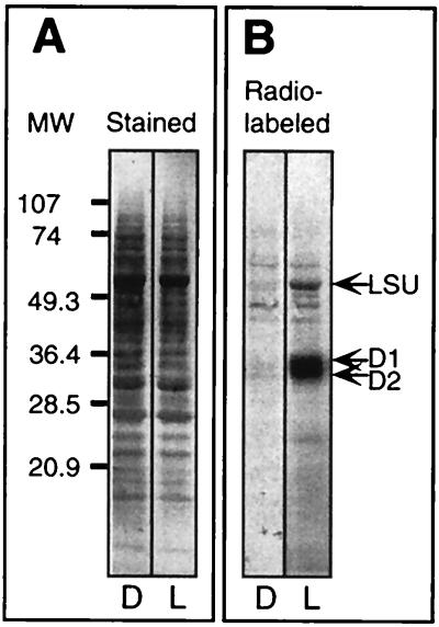
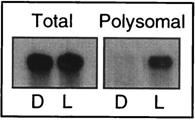
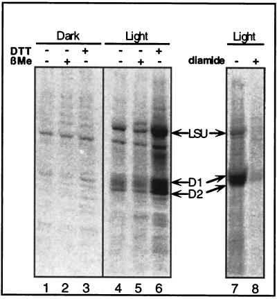
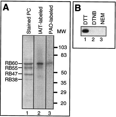
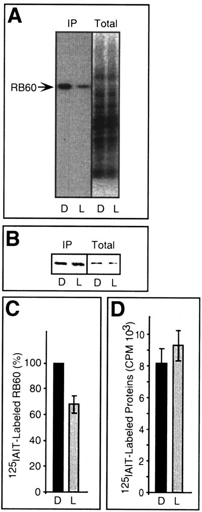
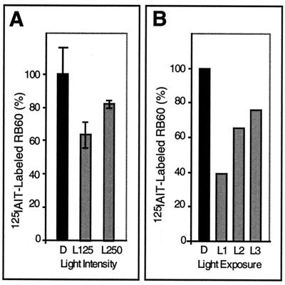
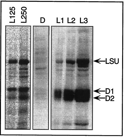
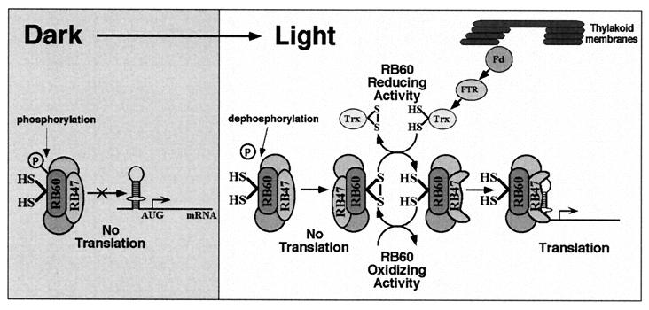
Light has been proposed to stimulate the translation of Chlamydomonas reinhardtii chloroplast psbA mRNA by activating a protein complex associated with the 5' untranslated region of this mRNA. The protein complex contains a redox-active regulatory site responsive to thioredoxin. We identified RB60, a protein disulfide isomerase-like member of the protein complex, as carrying the redox-active regulatory site composed of vicinal dithiol. We assayed in parallel the redox state of RB60 and translation of psbA mRNA in intact chloroplasts. Light activated the specific oxidation of RB60, on the one hand, and reduced RB60, probably via the ferredoxin-thioredoxin system, on the other. Higher light intensities increased the pool of reduced RB60 and the rate of psbA mRNA translation, suggesting that a counterbalanced action of reducing and oxidizing activities modulates the translation of psbA mRNA in parallel with fluctuating light intensities. In the dark, chemical reduction of the vicinal dithiol site did not activate translation. These results suggest a mechanism by which light primes redox-regulated translation by an unknown mechanism and then the rate of translation is determined by the reduction-oxidation of a sensor protein located in a complex bound to the 5' untranslated region of the chloroplast mRNA.


Figures

FIG. 1
FIG. 1
Light-regulated translation in mature chloroplasts isolated from C. reinhardtii cells. Intact C. reinhardtii cw15 chloroplasts were collected from a 45%/70% interface of discontinuous Percoll gradient by published methods (4, 22). Intact chloroplasts were incubated in the dark for 30 min, and an aliquot was transferred for 10 min into the light (150 μmol m−2 s−1). The translational activities of the dark-incubated (D) and light-incubated (L) chloroplasts were then assayed by labeling newly synthesized proteins for 10 min with [35S]methionine. The nascent polypeptides were allowed to complete synthesis by incubation for an additional 5 min in the presence of excess nonradioactive methionine. Chloroplasts were lysed, and extracted proteins were fractionated by SDS-PAGE and blotted onto nitrocellulose membranes. (A) Amido Black staining of chloroplast proteins. (B) Autoradiograph of the same blot as in panel A showing the 35S-labeled proteins. LSU, large subunit of ribulose bisphosphate carboxylase/oxygenase.
FIG. 2
FIG. 2
RNA blot showing light-regulated polysome association of psbA mRNA in mature chloroplasts isolated from C. reinhardtii cells. RNA was isolated from chloroplasts treated as for the experiment in Fig. 1. Equal amounts of total RNA and polysome-associated RNA were loaded on the gel. A 32P-labeled psbA cDNA was used to probe the blots.
FIG. 3
FIG. 3
Light- and redox-regulated translation in mature chloroplasts. Shown is an autoradiograph of an SDS-PAGE blot, showing 35S-labeled proteins from chloroplasts treated as for the experiment in Fig. 1, except that a dithiol reductant (DTT), a monothiol reductant (β-ME), or a thiol oxidant (diamide) was added as indicated in the figure.
FIG. 4
FIG. 4
Identification of the regulatory VDS-containing protein. (A) Lanes: 1, Coomassie blue-stained SDS-PAGE of a highly purified preparation of psbA 5′PC (Stained PC) isolated from C. reinhardtii cells using psbA RNA-affinity chromatography; 2, PhosphorImage of SDS-PAGE of psbA 5′PC labeled with [125I]IAIT (IAIT-labeled); 3, autoradiograph of SDS-PAGE of psbA 5′PC labeled with [14C]PAO (PAO-labeled). (B) Autoradiograph of SDS-PAGE of recombinant RB60 labeled with [125I]IAIT. Lanes: 1, protein reduced with DTT; 2, protein oxidized with dithionitrobenzoate (DTNB); 3, protein modified with n-ethylmaleimide (NEM).
FIG. 5
FIG. 5
In vivo characterization of the redox state of RB60 and total proteins in dark- and light-incubated chloroplasts. (A) Autoradiograph of immunoprecipitation assays of RB60 (IP) conducted with extracts from isolated chloroplasts treated for 10 min in the dark (D) or light (L), followed by [125I]IAIT labeling. Total [125I]IAIT-labeled proteins (without immunoprecipitation) (Total) represent 5% of the amount used for immunoprecipitation assays. (B) The same blot as in panel A, probed with anti-RB60 sera showing equal loading of RB60 protein. (C) Quantification of [125I]IAIT-labeled RB60 (as determined by PhosphorImager analysis and normalized for equal amounts of precipitated RB60 protein) in 10-min light- and dark-treated chloroplasts. Values are means of six independent experiments, and the light value is expressed as percentage of the corresponding dark value (in each experiment designated as 100% of [125I]IAIT-labeled RB60). (D) Quantification of total chloroplast proteins labeled with [125I]IAIT, showing that light does not affect the global protein thiol state in the chloroplast. Each value is an average of three replications.
FIG. 6
FIG. 6
The redox state of the pool of RB60 in chloroplasts incubated in different light regimes. (A) The redox state of the pool of RB60 was determined as for the experiment in Fig. 5A, except that immunoprecipitation assays were performed using proteins of chloroplasts incubated in the dark (D) and under 125 (L125) or 250 (L250) μmol of light m−2 s−1. Quantification was performed as for the experiment in Fig. 5C, except that each value is an average of three replications (one replication of the dark treatment was designated as 100% of [125I]IAIT-labeled RB60). (B) The redox state of the pool of RB60 was determined as for the experiment in Fig. 5A, except that immunoprecipitation assays were performed with proteins of chloroplasts incubated in the dark or in the light (150 μmol m−2 s−1) at decreasing chloroplast concentrations, resulting in an increasing average light exposure of the chloroplasts. L1, 10 μg; L2, 5 μg; L3, 2.5 μg.
FIG. 7
FIG. 7
Effect of different light regimes on protein synthesis in mature isolated chloroplasts. Shown is an autoradiograph of protein labeling performed as for the experiment in Fig. 1, under the same light conditions as for the experiment in Fig. 6. LSU, large subunit of ribulose bisphosphate carboxylase/oxygenase.
FIG. 8
FIG. 8
A working model for redox signaling in light-regulated translation. The redox state of the regulatory VDS of RB60 controls the RNA-binding capacity of psbA 5′PC, which binds the RNA through the RB47 protein. Oxidation of the regulatory VDS inactivates and reduction activates binding of the PC to the 5′UTR of psbA mRNA and consequently psbA mRNA translation. Light regulates translation via a prerequisite (priming) redox-independent pathway and a redox-dependent pathway. The priming pathway probably involves dephosphorylation and specific oxidation of the pool of RB60 (either by autooxidation or by an RB60-specific oxidizing factor), rendering RB60 receptive to the reductive signal transduced by the redox-dependent pathway. In the redox-dependent pathway, light energy is captured by the thylakoid-associated photosynthetic complexes to produce reducing potential. Photosynthetic reducing equivalents are transduced by ferredoxin (Fd), ferredoxin-thioredoxin reductase (FTR), and thioredoxin (Trx) to reduce the regulatory VDS of RB60. Hence, in the light, a counterbalanced action of reducing and oxidizing activities modulates the redox state of the pool of RB60 and consequently the translation of psbA mRNA, in parallel with fluctuating light intensities. Transfer to the dark activates phosphorylation and inactivates the oxidation of RB60, resulting in an inactive form of psbA 5′PC that is not redox responsive. The encircled P denotes an inorganic phosphate.

Similar articles

Cited by

References

    1. Arnon D. Copper enzymes in isolated chloroplasts. Polyphenol-oxidase in Beta vulgaris. Plant Physiol. 1949;24:1–15. - PMC - PubMed
    1. Barber J, Anderson B. Too much of a good thing: light can be bad for photosynthesis. Trends Biochem Sci. 1992;17:61–66. - PubMed
    1. Barkan A. Proteins encoded by a complex chloroplast transcription unit are each translated from both monocistronic and polycistronic mRNAs. EMBO J. 1988;9:2637–2644. - PMC - PubMed
    1. Belknap W R. Partial purification of intact chloroplasts from Chlamydomonas reinhardtii. Plant Physiol. 1983;72:1130–1132. - PMC - PubMed
    1. Berry J O, Breiding D E, Klessig D F. Light-mediated control of translational initiation of ribulose-1,5-bisphosphate carboxylase in amaranth cotyledons. Plant Cell. 1990;2:795–803. - PMC - PubMed

Publication types

MeSH terms

Substances

LinkOut - more resources