. 2000 Feb;20(4):1234-42.
doi: 10.1128/MCB.20.4.1234-1242.2000.

Novel WD-repeat protein Mip1p facilitates function of the meiotic regulator Mei2p in fission yeast

Affiliations

Novel WD-repeat protein Mip1p facilitates function of the meiotic regulator Mei2p in fission yeast

S Shinozaki-Yabana et al. Mol Cell Biol. 2000 Feb.

Abstract

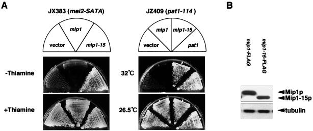
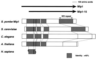
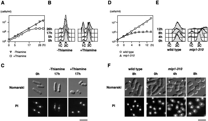
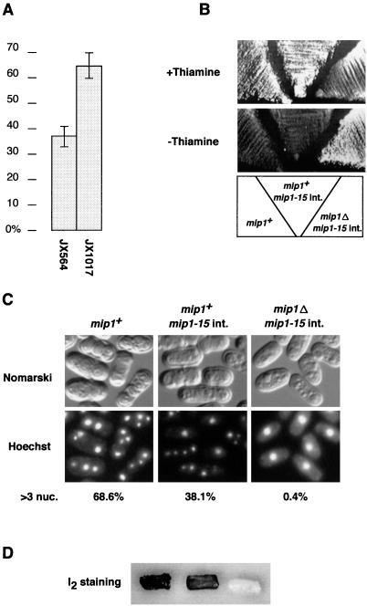
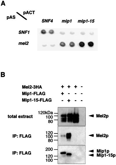
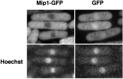
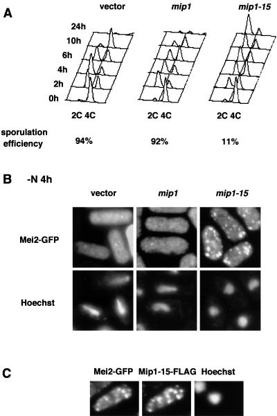
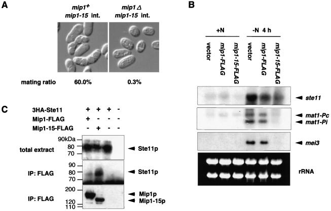
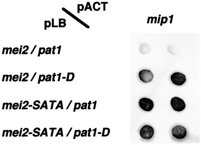
In fission yeast, the onset of meiosis is triggered by activation of the RNA-binding protein Mei2p. We screened for a high-copy-number suppressor of the ectopic meiosis induced by expression of an active form of Mei2p. Consequently we isolated a truncated form of a novel gene, named mip1, from a fission yeast genomic library. The mip1 gene encoded a protein of 1,313 amino acids which carried a WD-repeat motif in the C-terminal region and was apparently conserved among eukaryotes. Mip1p was cytoplasmic, and two-hybrid and immunoprecipitation analyses demonstrated that Mip1p was bound to Mei2p in vivo. Genetic evidence indicated that wild-type Mip1p was required for the function of Mei2p to induce meiosis and that the truncated form of it (Mip1-15p) dominantly interfered with Mei2p. Mip1p appeared to be involved also in conjugation, associating with Ste11p, which is a key transcription factor for sexual development. Furthermore, Mip1p was essential for cell growth, to which neither Mei2p nor Ste11p is relevant. These results suggest that Mip1p assists functional expression of a number of proteins required for proliferation and sexual development in fission yeast.


Figures

FIG. 1
FIG. 1
Suppression of ectopic meiosis of the nmt1-mei2-SATA and pat1-114 strains by overexpression of mip1-15. (A) JX383 (h nmt1-mei2-SATA) was transformed with either pREP41-mip1, pREP41-mip1-15, or the vector pREP41. JZ409 (h pat1-114) was also transformed with either pREP41-mip1, pREP41-mip1-15, pREP41-pat1, or the vector pREP41. Each transformant of JX383 was grown on a plate containing thiamine, restreaked on an SSA plates with or without 2 μM thiamine, and incubated at 32°C. Each transformant of JZ409 was grown at 26.5°C, spread on an SSA plate, and incubated at either 32 or 26.5°C. (B) To examine protein stability, the wild-type strain JY741 was transformed with either pREP41-mip1-FLAG or pREP41-mip1-15-FLAG, and total extracts were prepared from the transformants grown in thiamine-free MM for 16 h. Proteins extracted from 106 cells were separated by SDS-PAGE and subjected to Western blot analysis using an anti-FLAG antibody (upper panel) or the antitubulin antibody Tat1 (lower panel).
FIG. 2
FIG. 2
Schematic illustration of the structure of S. pombe Mip1 protein (SPAC57A7.11) and its S. cerevisiae (SCYHR186C), C. elegans (C10C5.6), and A. thaliana (T16O11.22) homologues. A fragmentary sequence derived from the human cancer genome anatomy project is also shown (ng92c11.s1). The regions highly conserved (>45% identity) between Mip1p and the homologues are hatched.
FIG. 3
FIG. 3
mip1+ is essential for cell growth. (A) Growth of mip1Δ cells transformed with pREP81-mip1-15. Cells were grown in MM to a concentration of about 105/ml; cell number was measured chronologically after the addition of 2 μM thiamine to the culture (□). Results for a control culture with no thiamine addition are also shown (▿). (B) Flow cytometry of the cells at 0, 5, 17, and 26 h after the addition of thiamine. (C) Nomarski micrographs of mip1Δ cells carrying the mip1-15 plasmid cultured with or without thiamine for the indicated time. Fluorescence micrographs of the same cell population stained with the DNA-binding dye propidium iodide (PI) are also shown. Bar, 10 μm. (D) Growth of mip1-310 cells. Cells were grown in YE medium to a concentration of about 105/ml at 26.5°C. They were shifted up to 36°C (0 h), and cell number was measured chronologically (▵). Results for a control wild-type strain are also shown (○). (E) Flow cytometry of the cells at 0, 4, 8, and 12 h after the shift. (F) Morphology of mip1-310 and control wild-type cells cultured at 36°C for the indicated time. Arrowheads indicate dividing cells. Fluorescence micrographs of the same cells stained with propidium iodide are also shown. Bar, 10 μm.
FIG. 4
FIG. 4
Mip1p is required for the entry to meiosis. (A) Lowered sporulation efficiency in diploid cells heterozygous for mip1. The wild-type strain JX1017 and the mip1+/mip1Δ strain JX564 were streaked on sporulation medium MEA and incubated at 30°C for 3 days. The sporulation frequency of each strain was scored under the microscope. The error bar for each data represents the standard deviation of three independent measurements. (B to D) Requirement of mip1+ for haploid meiosis induced by mei2-SATA. JW192 (nmt1-mei2-SATA adh1-mip1-15) and the isogenic mip1Δ strain JW193 were streaked on an SSA plate with or without thiamine and incubated at 32°C for 3 days (+Thiamine) or 5 days (−Thiamine) (B). The same strains were cultured in liquid MM at 30°C for 48 h. Cells were fixed with ethanol, stained with Hoechst 33342, and photographed (C). They were also spread in patches on an SSA plate, incubated at 30°C for 4 days, and stained with I2 vapor (D). Nuc., nuclei.
FIG. 5
FIG. 5
Mip1p and Mei2p form a complex in vivo. (A) Yeast two-hybrid analysis. The interaction between Mip1p and Mei2p and that between Mip1-15p and Mei2p are positive, as judged by the blue coloring of the host cells. The intensity of the blue color is reproduced in black and white here. The pair SNF1 and SNF4 represents a standard positive control. (B) Coimmunoprecipitation of Mip1p and Mei2p. Crude extracts were prepared from vegetative cells expressing tagged proteins as indicated and subjected to immunoprecipitation (IP) with an anti-FLAG antibody. Each precipitate was separated by SDS-PAGE and examined for the presence of Mei2 protein by Western blotting using an anti-HA antibody. The immunoblotting patterns of total crude extracts are also shown.
FIG. 6
FIG. 6
Subcellular localization of Mip1p tagged with GFP. Wild-type strain JY362 transformed with either pREP41-mip1-GFP or pREP41-GFP was grown in liquid MM and examined for GFP fluorescence without fixation. A fluorescence micrograph of the same cells stained with the DNA-binding dye Hoechst 33342 is also shown for each transformant.
FIG. 7
FIG. 7
Aggregation of Mei2p induced by Mip1-15p overproduction. (A) Flow cytometry of the wild-type diploid strain JY765 carrying pGFT81-mei2 and either pREP2-mip1-FLAG, pREP2-mip1-15-FLAG, or the vector pREP2. Each strain was grown to 4 × 10⁶ cells/ml in MM and shifted to MM-N to induce meiosis. Cells were harvested at intervals after the shift and subjected to flow cytometry. Sporulation efficiency of each strain was determined 24 h after the shift. (B) The same strains as in panel A were fixed by methanol at 4 h after the shift and observed for fluorescence of Mei2p-GFP. Fluorescence micrographs of cells stained with the DNA-binding dye Hoechst 33342 are also shown. (C) Cells producing Mei2p-GFP and Mip1-15p-FLAG were fixed by formaldehyde at 4 h after the shift. They were doubly immunostained with anti-GFP antibodies and a monoclonal anti-FLAG antibody and subjected to fluorescence microscopy.
FIG. 8
FIG. 8
mip1 is required for the mating process. (A) h90 mip1-15 integrant (int.) strains with or without the authentic mip1+ gene (JW195 or JW196) were streaked on sporulation medium SSA and incubated at 30°C for 4 days. The mating frequency of each strain was scored under the microscope. (B) Northern blot analysis of mip1-15-overexpressing cells. Total RNA was extracted from the same strains as analyzed in Fig. 7, either growing exponentially (0 h) or starved of nitrogen for 4 h. RNA (10 μg) was loaded in each lane after denaturation by formamide, electrophoresed, transferred to a nylon membrane, and hybridized with the indicated probes. rRNA in each sample, stained with ethidium bromide, are shown in the lower panel to verify nearly equal loading of RNA in each lane. (C) Coimmunoprecipitation of Mip1p and Ste11p. Crude extracts were prepared from cells expressing tagged proteins as indicated and growing mitotically. They were subjected to immunoprecipitation (IP) with an anti-FLAG antibody. The precipitates were subjected to SDS-PAGE and examined for the presence of Ste11 protein by Western blotting using an anti-HA antibody. Immunoblotting patterns of the crude extracts are also shown.
FIG. 9
FIG. 9
Interaction of Mip1p with unphosphorylated Mei2p, examined by yeast three-hybrid analysis. Either LexA-Mei2p or LexA-Mei2-SATAp was expressed together with either active Pat1p or kinase-negative Pat1-Dp from a single plasmid. GAD-Mip1p was expressed from another plasmid. For each combination, two independent transformants were assayed for β-galactosidase activity. The intensity of the blue color is reproduced in black and white here.

References

    1. Bai C, Elledge S J. Gene identification using the yeast two-hybrid system. Methods Enzymol. 1996;273:331–347. - PubMed
    1. Beach D, Rodgers L, Gould J. RAN1+ controls the transition from mitotic division to meiosis in fission yeast. Curr Genet. 1985;10:297–311. - PubMed
    1. Bresch C, Muller G, Egel R. Genes involved in meiosis and sporulation of a yeast. Mol Gen Genet. 1968;102:301–306. - PubMed
    1. Egel R. Mating-type genes, meiosis, and sporulation. In: Nasim A, et al., editors. Molecular biology of the fission yeast. San Diego, Calif: Academic Press; 1989. pp. 31–73.
    1. Francesconi S, Park H, Wang T S-F. Fission yeast with DNA polymerase δ temperature-sensitive alleles exhibits cell division cycle phenotype. Nucleic Acids Res. 1993;21:3821–3828. - PMC - PubMed

Publication types

MeSH terms

Associated data