. 2000 Feb;20(4):1254-62.
doi: 10.1128/MCB.20.4.1254-1262.2000.

Characterization of Schizosaccharomyces pombe Hus1: a PCNA-related protein that associates with Rad1 and Rad9

Affiliations

Characterization of Schizosaccharomyces pombe Hus1: a PCNA-related protein that associates with Rad1 and Rad9

T Caspari et al. Mol Cell Biol. 2000 Feb.

Abstract

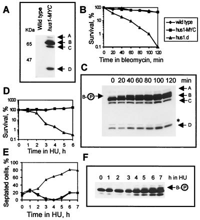
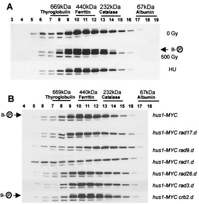
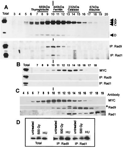
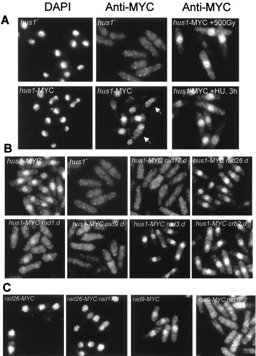
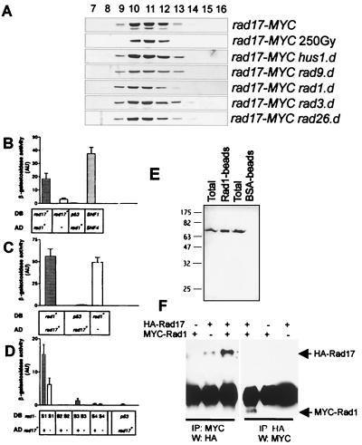
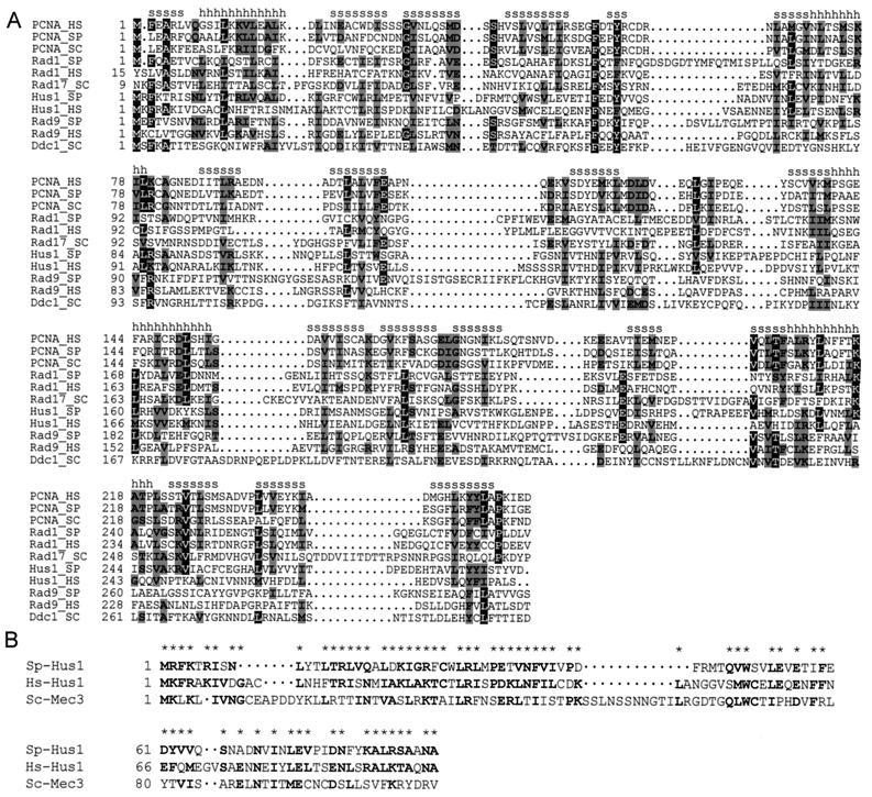
Hus1 is one of six checkpoint Rad proteins required for all Schizosaccharomyces pombe DNA integrity checkpoints. MYC-tagged Hus1 reveals four discrete forms. The main form, Hus1-B, participates in a protein complex with Rad9 and Rad1, consistent with reports that Rad1-Hus1 immunoprecipitation is dependent on the rad9(+) locus. A small proportion of Hus1-B is intrinsically phosphorylated in undamaged cells and more becomes phosphorylated after irradiation. Hus1-B phosphorylation is not increased in cells blocked in early S phase with hydroxyurea unless exposure is prolonged. The Rad1-Rad9-Hus1-B complex is readily detectable, but upon cofractionation of soluble extracts, the majority of each protein is not present in this complex. Indirect immunofluorescence demonstrates that Hus1 is nuclear and that this localization depends on Rad17. We show that Rad17 defines a distinct protein complex in soluble extracts that is separate from Rad1, Rad9, and Hus1. However, two-hybrid interaction, in vitro association and in vivo overexpression experiments suggest a transient interaction between Rad1 and Rad17.


Figures

FIG. 1
FIG. 1
Hus1-MYC exists in four forms. (A) Total cell extracts were prepared under denaturing conditions from wild-type and hus1-MYC cells and probed with anti-MYC antibody. The arrows mark the four forms of Hus1-MYC. (B) Wild-type cells, hus1-MYC cells, and hus1.d cells were incubated for 120 min in bleomycin-containing medium (40 μg/ml), and 500 cells were plated at the indicated time points to analyze cell viability. (C) At the same time points, 108 cells were removed and total cell extracts were probed with anti-MYC antibody. The asterisk marks an unspecific band. (D) Wild-type cells, hus1-MYC cells, and hus1.d cells were incubated in the presence of 10 mM HU, and cells were plated at the indicated time points to analyze cell viability. (E) The same strains were incubated in 10 mM HU for 7 h, and samples were fixed in methanol to monitor cell cycle arrest; (F) 108 cells were collected to prepare total extracts. The arrow indicates phosphorylated Hus1-B. Checkpoint-deficient hus1.d cultures show a strong increase in septated cells, because they enter a catastrophic mitosis in the presence of HU, in which the septum cuts through the nucleus.
FIG. 2
FIG. 2
Hus1-B forms a protein complex which is sensitive to loss of Rad1 and Rad9. (A) Soluble extracts were prepared from untreated hus1-MYC cells (0 Gy) and from hus1-MYC cells irradiated with 500 Gy or treated with 10 mM HU for 3.5 h and subjected to size exclusion chromatography. (B) Soluble extracts were prepared from hus1-MYC cells deleted for rad17, rad9, rad1, rad26, rad3, or crb2/rhp9 and analyzed by size exclusion chromatography. All Western blots were probed with anti-MYC antibodies. The arrows indicate phosphorylated Hus1-B.
FIG. 3
FIG. 3
Hus1-B interacts with Rad1 and Rad9 in a rad9-dependent manner, but most of Rad1 and Rad9 do not coelute with Hus1-B. (A) A soluble protein extract prepared from untreated hus1-MYC cells was subjected to size exclusion chromatography. After removal of 50 μl for Western analysis (top; Western blot probed with anti-MYC antibodies, total = 30 μg of starting extract), half of each fraction was incubated with the Rad9-specific antibody (middle) and the other half was incubated with the Rad1-specific antibody (bottom). Precipitated Hus1-MYC was visualized using the anti-MYC antibody. Immunoprecipitations (IP) were performed in the presence of saturating antibody concentrations. The total remaining material after incubation with both antibodies was largely unchanged, indicating that both antibodies precipitate only a small proportion of Hus1-B. (B) The same experiment was performed using an extract prepared from hus1-MYC rad9.d cells (total = 30 μg of starting extract). (C) Fractions obtained from untreated hus1-MYC cells were analyzed directly using the anti-MYC antibody (top), the Rad9-specific antibody (middle), and the Rad1-specific antibody (bottom). The asterisk marks an unspecific band. (D) Soluble protein extracts were prepared from untreated hus1-MYC cells and from hus1-MYC cells irradiated with a dose of 500 Gy or arrested in 10 mM HU for 3.5 h. Left, 30 μg of total starting protein; middle, Hus1-B precipitated with the Rad9-specific antibody from 500 μg of total protein; right, Hus1-B precipitated with the Rad1-specific antibody from 500 μg of total protein. Hus1-B was visualized using the anti-MYC antibody.
FIG. 4
FIG. 4
Rad17 is required for nuclear localization of Hus1 and Rad9. (A) Indirect immunofluorescence microscopy of wild-type cells (hus1+), untreated hus1-MYC cells, and hus1-MYC cells irradiated with a dose of 500 Gy or treated with HU for 3 h. 4′,6-Diamidino-2-phenylindole (DAPI) was used to stain the chromatin; anti-MYC antibodies were used to localize Hus1-MYC. The arrows mark a mitotic cell and a binucleated cell in G1/S phase. (B) Localization of Hus1-MYC in hus1-MYC cells deleted for rad1, rad3, rad9, rad17, rad26, and crb2/rhp9. (C) Localization of Rad26-MYC (left two panels) and Rad9-MYC (right two panels) in cells containing rad17+ and in cells deleted for rad17.
FIG. 5
FIG. 5
Rad17 forms a protein complex and interacts with Rad1. (A) Size exclusion chromatography of soluble extracts prepared from untreated rad17-MYC cells, rad17-MYC cells irradiated with a dose of 250 Gy, and rad17-MYC cells deleted for hus1, rad9, rad1, rad3, or rad26. Rad17-MYC was visualized using anti-MYC antibodies. (B) Two-hybrid interaction between Rad17 and Rad1. S. cerevisiae Y190 was cotransformed with a DB and an AD plasmid carrying inserts of wild-type or mutant genes or no insert, as indicated below each bar. Activity of the lacZ reporter was assayed with cell patches on nitrocellulose filters. Each bar represents six to nine independent measurements. Error bars indicate 95% confidence interval (AU, arbitrary units). rad17+ in DB vector and rad1+ in AD vector caused a significant increase in β-galactosidase activity. As a positive control, plasmids containing S. cerevisiae SNF1 and SNF4 genes were used. As negative control, the p53 gene was cotransformed with rad1. (C) Inverse orientation: rad1+ in DB vector and rad17+ in AD vector. This combination resulted in a strong background signal. (D) Activity of mutant rad1-S alleles in DB vector and rad17+ in AD vector. Together with AD-rad17+, DB-rad1-S1 and DB-rad1-S3 gave a signal that was above the background. (E) Whole-cell extracts from S. pombe cells overexpressing MYC-Rad17 were incubated with Rad1-coated or bovine serum albumin-coated beads. The washed beads were boiled in sample buffer, and MYC-Rad17 was visualized using anti-MYC antibodies after separation by SDS-PAGE on a 12.5% gel and Western blotting (Total, whole-cell lysate). (F) S. pombe cells were cotransformed with plasmids containing inserts encoding HA-Rad17, MYC-Rad1, or no insert, as indicated above each lane. After lysis, proteins were immunoprecipitated (IP) with anti-MYC or anti-HA antibodies. The precipitates were probed with the antibody that was not used for precipitation.
FIG. 6
FIG. 6
Rad9, Rad1, and Hus1 are all related to PCNA. Hus1Sp and Mec3Sc share a common domain. (A) Multiple alignment of PCNA, Rad1 (Rad17Sc), Hus1, and Rad9 (Ddc1Sc) from S. pombe (SP), S. cerevisiae (SC), and H. sapiens (HS). Identical residues are shown in white capital letters on black background, and conservative replacements are shown in black capital letters on grey background. (s, β-sheet; h, α-helix). (B) Multiple alignment of the N-terminal region of Hus1Sp, Hus1Hs, and Mec3Sc. Identical residues and conserved replacements are in bold letters and marked by asterisks.

References

    1. Al-Khodairy F, Carr A M. DNA repair mutants defining G2 checkpoint pathways in Schizosaccharomyces pombe. EMBO J. 1992;11:1343–1350. - PMC - PubMed
    1. Aravind L, Walker D R, Koonin E V. Conserved domains in DNA repair proteins and evolution of repair systems. Nucleic Acids Res. 1999;27:1223–1242. - PMC - PubMed
    1. Bähler J, Wu J Q, Longtine M S, Shah N G, McKenzie A, Steever A B, Wach A, Philippsen P, Pringle J R. Heterologous modules for efficient and versatile PCR-based gene targeting in Schizosaccharomyces pombe. Yeast. 1998;14:943–951. - PubMed
    1. Bentley N J, Holtzman D A, Flaggs G, Keegan K S, DeMaggio A, Ford J C, Hoekstra M, Carr A M. The Schizosaccharomyces pombe rad3 checkpoint gene. EMBO J. 1996;15:6641–6651. - PMC - PubMed
    1. Boddy M N, Furnari B, Mondesert O, Russell P. Replication checkpoint enforced by kinases Cds1 and Chk1. Science. 1998;280:909–912. - PubMed

Publication types

MeSH terms