. 2000 Feb;20(4):1278-90.
doi: 10.1128/MCB.20.4.1278-1290.2000.

NF-kappaB activation by double-stranded-RNA-activated protein kinase (PKR) is mediated through NF-kappaB-inducing kinase and IkappaB kinase

Affiliations

NF-kappaB activation by double-stranded-RNA-activated protein kinase (PKR) is mediated through NF-kappaB-inducing kinase and IkappaB kinase

M Zamanian-Daryoush et al. Mol Cell Biol. 2000 Feb.

Abstract

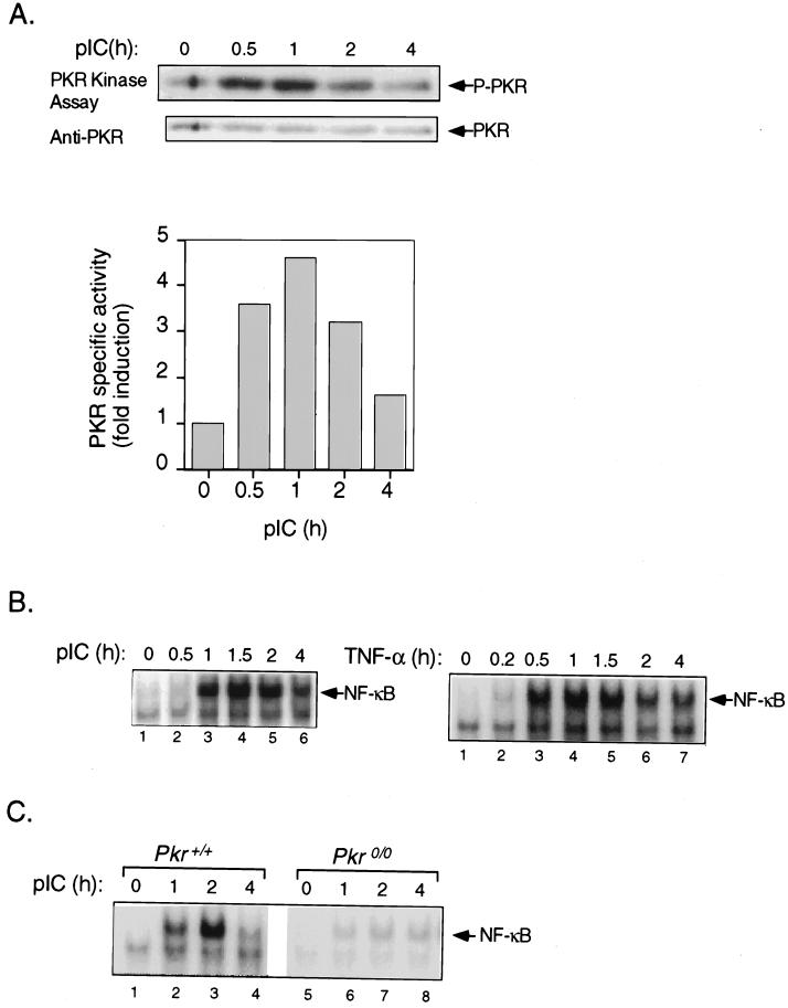
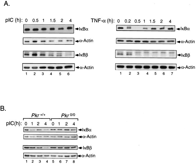
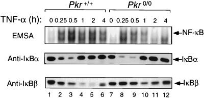
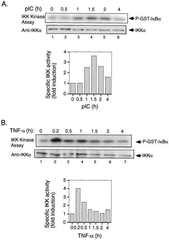
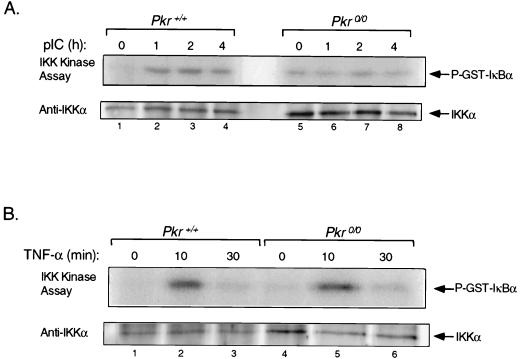
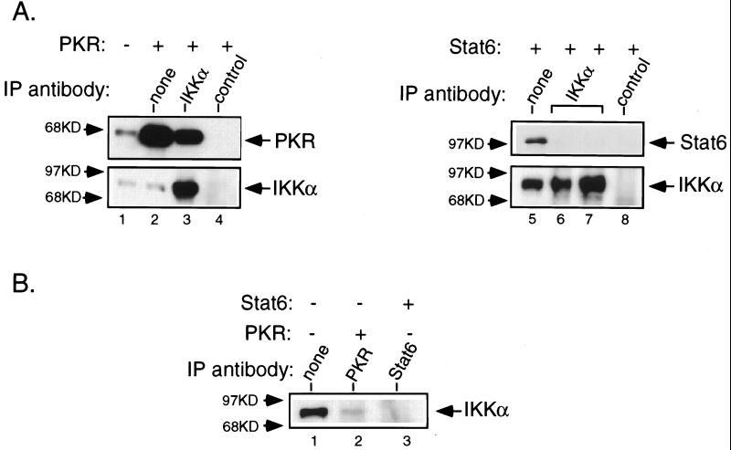
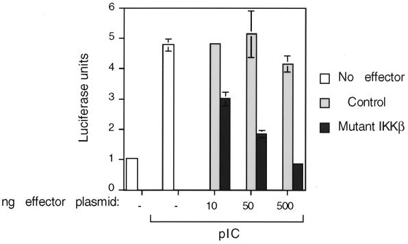
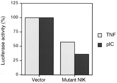
The interferon (IFN)-inducible double-stranded-RNA (dsRNA)-activated serine-threonine protein kinase (PKR) is a major mediator of the antiviral and antiproliferative activities of IFNs. PKR has been implicated in different stress-induced signaling pathways including dsRNA signaling to nuclear factor kappa B (NF-kappaB). The mechanism by which PKR mediates activation of NF-kappaB is unknown. Here we show that in response to poly(rI). poly(rC) (pIC), PKR activates IkappaB kinase (IKK), leading to the degradation of the inhibitors IkappaBalpha and IkappaBbeta and the concomitant release of NF-kappaB. The results of kinetic studies revealed that pIC induced a slow and prolonged activation of IKK, which was preceded by PKR activation. In PKR null cell lines, pIC failed to stimulate IKK activity compared to cells from an isogenic background wild type for PKR in accord with the inability of PKR null cells to induce NF-kappaB in response to pIC. Moreover, PKR was required to establish a sustained response to tumor necrosis factor alpha (TNF-alpha) and to potentiate activation of NF-kappaB by cotreatment with TNF-alpha and IFN-gamma. By coimmunoprecipitation, PKR was shown to be physically associated with the IKK complex. Transient expression of a dominant negative mutant of IKKbeta or the NF-kappaB-inducing kinase (NIK) inhibited pIC-induced gene expression from an NF-kappaB-dependent reporter construct. Taken together, these results demonstrate that PKR-dependent dsRNA induction of NF-kappaB is mediated by NIK and IKK activation.


Figures

FIG. 1
FIG. 1
PKR is essential for activation of NF-κB by pIC. (A) T98G cells were treated with pIC (100 μg/ml) for the times indicated in the figure, and PKR was immunoprecipitated using a monoclonal antibody. The immune complex was subjected to in vitro kinase assay as described in Materials and Methods and analyzed by SDS-PAGE followed by Western blotting for PKR protein. The blot was subsequently subjected to autoradiography to detect PKR autophosphorylation. P-PKR, phosphorylated PKR. (B) T98G cells were treated with pIC (100 μg/ml) or TNF-α (5 ng/ml) for the indicated times, and whole-cell extracts were analyzed for NF-κB DNA binding activity by EMSA using 18 μg of protein and a radiolabeled oligonucleotide probe containing a consensus NF-κB binding site. (C) Cell lines derived from Pkr+/+ and Pkr0/0 mouse embryo fibroblasts with isogenic background were treated with pIC (100 μg/ml), and whole-cell extracts were analyzed for NF-κB DNA binding activity by EMSA as described above for panel B.
FIG. 2
FIG. 2
pIC-induced activation of NF-κB targets IκBα and IκBβ for degradation, an effect mediated by PKR. (A) T98G cells were treated with pIC (100 μg/ml) or TNF-α (5 ng/ml) for the times indicated over the lanes, and equal protein amounts from whole-cell extracts were analyzed by Western blotting using antibodies against IκBα and IκBβ. Blots were stripped and reprobed with an antibody against α-actin to normalize for loading. (B) Pkr+/+ and Pkr0/0 cell lines were treated with pIC (100 μg/ml) for the indicated times. Western blot analyses were performed as described above for panel A.
FIG. 3
FIG. 3
PKR is required for sustained TNF-α signaling to NF-κB. Cell lines derived from Pkr+/+ and Pkr0/0 mouse embryo fibroblasts with isogenic background were treated with TNF-α (5 ng/ml) for the times indicated at the top of the figure. Whole-cell extracts were subjected to EMSA for NF-κB DNA binding activity and Western blotting for IκBα and IκBβ protein levels.
FIG. 4
FIG. 4
PKR is required for the enhanced activation of NF-κB by TNF-α and IFN-γ. Pkr+/+ and Pkr0/0 cell lines were treated with TNF-α (T) (0.025 ng/ml) or IFN-γ (I) (1,000 U/ml) or cotreated with both (T/I) for 1 h or not treated (−) (control). Whole-cell extracts were subjected to EMSA.
FIG. 5
FIG. 5
IKK is activated in response to pIC. (A) T98G cells were treated with pIC (100 μg/ml) for the indicated times. IKK complex was immunoprecipitated with an anti-IKKα monoclonal antibody, and the immune complex was subjected to in vitro kinase assay using GST-IκBα (amino acids 1 to 54) as a substrate. Following SDS-PAGE, the portion of the gel containing the substrate was dried and processed for autoradiography. The portion containing IKK was analyzed by Western blotting for IKKα protein. (B) T98G cells were treated with TNF-α (5 ng/ml) for the indicated times. IKK kinase assay and immunoblotting were performed as described above for panel A. P-GST-IκBα, phosphorylated GST-IκBα.
FIG. 6
FIG. 6
IKK activation in response to pIC is PKR dependent. (A) Cell lines derived from Pkr+/+ and knockout mice with isogenic background were treated with pIC (100 μg/ml) for the indicated times. IKK kinase assay and immunoblotting were performed as described in the legend to Fig. 5A. (B) Pkr+/+ and Pkr0/0 cell lines were treated with TNF-α (5 ng/ml) and processed for IKK kinase assay and immunoblotting as described for panel A.
FIG. 7
FIG. 7
PKR interacts with IKK complex in vivo. Cells (293T) were transiently transfected with 2 μg of plasmids expressing a catalytically inactive mutant of human PKR or human Stat6 as described in Materials and Methods. (A) IKK complex was immunoprecipitated from extracts (0.5 mg [lane 6] or 1 mg [lanes 2 to 4 and 7 to 8]) using an anti-IKKα antibody, and the immune complex was subjected to Western blotting for PKR or Stat6 as indicated. A rabbit IgG polyclonal antibody was used as the control. The blots were subsequently stripped and reprobed for IKKα. (B) PKR and Stat6 were immunoprecipitated from PKR- or Stat6-overexpressing extracts (1 mg), respectively, and the immunocomplexes were subjected to analysis by Western blotting for endogenous IKKα.
FIG. 8
FIG. 8
Dominant negative IKKβ inhibits pIC-stimulated NF-κB-dependent transcription in a dose-dependent manner. T98G cells were transiently cotransfected with an NF-κB-dependent luciferase reporter (5 NF-κB sites) and increasing amounts of a mutant IKKβ expression plasmid or the corresponding empty vector as the control. Approximately 36 h posttransfection, cells were treated with pIC (100 μg/ml for 8 h) and extracts were prepared and analyzed for luciferase activity. The graph reflects firefly luciferase units corrected for transfection efficiency by expression from a Renilla luciferase plasmid included as an internal control in the dual-luciferase assay system. The experiment was performed in duplicate and is representative of several separate experiments.
FIG. 9
FIG. 9
Dominant negative NIK inhibits pIC-stimulated NF-κB-dependent transcription T98G cells were transiently cotransfected with an NF-κB-dependent luciferase reporter (5 NF-κB sites) and 500 ng of a plasmid expressing a kinase-dead mutant of NIK or the corresponding empty vector as the control. Approximately 36 h posttransfection, cells were treated with pIC or TNF-α (100 μg/ml and 10 ng/ml, respectively, for 8 h) and extracts were prepared and analyzed for luciferase activity. The graph reflects firefly luciferase units corrected for transfection efficiency by expression from a Renilla luciferase plasmid included as an internal control in the dual-luciferase assay system. The experiment was performed in duplicate and is representative of several separate experiments.

References

    1. Balachandran S, Kim C N, Yeh W C, Mak T W, Bhalla K, Barber G N. Activation of the dsRNA-dependent protein kinase, PKR, induces apoptosis through FADD-mediated death signaling. EMBO J. 1998;17:6888–6902. - PMC - PubMed
    1. Baldwin A S., Jr The NF-κB and IκB proteins: new discoveries and insights. Annu Rev Immunol. 1996;14:649–683. - PubMed
    1. Barber G N, Wambach M, Thompson S, Jagus R, Katze M G. Mutants of the RNA-dependent protein kinase (PKR) lacking double-stranded RNA binding domain I can act as transdominant inhibitors and induce malignant transformation. Mol Cell Biol. 1995;15:3138–3146. - PMC - PubMed
    1. Beg A A, Finco T S, Nantermet P V, Baldwin A S., Jr Tumor necrosis factor and interleukin-1 lead to phosphorylation and loss of IκBα: a mechanism for NF-κB activation. Mol Cell Biol. 1993;13:3301–3310. - PMC - PubMed
    1. Beraud C, Henzel W J, Baeuerle P A. Involvement of regulatory and catalytic subunits of phosphoinositide 3-kinase in NF-κB activation. Proc Natl Acad Sci USA. 1999;96:429–434. - PMC - PubMed

Publication types

LinkOut - more resources