. 2000 Feb;20(4):1291-8.
doi: 10.1128/MCB.20.4.1291-1298.2000.

The p21(WAF1/CIP1) promoter is methylated in Rat-1 cells: stable restoration of p53-dependent p21(WAF1/CIP1) expression after transfection of a genomic clone containing the p21(WAF1/CIP1) gene

Affiliations

The p21(WAF1/CIP1) promoter is methylated in Rat-1 cells: stable restoration of p53-dependent p21(WAF1/CIP1) expression after transfection of a genomic clone containing the p21(WAF1/CIP1) gene

L A Allan et al. Mol Cell Biol. 2000 Feb.

Abstract

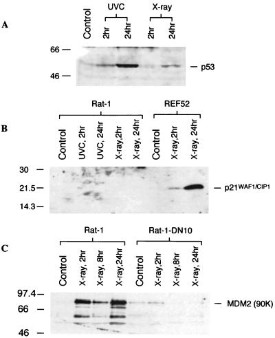
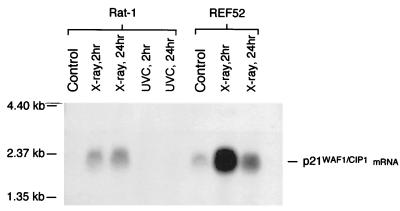
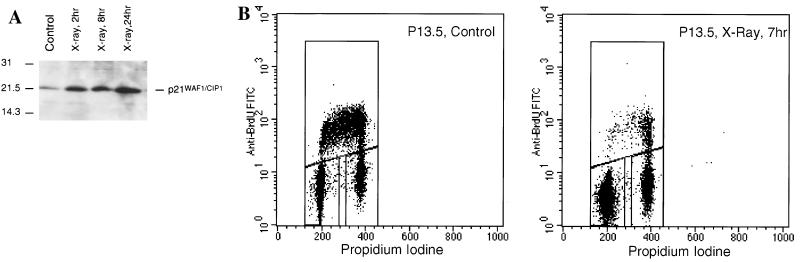
Rat-1 cells are used in many studies on transformation, cell cycle, and apoptosis. Whereas UV treatment of Rat-1 cells results in apoptosis, X-ray treatment does not induce either apoptosis or a cell cycle block. X-ray treatment of Rat-1 cells results in both an increase of p53 protein and expression of the p53-inducible gene MDM2 but not the protein or mRNA of the p53-inducible p21(WAF1/CIP1) gene, which in other cells plays an important role in p53-mediated cell cycle block. The lack of p21(WAF1/CIP1) expression appears to be the result of hypermethylation of the p21(WAF1/CIP1) promoter region, as p21(WAF1/CIP1) protein expression could be induced by growth of Rat-1 cells in the presence of 5-aza-2-deoxycytidine. Furthermore, sequence analysis of bisulfite-treated DNA demonstrated extensive methylation of cytosine residues in CpG dinucleotides in a CpG-rich island in the promoter region of the p21(WAF1/CIP1) gene. Stable X-ray-induced p53-dependent p21(WAF1/CIP1) expression and cell cycle block were restored to a Rat-1 clone after transfection with a P1 artificial chromosome (PAC) DNA clone containing a rat genomic copy of the p21(WAF1/CIP1) gene. The absence of expression of the p21(WAF1/CIP1) gene may contribute to the suitability of Rat-1 cells for transformation, cell cycle, and apoptosis studies.


Figures

FIG. 1
FIG. 1
Effect of X rays on viability and cell cycle profile in Rat-1 cells. (A) X-ray (12 Gy) treatment does not cause apoptosis in Rat-1 cells, shown by the absence of cells with a sub-G1 content (M1). In contrast, UVC treatment (20 J/m2) results in apoptosis, with more than 30% of the cells exhibiting a sub-G1 DNA content. (B) X-ray treatment has little effect on the cell cycle distribution in Rat-1 cells at either 7 or 24 h after irradiation. Although there appears to be a minor accumulation in G2, there is no G1 arrest and cells continue to cycle into S phase.
FIG. 2
FIG. 2
p53 expression and transcriptional activity after X irradiation. (A) X-ray (12 Gy) treatment stabilized p53 in Rat-1 cells 24 h after irradiation, although to a lesser extent than UVC. (B) Rat-1 cells fail to express p21WAF1/CIP1 protein after X irradiation or UVC treatment, whereas REF52 cells, which arrest in G1 after X irradiation, exhibit increasing induction of p21WAF1/CIP1 at 2 and 24 h postirradiation, respectively. (C) X-ray (12 Gy) treatment induces MDM2 in Rat-1 cells in a p53-dependent manner, as shown by the abrogation of this response by expression of a dominant negative p53 in Rat-1-DN10 cells. Sizes are indicated in kilodaltons.
FIG. 3
FIG. 3
Northern blot analysis of MDM2 mRNA after X-ray treatment. X-ray (12 Gy) treatment induces only very low levels of p21WAF1/CIP1 mRNA in Rat-1 cells at either 2 or 24 h postirradiation. This induced level reflects the background level of expression observed in REF52 cells, which show highly elevated levels of p21WAF1/CIP1 mRNA after X irradiation.
FIG. 4
FIG. 4
Quantitation of CpG frequency (observed/expected [obs/exp]) in a contig encompassing the promoter, exon 1, and the 5′ end of intron 1 of the rat p21WAF1/CIP1 gene. CpG frequency was computed over 800-bp intervals and shows a putative CpG island spanning bp 4800 to 7201 comprising the last 20 bp of exon 1 (located in the interval from bp 4800 to 5600) and the 5′ end of intron 1.
FIG. 5
FIG. 5
Effect of 5-AzaC treatment on p21WAF1/CIP1 expression in Rat-1 cells. Incubation of Rat-1 cells with 5-AzaC for 4 days resulted in high levels of p21WAF1/CIP1. X irradiation of 5-AzaC-treated Rat-1 cells showed little increase in p21WAF1/CIP1 expression.
FIG. 6
FIG. 6
Bisulfite sequencing of part of the putative CpG island in the p21WAF1/CIP1 gene in Rat-1 cells. A 120-bp sequence containing 15 CpGs out of a total of 330 bp and 37 CpGs analyzed shown in a comparison of bisulfite-treated Rat-1 DNA (Rat-1.bs, middle sequence) with bisulfite-treated REF52 DNA (REF52.bs, lower sequence) and untreated DNA (upper sequence). Rat-1 cells contain methylated cytosine (detected as cytosine) at all the CpG dinucleotides analyzed, while all other cytosines were unmethylated (detected as thymine [boxed]). Bisulfite-treated REF52 exhibited only unmethylated cytosines (detected as thymine [boxed]) even at the CpG dinucleotides in this region.
FIG. 7
FIG. 7
p21WAF1/CIP1 expression and a G1 arrest are restored after X-ray treatment of P13.5 cells, a clone of Rat-1 cells stably transfected with a 120-kb PAC containing a genomic copy of the rat p21WAF1/CIP1 gene. (A) A low level of background p21WAF1/CIP1 expression was observed in P13.5 cells which is significantly increased after X irradiation (12 Gy). Sizes are indicated in kilodaltons. (B) P13.5 cells exhibit a G1 arrest 7 h after X irradiation (12 Gy).
FIG. 8
FIG. 8
Expression and activity of p21WAF1/CIP1 in P13.5 cells are p53 dependent. (A) Induction of p21WAF1/CIP1 expression by X irradiation (12 Gy) in P13.5 cells is abrogated by expression of dominant negative p53 (P13.5-DN.A1 cells). Sizes are indicated in kilodaltons. (B) The cell cycle arrest exhibited by parental P13.5 cells at 7 h after X irradiation (12 Gy) is absent in P13.5-DN.A1 cells.

References

    1. Allan L A, Fried M. p53-dependent apoptosis or growth arrest induced by different forms of radiation in U2OS cells: p21WAF1/CIP1 repression in UV induced apoptosis. Oncogene. 1999;18:5403–5412. - PubMed
    1. Balbin M, Hannon G J, Pendas A M, Ferrando A A, Vizoso F, Fueyo A, Lopez-Otin C. Functional analysis of a p21WAF1,CIP1,SDI1 mutant (Arg94→Trp) identified in a human breast carcinoma. Evidence that the mutation impairs the ability of p21 to inhibit cyclin-dependent kinases. J Biol Chem. 1996;271:15782–15786. - PubMed
    1. Bissonnette N, Hunting D J. p21-induced cycle arrest in G1 protects cells from apoptosis induced by UV-irradiation or RNA polymerase II blockage. Oncogene. 1998;16:3461–3469. - PubMed
    1. Brugarolas J, Chandrasekaran C, Gordon J I, Beach D, Jacks T, Hannon G J. Radiation-induced cell cycle arrest compromised by p21 deficiency. Nature. 1995;377:552–557. - PubMed
    1. Canman C E, Kastan M B. Small contribution of G1 checkpoint control manipulation to modulation of p53-mediated apoptosis. Oncogene. 1998;16:957–966. - PubMed

MeSH terms