. 2000 Feb;20(4):1321-8.
doi: 10.1128/MCB.20.4.1321-1328.2000.

Regulatory interactions between the Reg1-Glc7 protein phosphatase and the Snf1 protein kinase

Affiliations

Regulatory interactions between the Reg1-Glc7 protein phosphatase and the Snf1 protein kinase

P Sanz et al. Mol Cell Biol. 2000 Feb.

Abstract

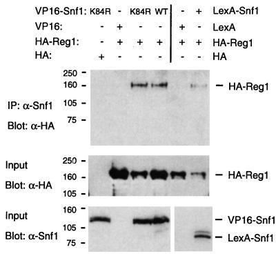
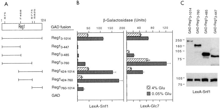
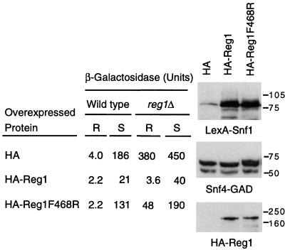
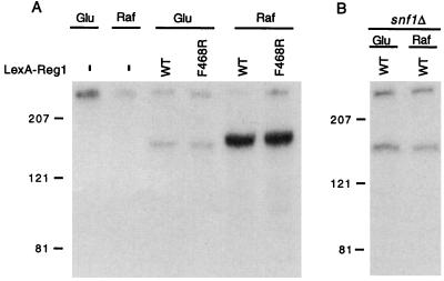
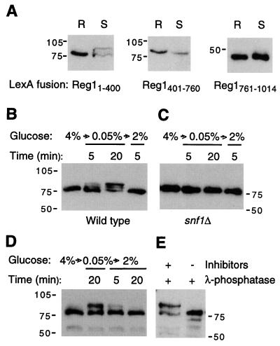
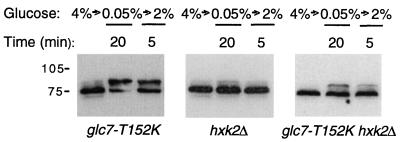
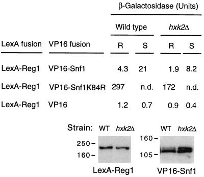
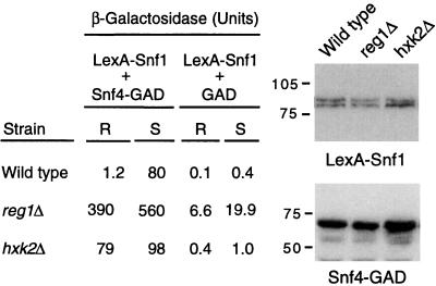
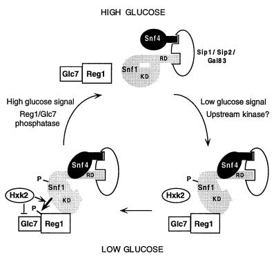
Protein phosphatase 1, comprising the regulatory subunit Reg1 and the catalytic subunit Glc7, has a role in glucose repression in Saccharomyces cerevisiae. Previous studies showed that Reg1 regulates the Snf1 protein kinase in response to glucose. Here, we explore the functional relationships between Reg1, Glc7, and Snf1. We show that different sequences of Reg1 interact with Glc7 and Snf1. We use a mutant Reg1 altered in the Glc7-binding motif to demonstrate that Reg1 facilitates the return of the activated Snf1 kinase complex to the autoinhibited state by targeting Glc7 to the complex. Genetic evidence indicated that the catalytic activity of Snf1 negatively regulates its interaction with Reg1. We show that Reg1 is phosphorylated in response to glucose limitation and that this phosphorylation requires Snf1; moreover, Reg1 is dephosphorylated by Glc7 when glucose is added. Finally, we show that hexokinase PII (Hxk2) has a role in regulating the phosphorylation state of Reg1, which may account for the effect of Hxk2 on Snf1 function. These findings suggest that the phosphorylation of Reg1 by Snf1 is required for the release of Reg1-Glc7 from the kinase complex and also stimulates the activity of Glc7 in promoting closure of the complex.


Figures

FIG. 1
FIG. 1
Coimmunoprecipitation of HA-Reg1 with VP16-Snf1 and LexA-Snf1. Protein extracts (250 μg) were prepared from FY250 snf1Δ cells expressing the indicated proteins. VP16 and LexA fusion proteins were immunoprecipitated (IP) with α-Snf1 polyclonal antibody, and the precipitated proteins were separated by SDS-PAGE and immunoblotted with α-HA monoclonal antibody (Blot) (upper panel). Proteins in the input crude extracts (1 μg) were also immunodetected with either α-HA (middle panel) or α-Snf1 (lower panel). Size standards are indicated in kilodaltons.
FIG. 2
FIG. 2
Two-hybrid interaction of Snf1 and Glc7 with different sequences of Reg1. (A) Reg1 sequences fused to GAD that were used in the two-hybrid analysis. GAD-Reg1 fusions were used because certain regions of Reg1 show self-activating activity. The positions of F468 and serines in potential Snf1 recognition sites (see text) are indicated. (B) CTY10-5d transformants expressing the GAD-Reg1 proteins and either LexA-Snf1 or LexA-Glc7 were grown to mid-log phase in selective SC–4% glucose medium; cells were then washed with water and shifted to SC–0.05% glucose medium for 3 h; values are average β-galactosidase activities of four to six transformants, and bars show standard deviations. (C) Western blots of proteins from transformants growing in 4% glucose and expressing LexA-Snf1 and the indicated GAD-Reg1 fusion; 10-μl aliquots of cell extracts prepared by the fast boiling method (see Materials and Methods) were immunoblotted with α-HA (top) or α-Snf1 (bottom). The production of GAD-Reg1424-1014, GAD-Reg1424-760, and GAD-Reg1760-1014 could not be assessed because these constructs lack the HA epitope tag; the production of GAD-Reg1 fusion proteins in transformants expressing LexA-Glc7 followed the same trend as transformants expressing LexA-Snf1 (data not shown). Size standards are indicated in kilodaltons.
FIG. 3
FIG. 3
Reg1 and Glc7 regulate the two-hybrid interaction of Snf1 and Snf4 within the kinase complex in response to glucose. Two-hybrid interaction between LexA-Snf1 and Snf4-GAD was measured in wild-type CTY10-5d and reg1Δ mutant transformants expressing the indicated HA-Reg1 fusion proteins. Values for cells growing in 4% glucose (R) and cells shifted to 0.05% glucose for 3 h (S) are average β-galactosidase activities of four to six transformants, with standard deviations lower than 15% in all cases. A Western blot of wild-type transformants growing in 4% glucose and expressing the indicated proteins is shown; 10-μl aliquots of crude extracts (boiling method) were immunodetected either with α-Snf1 (upper panel), α-Snf4 (middle panel), or α-HA (lower panel). Analysis of protein levels at the end of the 3-h shift did not reveal any dramatic changes, and the production of the different fusion proteins in reg1Δ transformants was similar to wild type (data not shown). Size standards are indicated in kilodaltons.
FIG. 4
FIG. 4
Snf1-dependent phosphorylation of Reg1 in response to glucose limitation. (A) Strain MCY3278 expressed no LexA fusion protein (−), wild-type (WT) LexA-Reg1, or LexA-Reg1F468R. (B) Strain FY250 snf1Δ expressed LexA-Reg1. Cultures were grown in 2% glucose, labeled with [32P]orthophosphate, collected by centrifugation, and resuspended in medium containing [32P]orthophosphate and either 2% glucose (Glu) or 2% raffinose (Raf) for another 20 min as described in Materials and Methods. Extracts were prepared, and proteins were immunoprecipitated with preconjugated α-LexA–agarose. Precipitates were subjected to SDS-PAGE in 6% acrylamide, and gels were dried for autoradiography. Size standards are indicated in kilodaltons.
FIG. 5
FIG. 5
N terminus of Reg1 is phosphorylated in low glucose in a Snf1-dependent manner. (A) FY250 cells expressing different regions of Reg1 fused to LexA were grown in selective SC–4% glucose medium (R). When they reached the mid-log phase, the cells were washed with water and shifted to SC–0.05% glucose medium for 3 h (S). Crude extracts (10 μl) prepared by the fast boiling method were subjected to immunoblot analysis with α-LexA. (B) FY250 cells expressing LexA-Reg11-400 were grown in selective SC–4% glucose medium and then were shifted to SC–0.05% glucose medium for 5 min or 20 min; after this time, 2% glucose was added to the medium, and cells were collected after 5 min. Crude extracts prepared by the boiling method (10 μl) were analyzed by immunoblotting with α-LexA. (C) FY250 snf1Δ cells expressing LexA-Reg11-400 were treated and analyzed as for panel B. (D) FY250 cells expressing HA-Reg11-443 were treated as for panel B except that an additional sample was taken 20 min after the addition of glucose; proteins in the crude extracts were immunodetected with α-HA. (E) glc7-T152K cells expressing HA-Reg11-443 were grown in selective SC–4% glucose medium and shifted to 0.05% glucose for 20 min; 2-μg aliquots of protein extract were treated with λ-phosphatase (50 U) in the presence or absence of phosphatase inhibitors (see Materials and Methods); HA-Reg11-443 was immunodetected with α-HA. Size standards are indicated in kilodaltons.
FIG. 6
FIG. 6
PP1 and hexokinase PII are involved in regulating the phosphorylation state of Reg1. Strains with the indicated genotypes (see Materials and Methods) were transformed with a plasmid expressing LexA-Reg11-400. Cells were grown and proteins were analyzed and immunodetected with α-LexA as for Fig. 5B. Size standards are indicated in kilodaltons.
FIG. 7
FIG. 7
Two-hybrid interaction of Reg1 and Snf1 in an hxk2Δ mutant. Wild-type CTY10-5d cells and an hxk2Δ mutant derivative of CTY10-5d expressing the indicated fusion proteins were treated as described for Fig. 2. Values for cells growing in 4% glucose (R) and cells shifted to 0.05% glucose for 3 h (S) are average β-galactosidase activities of four transformants; standard deviations were lower than 10% of the average values in all cases. A Western blot of wild-type (WT) and hxk2Δ transformants expressing LexA-Reg1 and VP16-Snf1 is shown; 10-μl aliquots of crude extracts (boiling method) were immunodetected with α-LexA (left) or α-Snf1 (right). Levels of VP16-Snf1K84R were also similar in both strains (data not shown). n.d., not determined. Size standards are indicated in kilodaltons.
FIG. 8
FIG. 8
Two-hybrid interaction of Snf1 and Snf4 in reg1Δ and hxk2Δ mutants. Wild-type CTY10-5d cells and reg1Δ and hxk2Δ derivatives of CTY10-5d were transformed with plasmids expressing LexA-Snf1 and Snf4-GAD or GAD alone. Cells were grown and treated as described in the legend of Fig. 2. Values for cells growing in 4% glucose (R) and cells shifted to 0.05% glucose for 3 h (S) are average β-galactosidase activities of four transformants; standard deviations were lower than 10% of the average values in all cases. A Western blot of the wild-type and reg1Δ and hxk2Δ transformants expressing the indicated fusion proteins is shown; 10-μl aliquots of crude extracts (boiling method) were immunodetected with α-Snf1 (top) or α-Snf4 (bottom). Size standards are indicated in kilodaltons.
FIG. 9
FIG. 9
Model for regulation of the Snf1 kinase complex in response to glucose. Cells growing in high glucose maintain the Snf1 complex predominantly in an autoinhibited conformation in which the regulatory domain (RD) binds to the catalytic kinase domain (KD). Low levels of glucose favor phosphorylation of the Snf1 kinase, possibly by an upstream kinase. The catalytic domain is released from autoinhibition and Snf4 binds to the regulatory domain, leading to an open and active conformation of the complex. The Reg1-Glc7 phosphatase complex then binds to Snf1, and Reg1 is phosphorylated by Snf1. Hxk2 either stimulates the binding and/or phosphorylation of Reg1 or inhibits the dephosphorylation of Reg1 by Glc7. Reg1-Glc7 facilitates the transition back to the autoinhibited state, presumably by dephosphorylating Snf1. The phosphorylation of Reg1 appears to stimulate closure of the complex. Reg1-Glc7 is released from its association with the kinase complex, and this release also requires phosphorylation of Reg1 by Snf1. Glc7 then dephosphorylates Reg1. In glucose-grown reg1, glc7-T152K, or hxk2 mutant cells, the Snf1 kinase, once activated, becomes trapped in the activated state. Each kinase complex contains one of the related proteins Sip1, Sip2, and Gal83.

References

    1. Alms G R, Sanz P, Carlson M, Haystead T A. Reg1p targets protein phosphatase 1 to dephosphorylate hexokinase PII in Saccharomyces cerevisiae: characterizing the effects of a phosphatase subunit on the yeast proteome. EMBO J. 1999;18:4157–4168. - PMC - PubMed
    1. Berben G, Dumont J, Gilliquet V, Bolle P A, Hilger F. The YDp plasmids: a uniform set of vectors bearing versatile gene disruption cassettes for Saccharomyces cerevisiae. Yeast. 1991;7:475–477. - PubMed
    1. Cannon J, Pringle J R, Fiechter A, Khalil M. Characterization of glycogen-deficient glc mutants of Saccharomyces cerevisiae. Genetics. 1994;136:485–503. - PMC - PubMed
    1. Carlson M. Glucose repression in yeast. Curr Opin Microbiol. 1999;2:202–207. - PubMed
    1. Celenza J L, Carlson M. Mutational analysis of the Saccharomyces cerevisiae SNF1 protein kinase and evidence for functional interaction with the SNF4 protein. Mol Cell Biol. 1989;9:5034–5044. - PMC - PubMed

Publication types

MeSH terms