. 2000 Feb 1;20(3):929-36.
doi: 10.1523/JNEUROSCI.20-03-00929.2000.

Somatostatin modulates voltage-gated K(+) and Ca(2+) currents in rod and cone photoreceptors of the salamander retina

Affiliations

Somatostatin modulates voltage-gated K(+) and Ca(2+) currents in rod and cone photoreceptors of the salamander retina

A Akopian et al. J Neurosci. .

Abstract

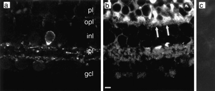
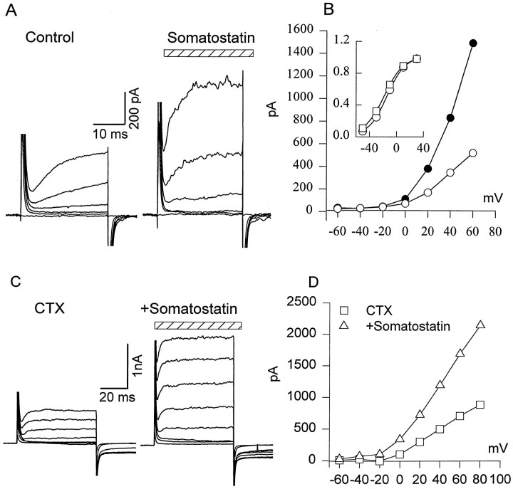
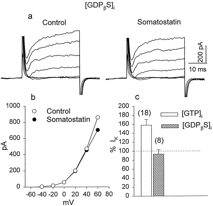
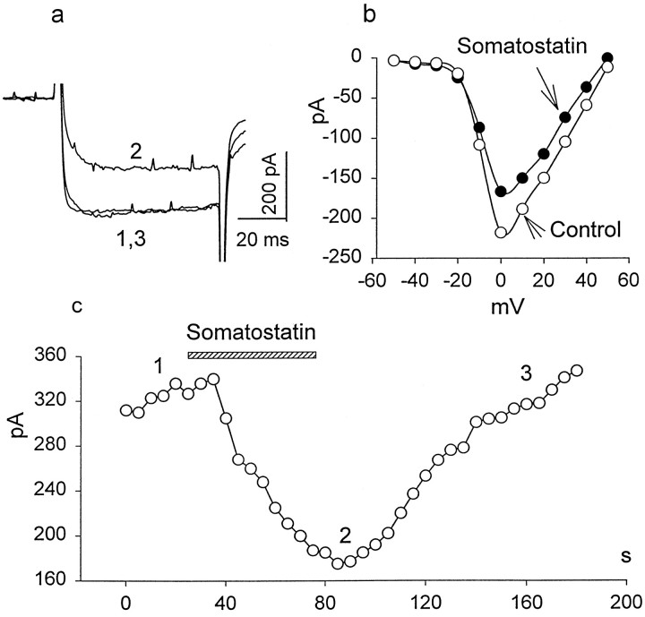
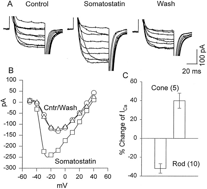
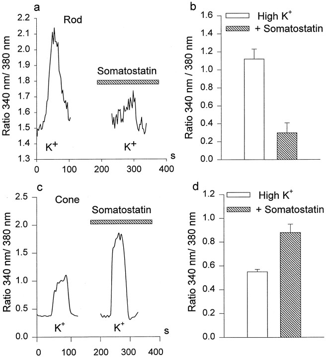
We investigated the cellular localization in the salamander retina of one of the somatostatin [or somatotropin release-inhibiting factor (SRIF)] receptors, sst(2A), and studied the modulatory action of SRIF on voltage-gated K(+) and Ca(2+) currents in rod and cone photoreceptors. SRIF immunostaining was observed in widely spaced amacrine cells, whose perikarya are at the border of the inner nuclear layer and inner plexiform layer. sst(2A) immunostaining was seen in the inner segments and terminals of rod and cone photoreceptors. Additional sst(2A) immunoreactivity was expressed by presumed bipolar and amacrine cells. SRIF, at concentrations of 100-500 nM, enhanced a delayed outwardly rectifying K(+) current (I(K)) in both rod and cone photoreceptors. SRIF action was blocked in cells pretreated with pertussis toxin (PTX) and was substantially reduced by intracellular GDP(beta)S. Voltage-gated L-type Ca(2+) currents in rods and cones were differently modulated by SRIF. SRIF reduced Ca(2+) current in rods by 33% but increased it in cones by 40%, on average. Both effects were mediated via G-protein activation and blocked by PTX. Ca(2+)-imaging experiments supported these results by showing that 500 nM SRIF reduced a K(+)-induced increase in intracellular Ca(2+) in rod photoreceptor terminals but increased it in those of cones. Our results suggest that SRIF may play a role in the regulation of glutamate transmitter release from photoreceptors via modulation of voltage-gated K(+) and Ca(2+) currents.


Figures

Fig. 1.
Fig. 1.
Retinal distribution of SRIF and somatostatin sst2A immunoreactivity. a, A SRIF-immunoreactive amacrine cell body whose processes extend into both distal and proximal portions of the IPL. b,sst2A receptor immunoreactivity that is located in both plexiform layers. Arrows indicate positive staining of photoreceptor bases. c, Absence of sst2Aimmunoreactivity when antibody was preabsorbed with a blocking peptide.Labels indicate retinal layers: gcl, ganglion cell layer; inl, inner nuclear layer;ipl, inner plexiform layer; opl, outer plexiform layer; and pl, photoreceptor layer. Scale bar:a–c, 5 μm.
Fig. 2.
Fig. 2.
Effect of SRIF on delayed outward K+ current (IK) in rods. A, SRIF increased voltage-activated K+ current. IK was evoked by holding cells at −70 mV and applying depolarizing steps from −60 to +40 mV in 20 mV increments in control external solution (left) and after a 4 min exposure to 0.5 μm SRIF (stripedhorizontalbar; right). B,I–V relationship of IKobtained from the experiment described in A is shown. Inset, Somatostatin increasedIK amplitude without substantially changing the current–voltage relation. ■, Control; ○, somatostatin. They-axis of the inset is normalized current. C, CTX reduced outward currents (left) but did not prevent a somatostatin-induced increase in IK. D, I–Vrelationship of outward current in the presence of CTX (■), or CTX + somatostatin (▵); n = 3.
Fig. 3.
Fig. 3.
Sensitivity to PTX of SRIF effect onIK. By the use of a voltage protocol similar to that in Figure 2, a family of IK currents was recorded in rods pretreated for 16–20 hr with PTX (400 ng/ml).a, Exposure to 0.5 μm SRIF for 5 min failed to enhance IK. b, There was no significant difference in the I–Vrelationship of IK recorded in control solution and in the presence of SRIF. c, The histogram summarizes the effects of SRIF on IK in control and PTX-pretreated rods. Numbers inparentheses in this and subsequent figures are the sample size.
Fig. 4.
Fig. 4.
The effect of GDPβS on SRIF-induced changes in IK. a, K+ currents were recorded with a patch pipette containing 500 μm GDPβS in control solution and after a 5 min exposure to 0.5 μm SRIF.b, The I–V relationship ofIK obtained from the experiments described in a indicates that SRIF failed to alterIK in the presence of GDPβS.c, The histogram summarizes SRIF-induced changes inIK when the internal solution contained GTP (openbar) and when GTP was replaced by GDPβS (hatchedbar) to block G-protein activation.
Fig. 5.
Fig. 5.
The inhibitory effect of SRIF on high voltage-activated Ca2+ currents in rods. The test solution contained 0 CaCl2 and 20 mmBaCl2 substituted for equimolar NaCl. a, A depolarizing step to 0 mV from a holding potential of −70 mV was applied to record whole-cell Ca2+ current in control external solution (1), after a 1 min exposure to 0.2 μm SRIF (2), and after a wash (3).b, The I–V relationship of Ca2+ currents evoked by depolarizing voltage steps from −50 to +50 mV in 10 mV increments in the absence (opencircles) and the presence of 0.2 μm SRIF (closedcircles) is shown. c, The time course of SRIF blockade was evaluated by applying 70 msec depolarizing pulses to a test potential of 0 mV from a holding potential of −70 mV each 5 sec. The time of SRIF application is shown by the hatchedhorizontalbar. Thenumbers correspond to the traces ina.
Fig. 6.
Fig. 6.
Excitatory effects of SRIF on the Ca2+ current in cones. A, Cones were held at −70 mV, and depolarizing pulses were applied from −40 to +40 mV in 10 mV steps. Responses were recorded in 20 mm Ba solution as described for Figure 5. Left, In control Ba Ringer's solution. Middle, In 0.5 μmSRIF. Right, After a wash in Ba Ringer's solution.B, Current–voltage plot of the data in Ais shown. C, A summary of the changes induced in the peak Ca current of rods and cones by SRIF is shown.Cntr, Control (○), wash (▵).
Fig. 7.
Fig. 7.
Effects of somatostatin on K+-induced Ca2+ accumulation in rods and cones. a, c, KCl (100 mm) was used to stimulate Ca2+ entry in the absence and the presence (hatchedhorizontalbar) of 0.5 μm somatostatin in rods (a) and cones (c). Somatostatin reduced Ca2+ accumulation in rods but increased it in cones. b, d, Histograms summarize the somatostatin-induced inhibition of Ca2+ accumulation in rods (b; n = 5) and its enhancement in cones (d; n = 5).Verticalbars show the mean values ± 1 SE.

References

    1. Akopian A, Witkovsky P. D2 dopamine receptor-mediated inhibition of a hyperpolarization-activated current in rod photoreceptors. J Neurophysiol. 1996;76:1828–1835. - PubMed
    1. Attwell D. The photoreceptor output synapse. Prog Retin Res. 1990;9:337–362.
    1. Bader CR, Bertrand D, Schwartz EA. Voltage-activated and Ca2+-activated currents studied in solitary rod inner segments from the salamander retina. J Physiol (Lond) 1982;331:253–284. - PMC - PubMed
    1. Barnes S, Hille B. Ionic channels of the inner segment of tiger salamander photoreceptors. J Gen Physiol. 1989;94:719–743. - PMC - PubMed
    1. Barnes S, Merchant V, Mahmud F. Modulation of transmission gain by protons at the photoreceptor output synapse. Proc Natl Acad Sci USA. 1993;90:10081–10085. - PMC - PubMed

Publication types

MeSH terms

LinkOut - more resources