. 2000 Feb 1;20(3):1001-8.
doi: 10.1523/JNEUROSCI.20-03-01001.2000.

The electrogenic sodium bicarbonate cotransporter: developmental expression in rat brain and possible role in acid vulnerability

Affiliations

The electrogenic sodium bicarbonate cotransporter: developmental expression in rat brain and possible role in acid vulnerability

R G Giffard et al. J Neurosci. .

Abstract

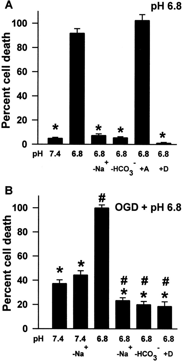
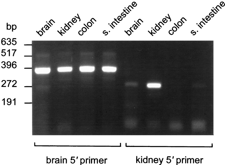
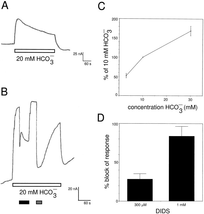
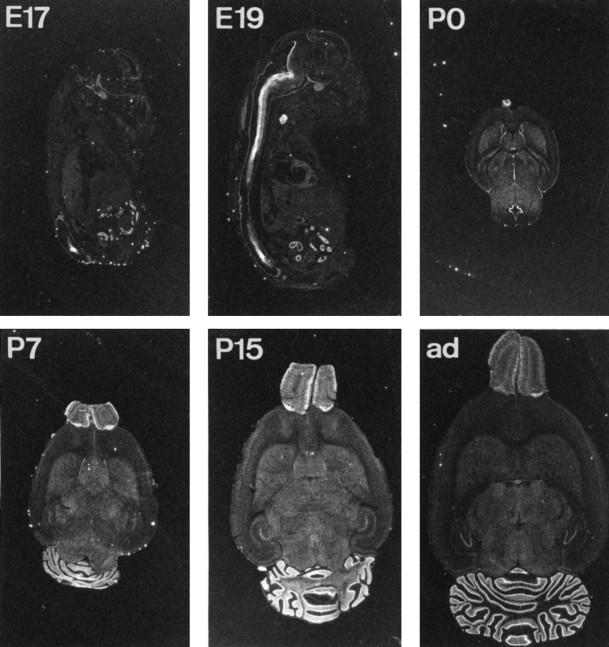
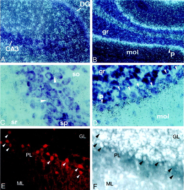
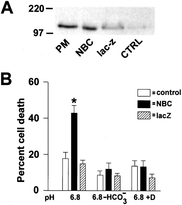
The electrogenic sodium bicarbonate cotransporter (NBC) is expressed in glial cells in the brain and plays an important role in the regulation of both intracellular and extracellular pH. Differential vulnerability to acidosis between neurons and glia has been noted and may contribute to infarction after cerebral ischemia. Ionic substitution studies and inhibition of injury by 4, 4'-di-isothiocyanostilbene-2,2'-disulfonic acid suggest that NBC is involved in astrocyte vulnerability to acidic injury. Recently two NBC cDNAs differing in 5'-untranslated and N-terminal coding sequence have been cloned from kidney and pancreas. We cloned one of these cDNAs from rat brain and demonstrate here that the clone is functional by expression in Xenopus oocytes. We determined the developmental and regional expression of NBC in the brain by in situ hybridization. Expression was observed in the spinal cord at embryonic day 17, whereas expression in brain was first seen at approximately postnatal day 0 (P0), increased at P15, and persisted in the adult brain. Expression was widespread throughout the cerebellum, cortex, olfactory bulb, and subcortical structures. Cellular resolution of the in situ hybridization signal and double labeling for glial fibrillary acidic protein were consistent with a glial localization for NBC. Expression of NBC in 3T3 cells that do not normally express this transporter rendered them vulnerable to acid injury. The expression profile suggests that this transporter is critical during the later stages of brain development and could be one of the factors contributing to the different patterns of injury seen in perinatal versus adult cerebral ischemia.


Figures

Fig. 1.
Fig. 1.
A, Astrocyte injury caused by acidosis alone requires extracellular Na+ and HCO3. Primary astrocyte cultures were kept in the indicated balanced salt solution (BSS5.5) for 24 hr; then injury was quantitated by release of LDH. Maximal LDH release was determined at the end of each experiment after freezing at −70°C and rapid thawing. Cell death was determined by counting the number of cells staining with trypan blue, expressed as a percentage of the total cells counted for the +DIDS condition because DIDS interfered with the LDH assay. Exposure to BSS5.5 at pH 7.4 or 6.8 was performed alone or with the addition of 0.1 mm amiloride or 1 mm DIDS (6.8 + A; 6.8 + D). Verticalbars represent means ± SEM forn = 12; * denotes significant (p < 0.05) difference from pH 6.8 by ANOVA and the Bonferroni test. B, Injury attributable to combined oxygen–glucose deprivation (OGD) at pH 6.8 has an ionic dependence similar to that of injury caused by acidosis alone. Primary astrocyte cultures were placed in an anoxic chamber and washed into the indicated BSS0 at pH 7.4 or 6.8, which had been equilibrated with anoxic gas. After 6 hr the cultures were washed into oxygenated BSS5.5 at pH 7.4 and placed in the normoxic incubator for 24 hr before LDH was measured or cells counted.D indicates addition of 1 mm DIDS. Significant difference (p < 0.05) from pH 6.8 alone is indicated by *; difference from 7.4 − Na is shown by #. Verticalbars are means ± SEM;n = 12–24.
Fig. 2.
Fig. 2.
Amplification of splice variant-specific fragments from cDNA. PCR was performed with primers specific for either the brain or kidney sequence, using cDNA from brain, kidney, small intestine (s. intestine), or colon. The kidney-specific product is 258 bp; the brain-specific product is 372 bp. A negative control that lacked cDNA did not produce any amplification bands (data not shown).
Fig. 3.
Fig. 3.
Expression of brain NBC in Xenopusoocytes leads to bicarbonate-inducible outward currents.A, An oocyte injected with NBC showed outward currents on application of bicarbonate, as indicated by the openhorizontalbar. B, The bicarbonate-induced outward current was markedly reduced when external sodium was reduced from 130 to 20 mm (blackhorizontalbar) or when DIDS (1 mm) was applied (grayhorizontalbar). C, The size of the outward current depends on the bicarbonate concentration. Amplitudes of the 3 and 30 mm bicarbonate-induced outward currents were normalized to that of the 10 mmbicarbonate-induced inward current (n = 3).D, Summary of the block of the bicarbonate-induced outward current by DIDS at 300 μm (n= 3) and 1 mm (n = 3) is shown.
Fig. 4.
Fig. 4.
Developmental expression of NBC in rat brain.In situ hybridization was performed on frozen sections from rat brains of the indicated ages. Control sections hybridized with probe to which a 200-fold excess of unlabeled probe was added showed essentially no signal (data not shown). ad,Adult.
Fig. 5.
Fig. 5.
Cellular resolution of hybridization signal. Emulsion-dipped sections from P15 rats were exposed for 6 weeks and then developed and counterstained. Regions from the hippocampus (A, C) and the cerebellum (B, D) are shown at two magnifications. Dark-field photomicrographs show the hybridization signal as whitedots(A, B), whereas bright field shows the hybridization signal as darkdots (C, D). A, C, Labeling in the hippocampus reveals a lack of association of the hybridization grains with neurons. Arrowheads in C indicate clusters of grains between pyramidal neuronal cell bodies.B, D, Labeling in the cerebellum is consistent with expression in astrocytes and in particular in Bergman glia. Few grains are observed directly over the Purkinje cells indicated by arrowheads in D. The fields were photographed with a 10× and 40× objective using a Zeissaxioplan microscope. DG, Dentate gyrus;gr, granular layer; mol, molecular layer;p, Purkinje cell layer; so, stratum oriens; sp, stratum pyramidale; sr,stratum radiatum. E, F, To confirm the identity of the hybridizing cells, double labeling was performed using anti-GFAP antibody to identify astrocytes on the same section that was hybridized with a digoxigenin-labeled probe for NBC. An area from the cerebellum is shown; arrowheads indicate individual cells showing both cytoplasm immunoreactivity for GFAP (E) and a hybridization signal to the riboprobe for NBC (F). GL, granular layer;ML, molecular layer; PL, Purkinje layer. Similar sections stained only for GFAP showed the same pattern of staining (data not shown).
Fig. 6.
Fig. 6.
A, Little NBC is detected by immunoblot in 3T3 cells (CTRL), whereas retroviral expression of NBC induces a large increase in the immunoreactive band for NBC. Purified brain plasma membrane (PM) is shown in the firstlane as a positive control, followed by NBC-expressing 3T3 cells (NBC), β-galactosidase-expressing 3T3 cells (lacZ), and control 3T3 cells. Equal amounts of protein were loaded.B, Overexpression of NBC renders 3T3 cells vulnerable to acid injury. Untransfected and control β-galactosidase-expressing 3T3 cultures exposed to BSS5.5 at pH 6.8 for 24 hr showed little injury, whereas 3T3 cells overexpressing NBC suffered ∼40% cell death. Substituting for bicarbonate or adding 1 mmDIDS significantly reduced the injury of NBC-overexpressing cells. * indicates significant difference from control and lacZ;p < 0.05; n = 10–16.

Similar articles

Cited by

References

    1. Abuladze N, Lee I, Newman D, Hwang J, Boorer K, Pushkin A, Kurtz I. Molecular cloning, chromosomal localization, tissue distribution, and functional expression of the human pancreatic sodium bicarbonate cotransporter. J Biol Chem. 1998;273:17689–17695. - PubMed
    1. Bing OH, Brooks WW, Messer JV. Heart muscle viability following hypoxia: protective effect of acidosis. Science. 1973;180:1297–1298. - PubMed
    1. Burnham CE, Amlal H, Wang Z, Shull GE, Soleimani M. Cloning and functional expression of a human kidney Na+:HCO3-cotransporter. J Biol Chem. 1997;272:19111–19114. - PubMed
    1. Catania MV, Tolle TR, Monyer H. Differential expression of AMPA receptor subunits in NOS-positive neurons of cortex, striatum, and hippocampus. J Neurosci. 1995;15:7046–7061. - PMC - PubMed
    1. Collins A, Somlyo AV, Hilgemann DW. The giant cardiac membrane patch method: stimulation of outward Na(+)-Ca2+ exchange current by MgATP. J Physiol (Lond) 1992;454:27–57. - PMC - PubMed

Publication types

MeSH terms

Associated data