. 2000 Feb 1;20(3):1179-89.
doi: 10.1523/JNEUROSCI.20-03-01179.2000.

Opioids suppress IPSCs in neurons of the rat medial septum/diagonal band of Broca: involvement of mu-opioid receptors and septohippocampal GABAergic neurons

Affiliations

Opioids suppress IPSCs in neurons of the rat medial septum/diagonal band of Broca: involvement of mu-opioid receptors and septohippocampal GABAergic neurons

M Alreja et al. J Neurosci. .

Abstract

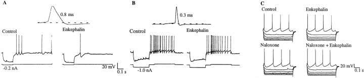
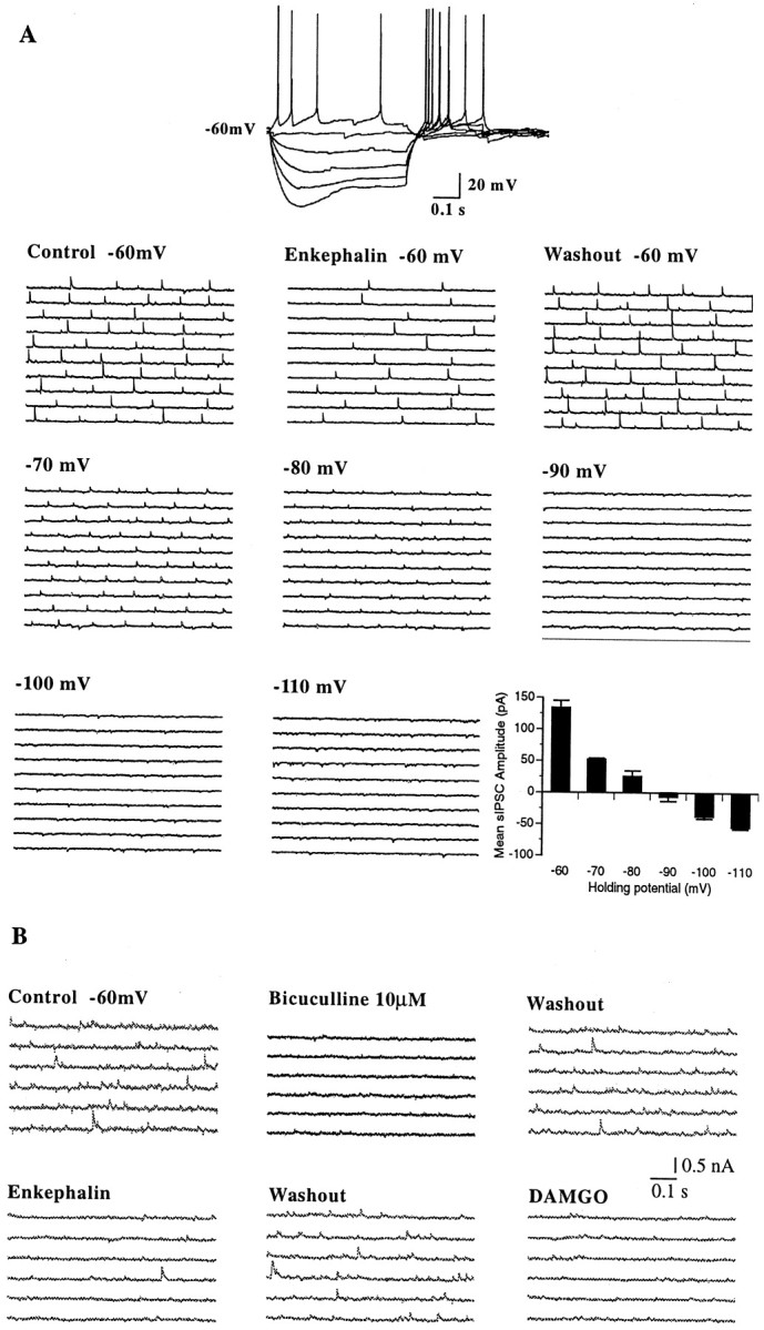
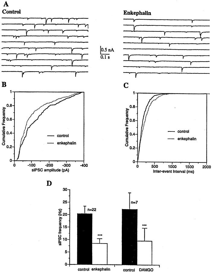
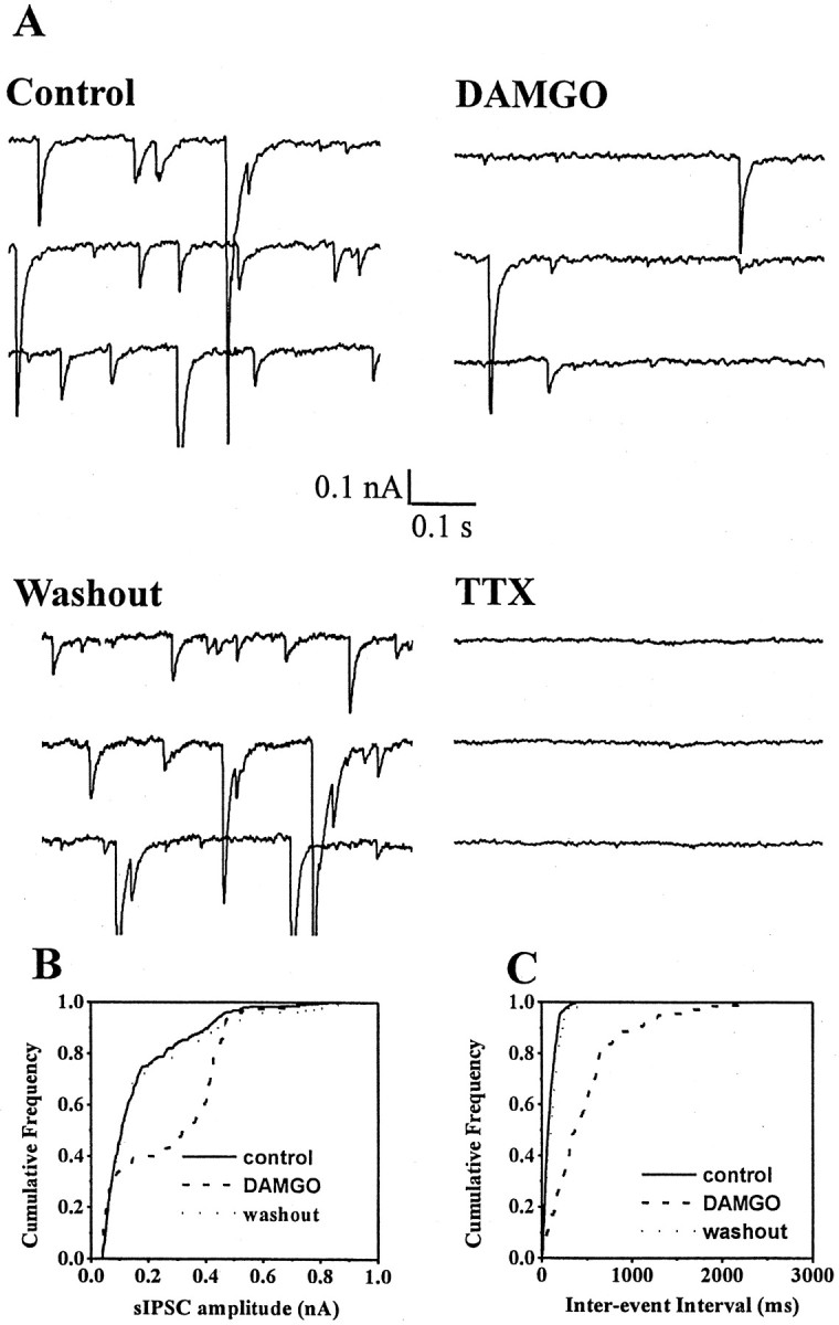
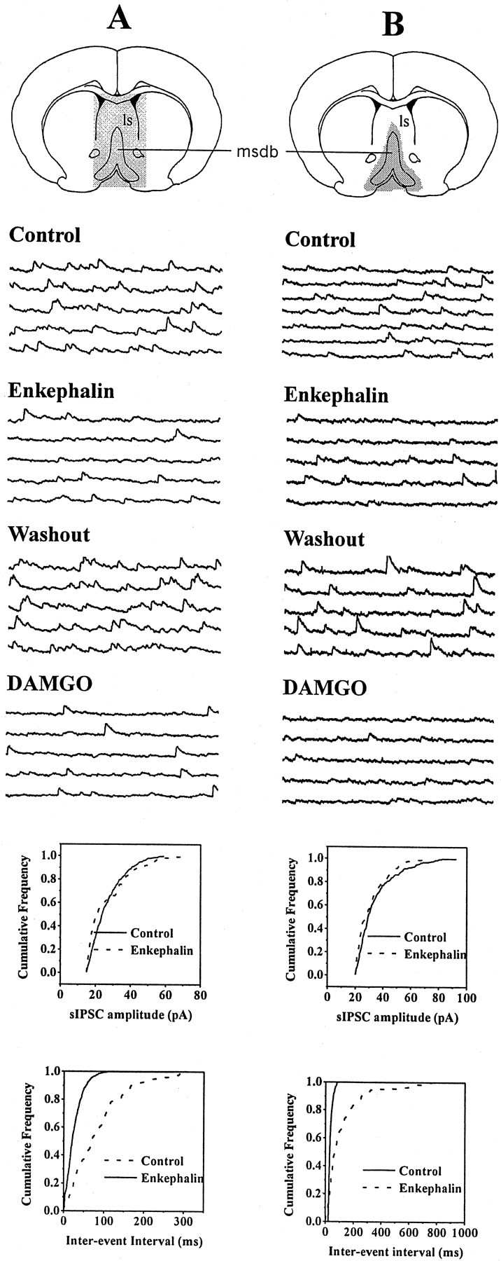
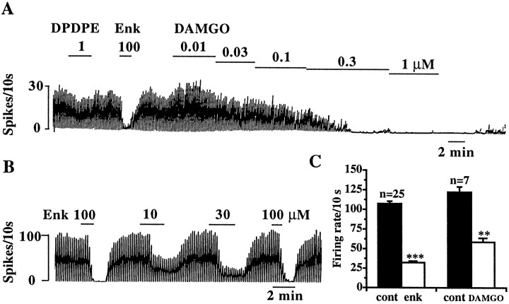
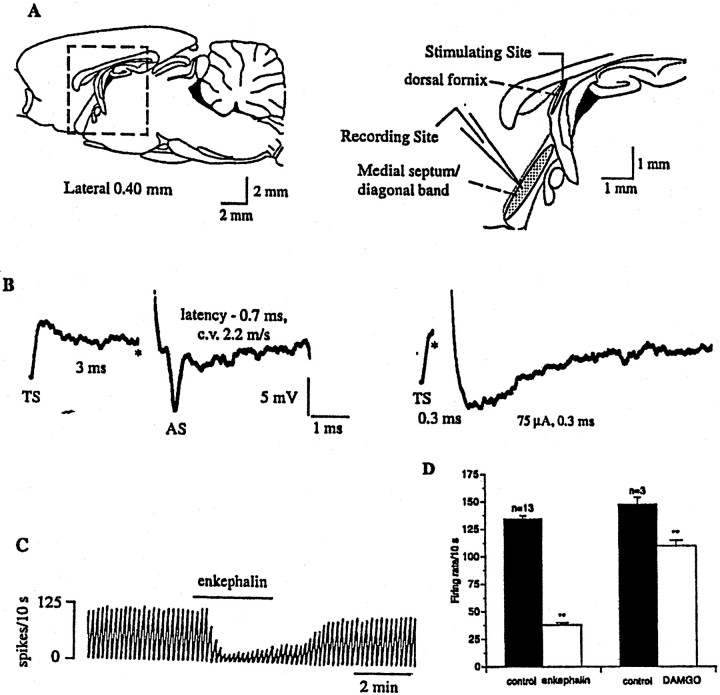
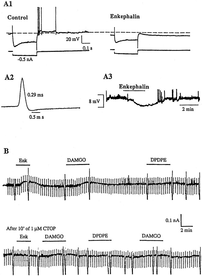
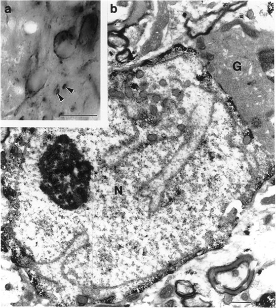
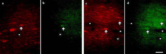
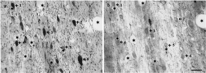
The medial septum/diagonal band region (MSDB), which provides a major cholinergic and GABAergic input to the hippocampus, expresses a high density of opioid receptors. Behaviorally, intraseptal injections of opioids produce deficits in spatial memory, however, little is known about the electrophysiological effects of opioids on MSDB neurons. Therefore, we investigated the electrophysiological effects of opioids on neurons of the MSDB using rat brain slices. In voltage-clamp recordings with patch electrodes, bath-applied met-enkephalin, a nonselective opioid receptor agonist, decreased the number of tetrodotoxin and bicuculline-sensitive inhibitory synaptic currents in cholinergic- and GABA-type MSDB neurons. A similar effect occurred in brain slices containing only the MSDB, suggesting that opioids decrease GABA release primarily by inhibiting spontaneously firing GABAergic neurons located within the MSDB. Accordingly, in extracellular recordings, opioid-sensitive, spontaneously firing neurons could be found within the MSDB. Additionally, in intracellular recordings a subpopulation of GABA-type neurons were directly inhibited by opioids. All effects of met-enkephalin were mimicked by a mu receptor agonist, but not by delta or kappa agonists. In antidromic activation studies, mu-opioids inhibited a subpopulation of septohippocampal neurons with high conduction velocity fibers, suggestive of thickly myelinated GABAergic fibers. Consistent with the electrophysiological findings, in double-immunolabeling studies, 20% of parvalbumin-containing septohippocampal GABA neurons colocalized the mu receptor, which at the ultrastructural level, was found to be associated with the neuronal cell membrane. Thus, opioids, via mu receptors, inhibit a subpopulation of MSDB GABAergic neurons that not only make local connections with both cholinergic and noncholinergic-type MSDB neurons, but also project to the hippocampus.


Figures

Fig. 1.
Fig. 1.
Bath applications of met-enkephalin, a nonselective opioid agonist, decreases synaptic activity in both cholinergic and noncholinergic-type MSDB neurons. A,B, Intracellular recordings with KCl-containing electrodes show cholinergic and noncholinergic-type MSDB neurons.A, A spontaneously firing broad-spiked cholinergic-type MSDB neuron (spike duration, 0.8 msec). Note the prominent slow afterhyperpolarization, which is characteristic of septal cholinergic neurons. Note that bath-applied met-enkephalin (100 μm) decreased depolarizing synaptic potentials in this neuron.B, Met-enkephalin also decreased the number of depolarizing synaptic potentials in a quiescent, sharp-spiked, noncholinergic-type (presumably GABAergic) MSDB neuron (spike duration, 0.3 msec). Note the prominent fast afterhyperpolarization and the depolarizing sag (in response to the hyperpolarizing pulse) that are characteristic of MSDB GABAergic neurons. C, The nonselective opioid antagonist naloxone blocked the inhibitory effect of enkephalin on spontaneously occurring synaptic potentials.
Fig. 2.
Fig. 2.
Met-enkephalin-sensitive synaptic activity reverses polarity near the chloride equilibrium potential and is blocked by the GABAA receptor antagonist bicuculline.A, Whole-cell current-clamp recording from a GABA-type MSDB neuron with a K-gluconate-containing patch electrode. Note the fast afterhyperpolarization and the depolarizing sag. Consecutive 0.5 sec traces show spontaneously occurring IPSCs recorded under different experimental conditions. The first sets of traces show IPSCs recorded at −60 mV under control conditions, with met-enkephalin and after washout of met-enkephalin. Subsequent sets of traces show IPSCs recorded under control conditions at different holding potentials (ranging from −60 to −120 mV). Note the changes in amplitude and polarity of the synaptic currents at different holding potentials. Thebar chart summarizes the data shown above. The mean IPSC amplitude under control conditions is plotted against different holding potentials. Note that the mean amplitude of the met-enkephalin-sensitive IPSCs decreased at more negative holding potentials and reversed polarity between −80 and −90 mV, which is close to the calculated ECl- of −80 mV. This experiment was done in presence of 20 μm CNQX and 50 μm AP-5 to block excitatory synaptic currents.B, In this cell, bicuculline blocked the spontaneously occurring IPSCs in a reversible manner. Subsequent sets of traces show the inhibitory effect of met-enkephalin. Note that the μ-selective opioid receptor agonist DAMGO mimicked the effect of met-enkephalin. All drugs were applied consecutively to the same cell.
Fig. 3.
Fig. 3.
Opioids alter both the frequency and amplitude distribution of sIPSCs. A, Whole-cell voltage-clamp recording from an MSDB neuron with a CsCl-containing patch electrode. EPSCs were blocked using glutamate receptor antagonists (see Materials and Methods). Consecutive 0.5 sec traces show spontaneously occurring IPSCs recorded at −90 mV before and after bath application of met-enkephalin. Note the reversed polarity of the IPSCs.B, C, Cumulative amplitude and frequency distributions of sIPSCs constructed from data shown inA. Note that both the amplitude and frequency distribution were statistically different under the two experimental conditions (p < 0.0001 for amplitude distribution and p < 0.005 for frequency distribution, 274 events analyzed for control and 222 events for met-enkephalin). This presumably reflects loss of action potential-dependent IPSCs after hyperpolarization of GABAergic neurons.D, Bar chart summarizes the effect of met-enkephalin and the μ agonist DAMGO on the frequency of sIPSCs recorded with K gluconate or CsCl-containing patch electrodes. Both met-enkephalin and DAMGO produced a significant decrease in the frequency of sIPSCs (p < 0.001, Student's ttest).
Fig. 4.
Fig. 4.
TTX blocks spontaneously occurring IPSCs in MSDB neurons. A, Whole-cell voltage-clamp recording from an MSDB neuron with a CsCl-containing patch electrode. EPSCs were blocked using glutamate receptor antagonists (see Materials and Methods). Consecutive 0.5 sec traces show spontaneously occurring IPSCs recorded at −90 mV before and after bath application of DAMGO and in presence of TTX. Note that DAMGO blocked spontaneously occurring IPSCs and that a subsequent application of TTX blocked the IPSCs. B,C, Cumulative amplitude and frequency distributions of sIPSCs constructed from data shown in A. Note that both the amplitude and frequency distribution were statistically different in the presence of DAMGO (p < 0.0001 for amplitude distribution and p < 0.005 for frequency distribution as compared to control). This presumably reflects loss of action potential-dependent IPSCs after hyperpolarization of GABAergic neurons. The distributions recorded under control and washout conditions were not statistically different from each other.
Fig. 5.
Fig. 5.
Opioid-sensitive IPSCs originate from within the MSDB. A, Consecutive 1 sec traces show spontaneously occurring IPSCs recorded at a holding potential of −60 mV under different experimental conditions. This whole-cell recording was made in a slice preparation that contained the MSDB as well as other neighboring brain structures (stippled area), such as the lateral septum (ls). Note that met-enkephalin decreased the number of sIPSCs. Also note that this effect was mimicked by the selective μ-opioid receptor agonist DAMGO, suggesting involvement of μ-opioid receptors. B, This whole-cell recording was made from a brain slice that contained only the MSDB (stippled area). Note that the spontaneously occurring IPSCs were inhibited both by met-enkephalin and DAMGO. Because the slice preparation contained only the MSDB, it can be concluded that met-enkephalin and DAMGO suppress sIPSCs that originate from within the MSDB. The amplitude and frequency distributions were statistically different under control and test conditions.
Fig. 6.
Fig. 6.
Opioids inhibit a subpopulation of spontaneously firing MSDB neurons. A, B, Extracellular recording from a spontaneously firing MSDB neuron showing the inhibitory effect of met-enkephalin. Note that the μ agonist DAMGO mimicked the inhibitory effect of met-enkephalin in a concentration-dependent manner and that DPDPE had very little effect. C, Summarizes the effect of met-enkephalin (100 μm) and DAMGO (100 nm) on spontaneously firing MSDB neurons. The smaller effect of DAMGO as compared to met-enkephalin was caused by the submaximal concentration used (A). An inhibitory effect of opioids was observed in 57% of spontaneously firing MSDB neurons tested.
Fig. 7.
Fig. 7.
Opioids inhibit a subpopulation of spontaneously firing, antidromically activated septohippocampal neurons.A, Sagittal section through the rat brain, showing the septal area. The boxed area is enlarged (right) and shows the MSDB, which was the recording site. For antidromic activation of septohippocampal neurons, the stimulating electrode was placed in the dorsal fornix because it conveys both cholinergic and GABAergic MSDB fibers to the hippocampus.B, Extracellular recording from a spontaneously firing antidromically activated septohippocampal neuron. A spontaneous spike was used to trigger the oscilloscope (TS), the dorsal fornix was stimulated (*) 3 msec later, and an antidromically activated spike (AS) was obtained after a latency of 0.7 msec (left trace). This cell was classified as GABA-type based on the calculated conduction velocity of 2.2 m/sec. Theright trace shows a positive collision test, wherein the cell could not be activated antidromically when the dorsal fornix was stimulated 0.3 msec after the triggering spike (approximately half the antidromic latency). This and other cells were also confirmed to be antidromically activating using additional criteria (see Materials and Methods). Stimulation current, 75 μA, 0.3 msec. C, Chart record showing the effect of met-enkephalin on the firing rate of a spontaneously firing septohippocampal neuron (same cell as shown inB, above). D, Summarizes the effect of a near-maximal concentration of met-enkephalin (100 μm) and a submaximal concentration of DAMGO (100 nm) on antidromically activated SHNs. The smaller effect of DAMGO as compared to met-enkephalin was attributable to the submaximal concentration used (Fig. 6). An inhibitory effect of opioids was observed in 76% of spontaneously firing SHNs tested.
Fig. 8.
Fig. 8.
Opioids inhibit a subpopulation of GABA-type MSDB neurons and induce a direct, μ receptor-mediated outward current. A1, A2, Whole-cell current-clamp recording from a GABA-type MSDB (spike duration, 0.29 msec). Also note the pronounced depolarizing sag in response to the hyperpolarizing pulses (0.5 nA steps). A3 shows the resting membrane potential recorded from the same cell. Note that the met-enkephalin effect reversed on washout. B1 and B2 show effect of different opioid receptor agonists in another GABA-type neuron that was voltage-clamped at −60 mV (characteristics not shown). Met-enkephalin produced a 50 pA outward current. This effect was mimicked by the μ-opioid receptor agonist DAMGO, but not by the δ-receptor agonist DPDPE. The κ receptor agonist U50488H also had no effect on this cell (data not shown). Pretreatment with the μ receptor antagonist CTOP blocked the inhibitory effects of met-enkephalin and DAMGO. Input conductance was measured by stepping the membrane potential to −70 mV for 1 sec every 20 sec. Note that both met-enkephalin and DAMGO increased the input conductance of the cell. The taller intermittent deflections indicate the time at which the cell membrane was stepped to −120 mV. In this cell, the opioid-induced outward current reversed near EK (−92 mV; data not shown). Both the outward current and the accompanying conductance change were blocked by the opioid antagonist CTOP. An inhibitory effect of opioids was observed in 24.3% of GABA-type neurons tested; this effect persisted in the presence of TTX in the three cells tested.
Fig. 9.
Fig. 9.
Light (a) and electron (b) micrographs demonstrate the result of immunostaining for μ-opioid receptor in the medial septum. Immunoreactivity for μ-opioid receptor is present in somata and large processes (arrowheads). The electron micrograph shows a μ-opioid receptor-immunoreactive neuron (N) characterized by an infolded nucleus. A neighboring glia cell (G) is free of reaction product. Note that in the immunostained neuron, the reaction product is mostly associated with the inner cell membrane. Scale bars: a, 20 μm;b, 1 μm.
Fig. 10.
Fig. 10.
Colocalization of μ-opioid receptor with parvalbumin-containing MSDB neurons using the technique of double immunofluorescence. Two pairs of color light micrographs (a and b; c andd) demonstrate three neurons (arrowheads) immunoreactive for both parvalbumin (immunolabeled with Texas Red;a and c) and μ-opioid receptor (immunostained with FITC; b and d) in the medial septum. Asterisks label the same capillaries. Scale bar, 20 μm.
Fig. 11.
Fig. 11.
Colocalization of μ-opioid receptor with parvalbumin-containing MSDB neurons using mirror colocalization technique. Light micrographs show the result of a mirror colocalization experiment for parvalbumin (a) and μ-opioid receptor (b) on a pair of consecutive vibratome sections of the medial septal area. A large population (numbered arrows) of parvalbumin-immunoreactive neurons exhibits immunoreactivity for μ-opioid receptor. Asteriskslabel identical capillaries. Scale bar, 20 μm.

References

    1. Alreja M. Excitatory actions of serotonin on GABAergic neurons of the medial septum and diagonal band of Broca. Synapse. 1996;22:15–27. - PubMed
    1. Alreja M, Aghajanian GK. Intracellular diffusion of macromolecules through patch pipettes in brain slices. In: Schurr A, Rigor BM, editors. Brain slices in basic and clinical research. CRC; Boca Raton, FL: 1995. pp. 117–130.
    1. Alreja M, Liu W. Noradrenaline induces IPSCs in rat medial septal/diagonal band neurons: involvement of septohippocampal GABAergic neurons. J Physiol (Lond) 1996;494:201–215. - PMC - PubMed
    1. Baldino F, Jr, Beckman AL. An analysis of the origins of the cholinergic and non-cholinergic septal projections to the hippocampal formation of the rat. Brain Res. 1982;232:247–252.
    1. Bloom FE, Rossier J, Battenberg ELF, Bayon A, French E, Henriksen SJ, Siggins GR, Segal D, Browne R, Ling N, Guillemin R. β-endorphin: cellular localization, electrophysiological and behavioral effects. In: Costa E, Trabucchi M, editors. Advances in biochemical psychopharmacology. Raven; New York: 1978. pp. 89–109. - PubMed

Publication types

MeSH terms