. 2000 Jan 15;14(2):187-97.

A mechanism of suppression of TGF-beta/SMAD signaling by NF-kappa B/RelA

Affiliations

A mechanism of suppression of TGF-beta/SMAD signaling by NF-kappa B/RelA

M Bitzer et al. Genes Dev. .

Abstract

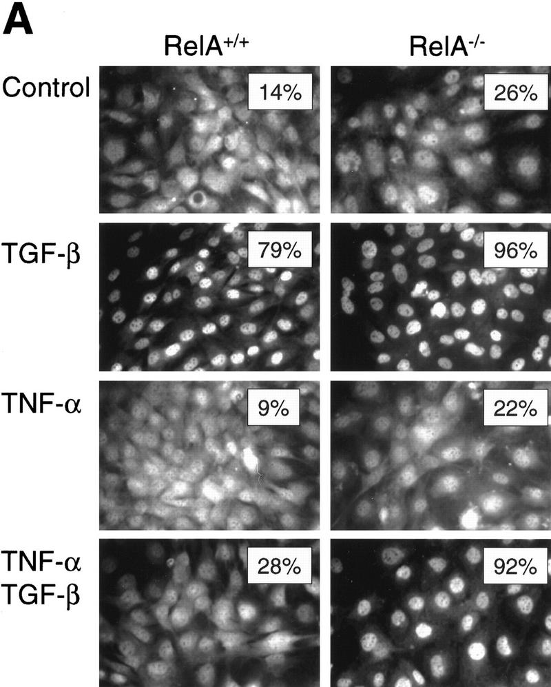
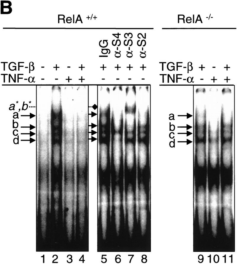
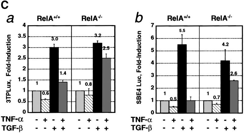
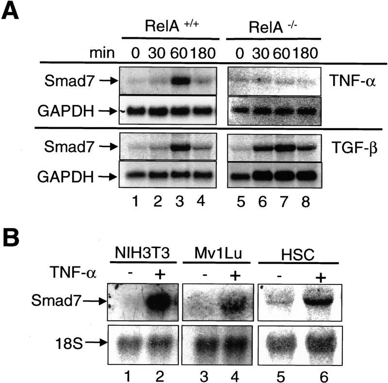
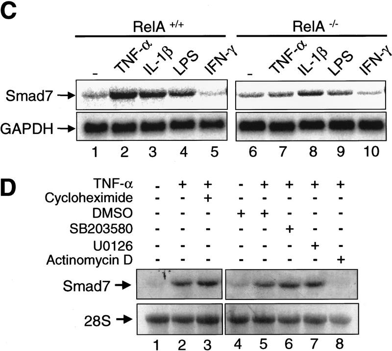
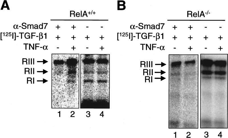
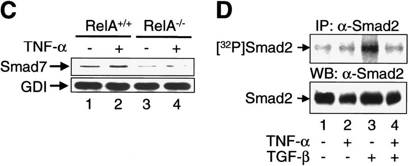
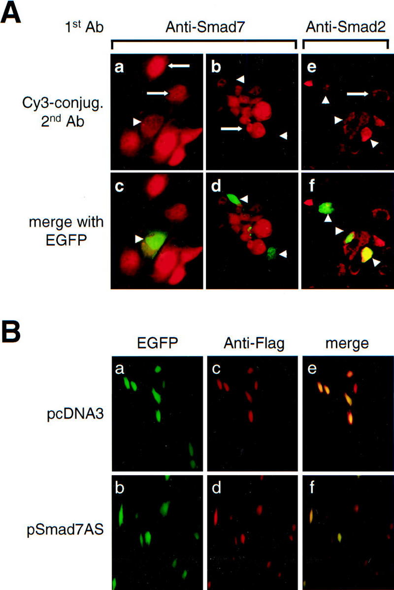
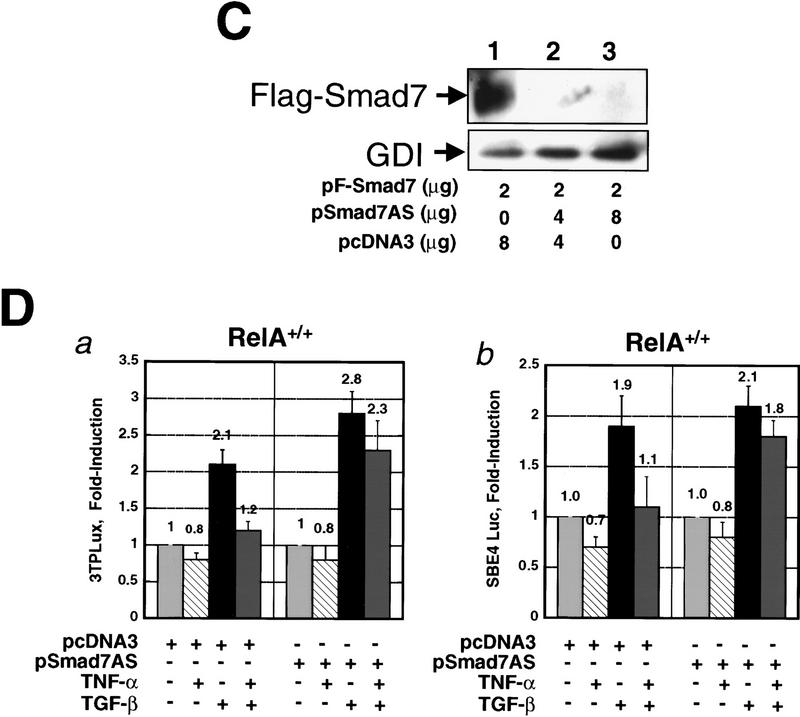
A number of pathogenic and proinflammatory stimuli, and the transforming growth factor-beta (TGF-beta) exert opposing activities in cellular and immune responses. Here we show that the RelA subunit of nuclear factor kappaB (NF-kappaB/RelA) is necessary for the inhibition of TGF-beta-induced phosphorylation, nuclear translocation, and DNA binding of SMAD signaling complexes by tumor necrosis factor-alpha (TNF-alpha). The antagonism is mediated through up-regulation of Smad7 synthesis and induction of stable associations between ligand-activated TGF-beta receptors and inhibitory Smad7. Down-regulation of endogenous Smad7 by expression of antisense mRNA releases TGF-beta/SMAD-induced transcriptional responses from suppression by cytokine-activated NF-kappaB/RelA. Following stimulation with bacterial lipopolysaccharide (LPS), or the proinflammatory cytokines TNF-alpha and interleukin-1beta (IL-1beta, NF-kappaB/RelA induces Smad7 synthesis through activation of Smad7 gene transcription. These results suggest a mechanism of suppression of TGF-beta/SMAD signaling by opposing stimuli mediated through the activation of inhibitory Smad7 by NF-kappaB/RelA.


Figures

Figure 1
Figure 1
The RelA subunit of NF-κB is required for inhibition of TGF-β/SMAD signaling by TNF-α. (A) Smad2 immunofluorescence in RelA+/+ and RelA−/− fibroblasts left untreated (Control), or treated with either 0.1 ng/ml TGF-β, 0.2 ng/ml TNF-α, or both (TNF-α and TGF-β). (Insets) The percentage of cells with exclusively nuclear staining for Smad2. (B) EMSA with a radiolabeled oligonucleotide probe containing a consensus SMAD protein binding site (Zawel et al. 1998). Nuclear protein extracts derived from both RelA+/+ (lane 2) and RelA−/− (lane 9) fibroblasts, treated with TGF-β, contained four distinct DNA-binding protein complexes marked as a, b, c, and d. Pretreatment of cells with TNF-α prior to TGF-β-treatment prevents formation of DNA-binding protein complexes in RelA+/+ (lane 4), but not in RelA−/− fibroblasts (lane 11). Antibody interference analysis showing the effect of preincubation of nuclear extracts from TGF-β-treated RelA+/+ fibroblasts with nonimmune IgG (lane 5), anti-Smad4 (lane 6), anti-Smad3 (lane 7), and anti-Smad2 (lane 8). (Lane 7) a*, b* denotes supershifted complex(es). (C) Transient transcriptional response assays with the TGF-β-responsive reporter constructs 3TPLux (histogram a), or SBE4-Luc (histogram b) (Zawel et al. 1998). RelA+/+ (a and b, left) and RelA−/− fibroblasts (a and b, right) were treated as indicated 20 hr after transfection. Luciferase activities were expressed as ratios (Fold-induction) of normalized luciferase activities in treated cells and untreated cells. Histograms represent mean ±S.D. of at least three experiments.
Figure 1
Figure 1
The RelA subunit of NF-κB is required for inhibition of TGF-β/SMAD signaling by TNF-α. (A) Smad2 immunofluorescence in RelA+/+ and RelA−/− fibroblasts left untreated (Control), or treated with either 0.1 ng/ml TGF-β, 0.2 ng/ml TNF-α, or both (TNF-α and TGF-β). (Insets) The percentage of cells with exclusively nuclear staining for Smad2. (B) EMSA with a radiolabeled oligonucleotide probe containing a consensus SMAD protein binding site (Zawel et al. 1998). Nuclear protein extracts derived from both RelA+/+ (lane 2) and RelA−/− (lane 9) fibroblasts, treated with TGF-β, contained four distinct DNA-binding protein complexes marked as a, b, c, and d. Pretreatment of cells with TNF-α prior to TGF-β-treatment prevents formation of DNA-binding protein complexes in RelA+/+ (lane 4), but not in RelA−/− fibroblasts (lane 11). Antibody interference analysis showing the effect of preincubation of nuclear extracts from TGF-β-treated RelA+/+ fibroblasts with nonimmune IgG (lane 5), anti-Smad4 (lane 6), anti-Smad3 (lane 7), and anti-Smad2 (lane 8). (Lane 7) a*, b* denotes supershifted complex(es). (C) Transient transcriptional response assays with the TGF-β-responsive reporter constructs 3TPLux (histogram a), or SBE4-Luc (histogram b) (Zawel et al. 1998). RelA+/+ (a and b, left) and RelA−/− fibroblasts (a and b, right) were treated as indicated 20 hr after transfection. Luciferase activities were expressed as ratios (Fold-induction) of normalized luciferase activities in treated cells and untreated cells. Histograms represent mean ±S.D. of at least three experiments.
Figure 2
Figure 2
RelA is required for transcriptional activation of Smad7 by TNF-α. (A) Northern blot analysis of Smad7 transcript levels in RelA+/+ and RelA−/− fibroblasts treated with TNF-α (10 ng/ml) or TGF-β (1 ng/ml) for the indicated time periods. The same blots were probed with GAPDH to control for RNA loading. (B) Smad7 mRNA in murine fibroblasts (NIH-3T3), Mv1Lu, and HSC. Cells were exposed to TNF-α for 1 hr. 18S rRNA was probed to control for loading. (C) RelA+/+ and RelA−/− fibroblasts were incubated with TNF-α (10 ng/ml), IL-1β (1 ng/ml), LPS (10 μg/ml), and IFN-γ (250 U/ml) for 1 hr, respectively. (D) RelA+/+ fibroblasts were incubated with TNF-α (10 ng/ml) for 1 hr in the absence or presence of various compounds including cycloheximide (10 μg/ml), SB203580 (10 μm), U0126 (10 μm), actinomycin D (10 μg/ml), or DMSO alone as control (28S rRNA as loading control).
Figure 2
Figure 2
RelA is required for transcriptional activation of Smad7 by TNF-α. (A) Northern blot analysis of Smad7 transcript levels in RelA+/+ and RelA−/− fibroblasts treated with TNF-α (10 ng/ml) or TGF-β (1 ng/ml) for the indicated time periods. The same blots were probed with GAPDH to control for RNA loading. (B) Smad7 mRNA in murine fibroblasts (NIH-3T3), Mv1Lu, and HSC. Cells were exposed to TNF-α for 1 hr. 18S rRNA was probed to control for loading. (C) RelA+/+ and RelA−/− fibroblasts were incubated with TNF-α (10 ng/ml), IL-1β (1 ng/ml), LPS (10 μg/ml), and IFN-γ (250 U/ml) for 1 hr, respectively. (D) RelA+/+ fibroblasts were incubated with TNF-α (10 ng/ml) for 1 hr in the absence or presence of various compounds including cycloheximide (10 μg/ml), SB203580 (10 μm), U0126 (10 μm), actinomycin D (10 μg/ml), or DMSO alone as control (28S rRNA as loading control).
Figure 3
Figure 3
TNF-α induces the association of inhibitory Smad7 and TGF-β receptor complexes, and inhibits TGF-β-induced phosphorylation of substrate SMADs. (A) Coimmunoprecipitation of affinity-labeled TGF-β receptors with anti-Smad7 antibody from whole-cell lysates of untreated (lane 1), or TNF-α-treated RelA+/+ fibroblasts (lane 2). Abundance of affinity-labeled TGF-β receptor complexes in the same lysates run on SDS–polyacrylamide gels (7.5%) prior to immunoprecipitation (lanes 3,4). RI, RII, and RIII denote TGF-β type I receptors, type II receptors, and TGF-β-binding protein betaglycan affinity labeled with [125I]TGF-β1, respectively. (B) Same experiment as described in A was performed with RelA−/− fibroblasts and samples were run on 10% SDS–polyacrylamide gels. (C) Western blot analysis of aliquots of affinity-labeled cell lysates showing Smad7 protein levels in untreated and TNF-α-treated RelA+/+ or RelA−/− fibroblasts. The same blot was probed for GDP dissociation inhibitor (GDI) to control for equal protein loading. (D) Immunoprecipitation of endogenous phospho-Smad2 [32P] Smad2 in metabolically labeled RelA+/+ fibroblasts following cytokine treatments as indicated. Western blotting of total endogenous Smad2 protein in cell lysates prior to immunoprecipitation.
Figure 3
Figure 3
TNF-α induces the association of inhibitory Smad7 and TGF-β receptor complexes, and inhibits TGF-β-induced phosphorylation of substrate SMADs. (A) Coimmunoprecipitation of affinity-labeled TGF-β receptors with anti-Smad7 antibody from whole-cell lysates of untreated (lane 1), or TNF-α-treated RelA+/+ fibroblasts (lane 2). Abundance of affinity-labeled TGF-β receptor complexes in the same lysates run on SDS–polyacrylamide gels (7.5%) prior to immunoprecipitation (lanes 3,4). RI, RII, and RIII denote TGF-β type I receptors, type II receptors, and TGF-β-binding protein betaglycan affinity labeled with [125I]TGF-β1, respectively. (B) Same experiment as described in A was performed with RelA−/− fibroblasts and samples were run on 10% SDS–polyacrylamide gels. (C) Western blot analysis of aliquots of affinity-labeled cell lysates showing Smad7 protein levels in untreated and TNF-α-treated RelA+/+ or RelA−/− fibroblasts. The same blot was probed for GDP dissociation inhibitor (GDI) to control for equal protein loading. (D) Immunoprecipitation of endogenous phospho-Smad2 [32P] Smad2 in metabolically labeled RelA+/+ fibroblasts following cytokine treatments as indicated. Western blotting of total endogenous Smad2 protein in cell lysates prior to immunoprecipitation.
Figure 4
Figure 4
Down-modulation of Smad7 releases TGF-β/SMAD signaling from suppression by TNF-α. (A) Indirect immunofluorescence labeling with anti-Smad7 antibody (a,b,c,d) and anti-Smad2 antibody (e,f) in RelA+/+ fibroblasts cotransfected with constructs expressing Smad7 antisense mRNA (pSmad7AS) and pEGFP. (a,b,e) Red fluorescence signal for Cy3-conjugated secondary anti-rabbit (a,b) and anti-mouse IgG (e) only; (c,d,f) identical fields with red and green (GFP) fluorescence merged to identify transfected cells. (Arrows) Staining for endogenous Smad7 (a,b) and Smad2 (c) in untransfected cells. (Arrowheads) Transfected cells that are identified by green fluorescence (EGFP) and reveal decreased expression of Smad7 (a,b), but normal expression of Smad2 (e), respectively. (B) EGFP fluorescence (a,b), indirect immunofluorescence staining for anti-Flag (c,d), and merged fluorescence image (e,f) in RelA+/+ fibroblasts cotransfected either with Flag-tagged Smad3 (pFSmad3) and empty vector (pcDNA3) (a,c,e), or with Flag-tagged Smad3 (pFSmad3) and Smad7 antisense mRNA expression vector (pSmad7AS) (b,d,f). (C) Anti-Flag immunoblotting of Flag Smad7 in COS cells cotransfected with Flag-Smad7 plasmid (pF-Smad7) and empty vector DNA (pcDNA3) (lane 1), or with increasing amounts of Smad7 antisense plasmid (pSmad7AS) (lanes 2,3). (D) RelA+/+ fibroblasts were cotransfected with either 3TPLux reporter vector (histogram a), or SBE4 Luc reporter vector (histogram b), together with either empty control vector pcDNA3, or the same vector expressing Smad7 antisense mRNA (pSmad7AS) as indicated. Cells were incubated with or without TNF-α for 45 min prior to treatment with or without TGF-β1 for an additional 4 hr. Results are expressed as ratios (Fold-induction) of normalized luciferase activities in treated and untreated cells. Bars, mean ± s.d. of three independent experiments, respectively.
Figure 4
Figure 4
Down-modulation of Smad7 releases TGF-β/SMAD signaling from suppression by TNF-α. (A) Indirect immunofluorescence labeling with anti-Smad7 antibody (a,b,c,d) and anti-Smad2 antibody (e,f) in RelA+/+ fibroblasts cotransfected with constructs expressing Smad7 antisense mRNA (pSmad7AS) and pEGFP. (a,b,e) Red fluorescence signal for Cy3-conjugated secondary anti-rabbit (a,b) and anti-mouse IgG (e) only; (c,d,f) identical fields with red and green (GFP) fluorescence merged to identify transfected cells. (Arrows) Staining for endogenous Smad7 (a,b) and Smad2 (c) in untransfected cells. (Arrowheads) Transfected cells that are identified by green fluorescence (EGFP) and reveal decreased expression of Smad7 (a,b), but normal expression of Smad2 (e), respectively. (B) EGFP fluorescence (a,b), indirect immunofluorescence staining for anti-Flag (c,d), and merged fluorescence image (e,f) in RelA+/+ fibroblasts cotransfected either with Flag-tagged Smad3 (pFSmad3) and empty vector (pcDNA3) (a,c,e), or with Flag-tagged Smad3 (pFSmad3) and Smad7 antisense mRNA expression vector (pSmad7AS) (b,d,f). (C) Anti-Flag immunoblotting of Flag Smad7 in COS cells cotransfected with Flag-Smad7 plasmid (pF-Smad7) and empty vector DNA (pcDNA3) (lane 1), or with increasing amounts of Smad7 antisense plasmid (pSmad7AS) (lanes 2,3). (D) RelA+/+ fibroblasts were cotransfected with either 3TPLux reporter vector (histogram a), or SBE4 Luc reporter vector (histogram b), together with either empty control vector pcDNA3, or the same vector expressing Smad7 antisense mRNA (pSmad7AS) as indicated. Cells were incubated with or without TNF-α for 45 min prior to treatment with or without TGF-β1 for an additional 4 hr. Results are expressed as ratios (Fold-induction) of normalized luciferase activities in treated and untreated cells. Bars, mean ± s.d. of three independent experiments, respectively.

Similar articles

Cited by

References

    1. Arsura M, Wu M, Sonenshein GE. TGF beta 1 inhibits NF-kappa B/Rel activity inducing apoptosis of B cells: Transcriptional activation of I kappa B alpha. Immunity. 1996;5:31–40. - PubMed
    1. Arsura M, FitzGerald MJ, Fausto N, Sonenshein GE. Nuclear factor-kappaB/Rel blocks transforming growth factor beta1-induced apoptosis of murine hepatocyte cell lines. Cell Growth Differ. 1997;8:1049–1059. - PubMed
    1. Baeuerle PA, Henkel T. Function and activation of NF-kappa B in the immune system. Annu Rev Immunol. 1994;12:141–179. - PubMed
    1. Baldwin AS., Jr The NF-kappa B and I kappa B proteins: New discoveries and insights. Annu Rev Immunol. 1996;14:649–683. - PubMed
    1. Ballermann BJ, Dardik A, Eng E, Liu A. Shear stress and the endothelium. Kidney Int Suppl. 1998;67:S100–S108. - PubMed

Publication types

MeSH terms

LinkOut - more resources