. 2000 Jan 15;14(2):232-44.

Antiapoptotic activity of Stat5 required during terminal stages of myeloid differentiation

Affiliations

Antiapoptotic activity of Stat5 required during terminal stages of myeloid differentiation

M Kieslinger et al. Genes Dev. .

Abstract

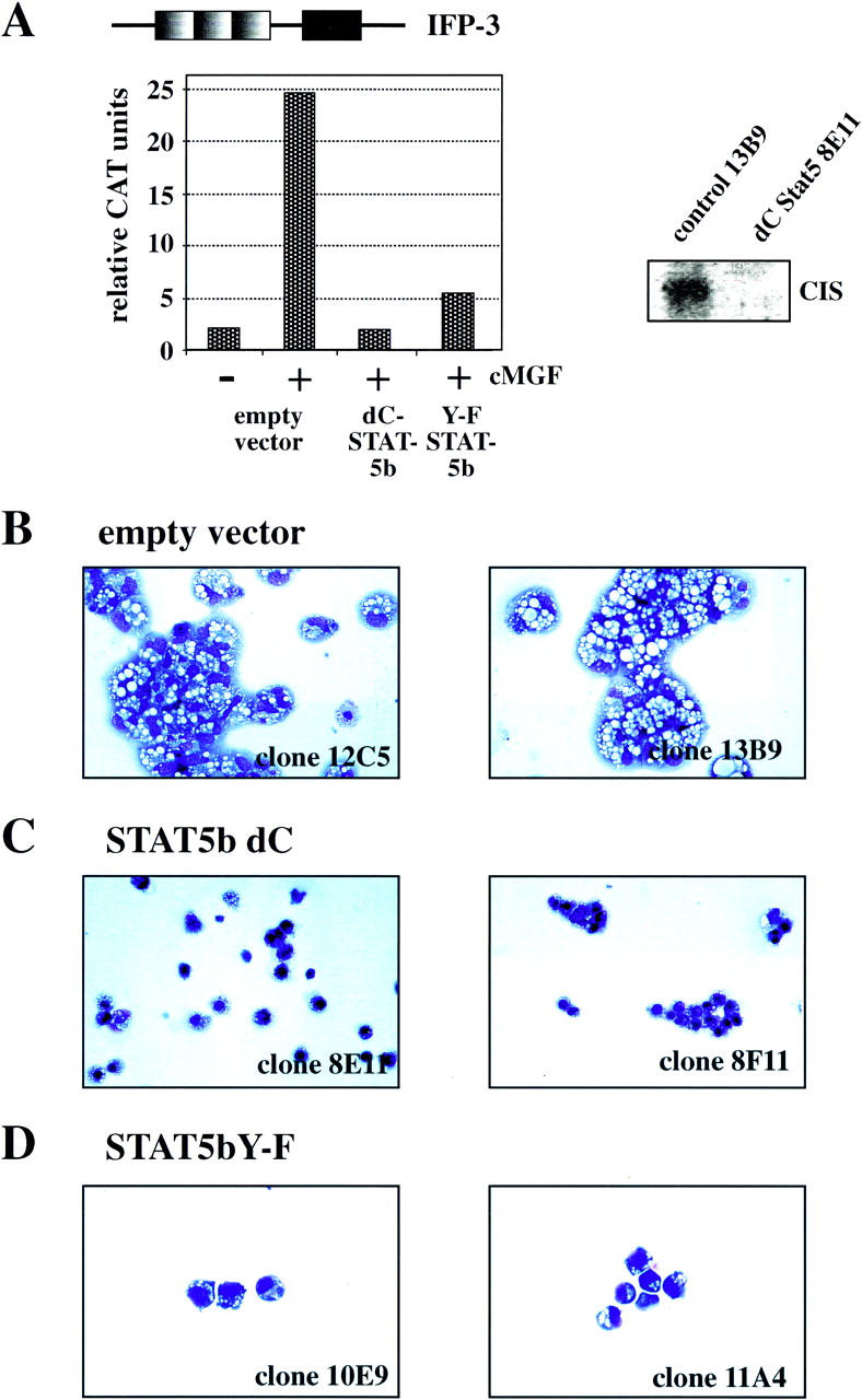
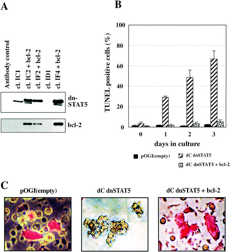
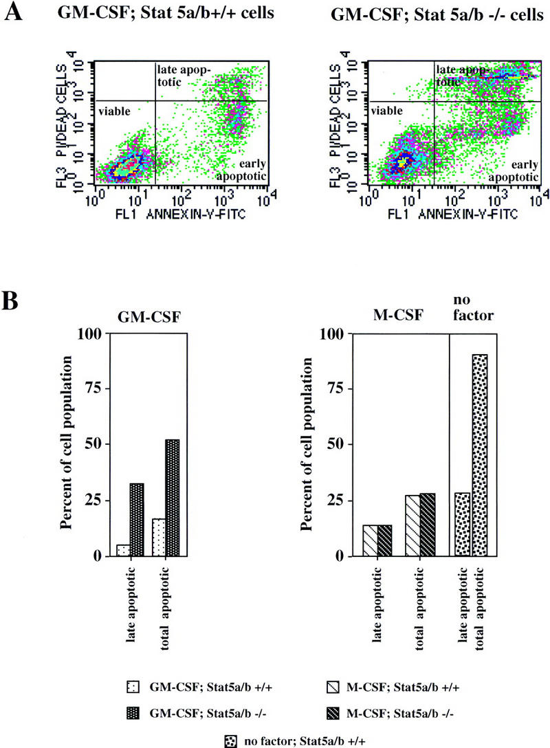
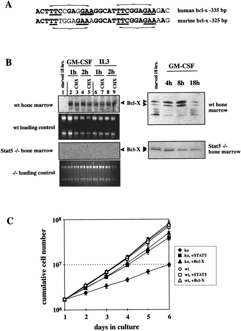
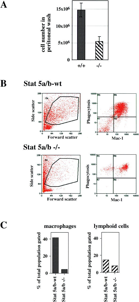
Stat5 is activated by multiple receptors of hematopoietic cytokines. To study its role during hematopoiesis, we have generated primary chicken myeloblasts expressing different dominant-negative (dn) alleles of Stat5. This caused a striking inability to generate mature cells, due to massive apoptosis during differentiation. Bcl-2 was able to rescue differentiating cells expressing dnStat5 from apoptosis, suggesting that during cytokine-dependent differentiation the main function of the protein is to ensure cell survival. Our findings with dnStat5-expressing chicken myeloblasts were confirmed with primary hematopoietic cells from Stat5a/Stat5b-deficient mice. Bone marrow cells from these animals displayed a strong increase in apoptotic cell death during GM-CSF-dependent functional maturation in vitro. The antiapoptotic protein Bcl-x was induced by GM-CSF and IL-3 in a Stat5-dependent fashion. Ectopic expression of Bcl-x rescued Stat5-deficient bone marrow cells from apoptosis, indicating that Stat5 promotes the survival of myeloid progenitor cells through its ability to induce transcription of the bcl-x gene. Finally, the recruitment of myeloid cells to inflammatory sites was found strongly impeded in Stat5-deficient mice. Taken together, our findings suggest that Stat5 may promote cytokine-dependent survival and proliferation of differentiating myeloid progenitor cells in stress or pathological situations, such as inflammation.


Figures

Figure 1
Figure 1
Impaired differentiation and increased cell death in dnStat5-expressing tsE26 myeloblasts after shift to 42°C. (A, left) Stat5dC and Stat5Y-F function as dominant-negative Stat5 versions. Clones verified for high dnStat5 expression by Western blot were transfected with a reporter construct containing three IFP-53 GAS sites (top) as described earlier (Woldman et al. 1997). (Right) Northern blot analysis for CIS mRNA expression, using a control clone and a Stat5dC-expressing clone after stimulation with cMGF. (BD) Cells from two representative clones expressing empty vector (B), Stat5dC (C), or Stat5Y-F (D) were induced to differentiate at 42°C in the presence of cMGF and IGF-1 for 4 days, cytocentrifuged onto slides, stained with May-Gruenwald-Giemsa solution, and photographed. Note terminally differentiated macrophages with a highly excentric nucleus in B but dead cells (C) or small, partially differentiated cells (D) in the cultures expressing dominant-negative versions of Stat5.
Figure 2
Figure 2
dnStat5-expressing tsE26 myeloblasts undergo apoptosis during differentiation at 42°C. (A) Combined TUNEL/propidium iodide staining and FACS analysis of representative tsE26 myeloblast clones expressing either empty vector (left) or Stat5dC (right) 3 days after differentiation induction at 42°C. The gate setting for the TUNEL-positive cell population is indicated. (B) Five tsE26 clones each, expressing empty vector (black bars), Stat5Y-F (dotted bars), or Stat5dC (hatched bars) were induced to differentiate at 42°C. TUNEL-positive, apoptotic cells accumulating during differentiation were quantitated daily by TUNEL assay as shown in A. s.d.s are indicated by error bars.
Figure 3
Figure 3
Bcl-2 expression rescues Stat5dC-expressing tsE26 myeloblasts clones from apoptosis during differentiation induction at 42°C. (A) Lysates of several tsE26 clones coinfected with dnStat5dC- and Bcl-2 expressing retroviruses (see Materials and Methods) as well as several control clones were analyzed for myc-tagged Stat5dC and Bcl-2 protein expression by Western blot. The lower molecular weight band detected by the myc antibody in some of the clones is a degradation product. (B) Several clones expressing either empty vector (black bars), dnStat5dC (hatched bars), or dnStat5C plus Bcl-2 (speckled bars) were induced to differentiate at 42°C and analyzed daily for apoptotic cells by TUNEL assay as described in the legend to Fig. 2. (C) Representative clones expressing empty vector (left), dnStat5dC (middle), or dnStat5dC + Bcl2 (right) were induced to differentiate for 3 days at 42°C and allowed to ingest Texas Red-labeled bacteria for 1 hr. Note large cells having phagocytosed bacteria (red fluorescence) in the control and dnStat5dC + Bcl2 expressing cells, whereas cells expressing dnStat5dC alone were nonphagocytic and resembled immature myeloblasts with an apoptotic morphology.
Figure 4
Figure 4
Cytokine-dependent proliferation/differentiation of primary myeloid fetal liver cells is impaired in Stat5a/b−/− mice. (A) Fetal liver cells (E12.5) from wild-type (Stat5a/b+/+) and Stat5a/b−/− mice were precultured in StemPro medium containing expansion factor mix; aliquots were infected with a Stat5a-expressing retrovirus (exo Stat5; see Materials and Methods). The cells were then switched to medium containing either GM-CSF (left) or IL3, IL-6, and SCF (right) and subjected to daily counting. Cumulative cell numbers are shown (mean values plus s.d. from three independent determinations). (B) Fetal liver cells from wild-type and Stat5a/b−/− mice were precultured and virus-infected as in A but switched to M-CSF instead of GM-CSF. Cell numbers were normalized to those obtained 3 days after switching to GM-CSF, to correct for the adaptation required from Stat5-dependent growth in expansion factor mix to Stat5-independent proliferation in M-CSF. (□) Stat5a/b−/−; (█) Stat5a/b−/−, exo Stat5; (○) Stat5a/b+/+; (●) Stat5a/b+/+, exo Stat5.
Figure 5
Figure 5
Impairment of cytokine-dependent myeloid cell proliferation/differentiation in Stat5a/b−/− mice is due to increased apoptosis. Cells from wild-type (Stat5a/b+/+) and Stat5a/b−/− mice were cultivated as described in the legend of Fig. 4 and analyzed for apoptotic cells by Annexin V staining and FACS analysis 4 days after switch to GM-CSF, M-CSF, or no factor. (A) Annexin V staining of Stat5a/b+/+ and Stat5a/b−/− cells treated with GM-CSF. This method distinguishes between early (bottom right gate) and late stages of apoptosis (top right gate). (B) Quantitative determination of late apoptotic (bottom right gate) and total apoptotic (bottom plus top right gate) in Stat5a/b+/+ and Stat5a/b−/− cells treated as in A. Note the strong up-regulation of late and total apoptotic cells from Stat5a/b−/− when treated with GM-CSF, no difference between Stat5a/b−/− and wild-type cells in response to M-CSF, and massive apoptosis of wild-type cells after factor withdrawal.
Figure 6
Figure 6
Bcl-x is a Stat5 target gene and mediates survival of differentiating myeloid progenitor cells. (A) Sequence comparison of putative, highly conserved Stat binding regions in the promoters of the human and murine bcl-x genes. (Bold) Homologous regions; (underlined) essential sequence elements of the Stat binding sites (indicated by horizontal brackets). (B) Bone marrow cells from wild-type or Stat5a/b-deficient mice were precultured as described for Fig. 4, withdrawn from factors for 18 hr and restimulated with GM-CSF or IL-3 [minus or plus cycloheximide (CHX) as indicated]. Total RNA was subsequently analyzed by Northern blotting (left) using a Bcl-x probe, and protein extracts were subjected to Western blotting with Bcl-x antibodies. (C) Bone marrow cells from wild-type or Stat5a/b-deficient mice were infected with retrovirus expressing Stat5 or Bcl-x while proliferating in expansion factor mix (see Materials and Methods). The infected cells were transferred to GM-CSF-containing medium and cumulative cell numbers were determined at the times indicated.
Figure 7
Figure 7
Recruitment of inflammatory myeloid cells to the peritoneal cavity of wild-type and Stat5a/b-deficient mice. Wild-type and Stat5a/b-deficient mice (three each) were injected intraperitoneally with thioglycollate (4% wt/vol). Three days later, inflammatory cells were collected by peritoneal lavage and analyzed. (A) Total cell numbers isolated from the peritoneal cavity (see Materials and Methods). (B) Analysis of peritoneal cells by flow cytometry: Cells were gated for the myeloid cell population (left) and analyzed for phagocytosis of Texas Red-labeled bacteria plus expression of the Mac-1 surface marker (right). (C, left) Total numbers of Mac-1/phagocytosis-positive myeloid cells (macrophages, calculated from top right quadrants in B). (Right) Total numbers of lymphoid-gated cells (low forward and side scatter, not shown).

Similar articles

Cited by

References

    1. Azam M, Erdjument-Bromage H, Kreider BL, Xia M, Quelle F, Basu R, Saris C, Tempst P, Ihle JN, Schindler C. Interleukin-3 signals through multiple isoforms of Stat5. EMBO J. 1995;14:1402–1411. - PMC - PubMed
    1. Barahmand-pour F, Meinke A, Eilers A, Gouilleux F, Groner B, Decker T. Colony-stimulating factors and interferon-gamma activate a protein related to MGF-Stat 5 to cause formation of the differentiation-induced factors in myeloid cells. FEBS Letters. 1995;360:29–33. - PubMed
    1. Bedi A, Sharkis SJ. Mechanisms of cell commitment in myeloid cell differentiation. Curr Opin Hematol. 1995;2:12–21. - PubMed
    1. Beug H, Leutz A, Kahn P, Graf T. Ts mutants of E26 leukemia virus allow transformed myeloblasts, but not erythroblasts or fibroblasts, to differentiate at the nonpermissive temperature. Cell. 1984;39:579–588. - PubMed
    1. Beug H, Blundell PA, Graf T. Reversibility of differentiation and proliferative capacity in avian myelomonocytic cells transformed by tsE26 leukemia virus. Genes & Dev. 1987;1:277–286. - PubMed

Publication types

MeSH terms