. 2000 Jan 15;14(2):245-54.

A forkhead gene, FoxE3, is essential for lens epithelial proliferation and closure of the lens vesicle

Affiliations

A forkhead gene, FoxE3, is essential for lens epithelial proliferation and closure of the lens vesicle

A Blixt et al. Genes Dev. .

Abstract

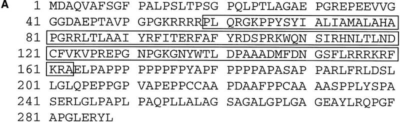
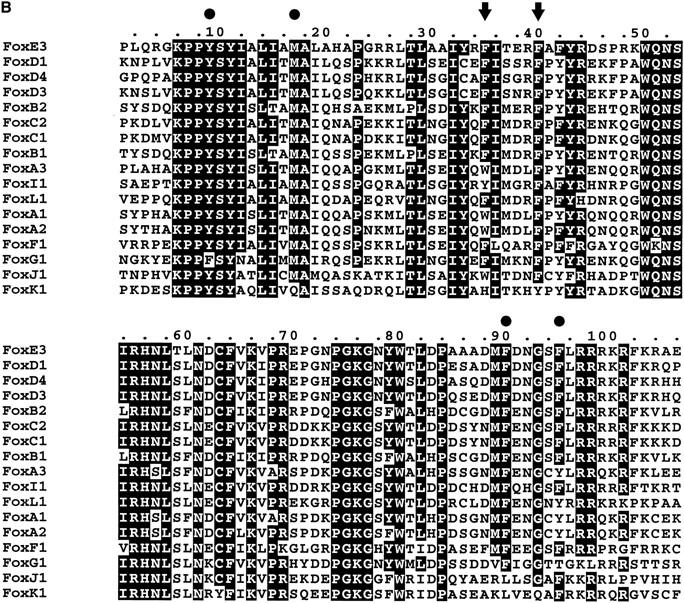
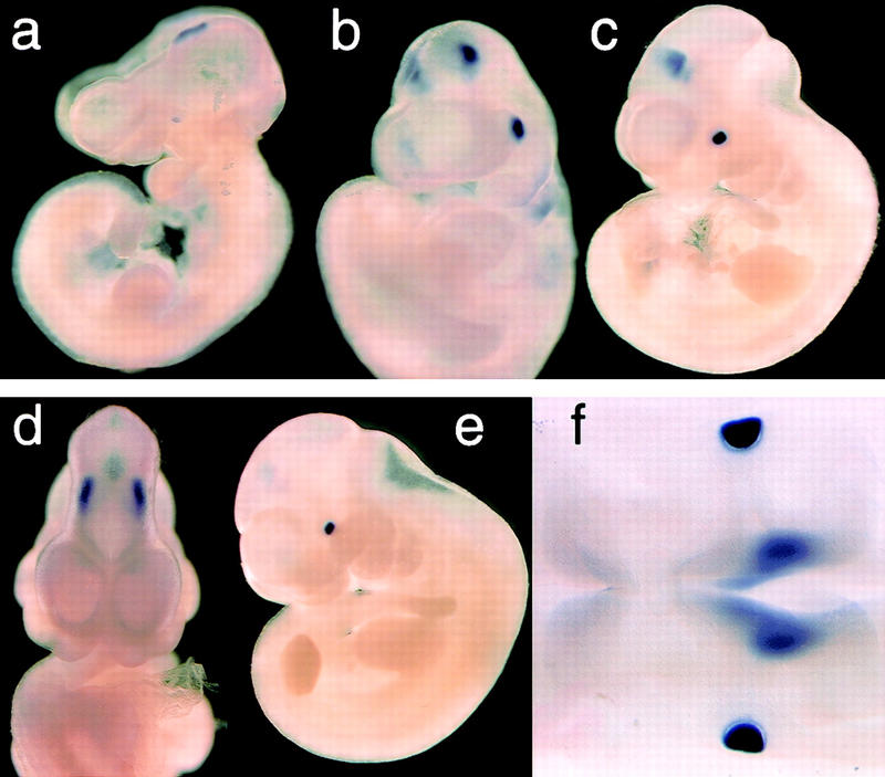
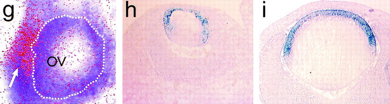
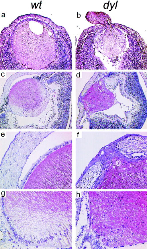
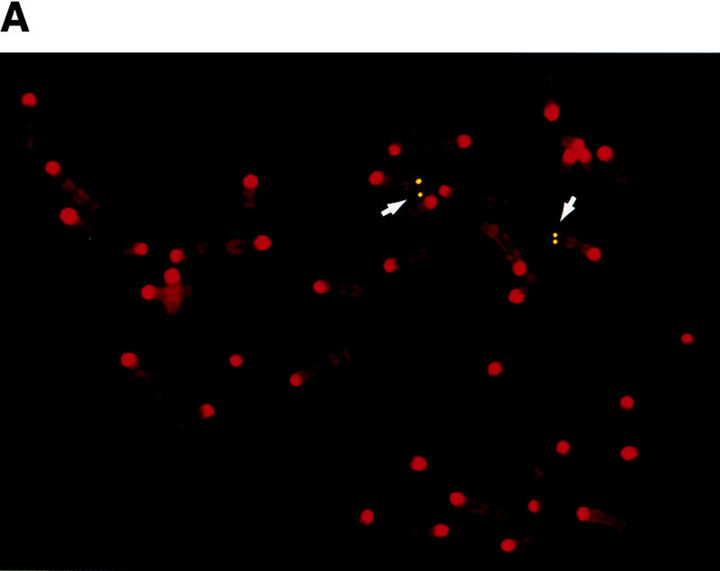
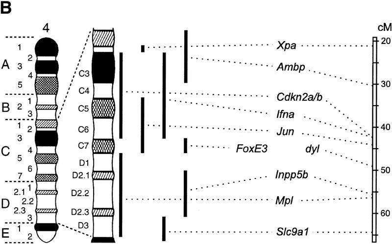
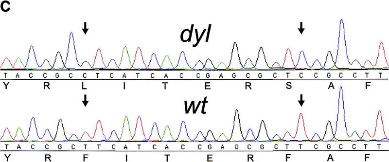
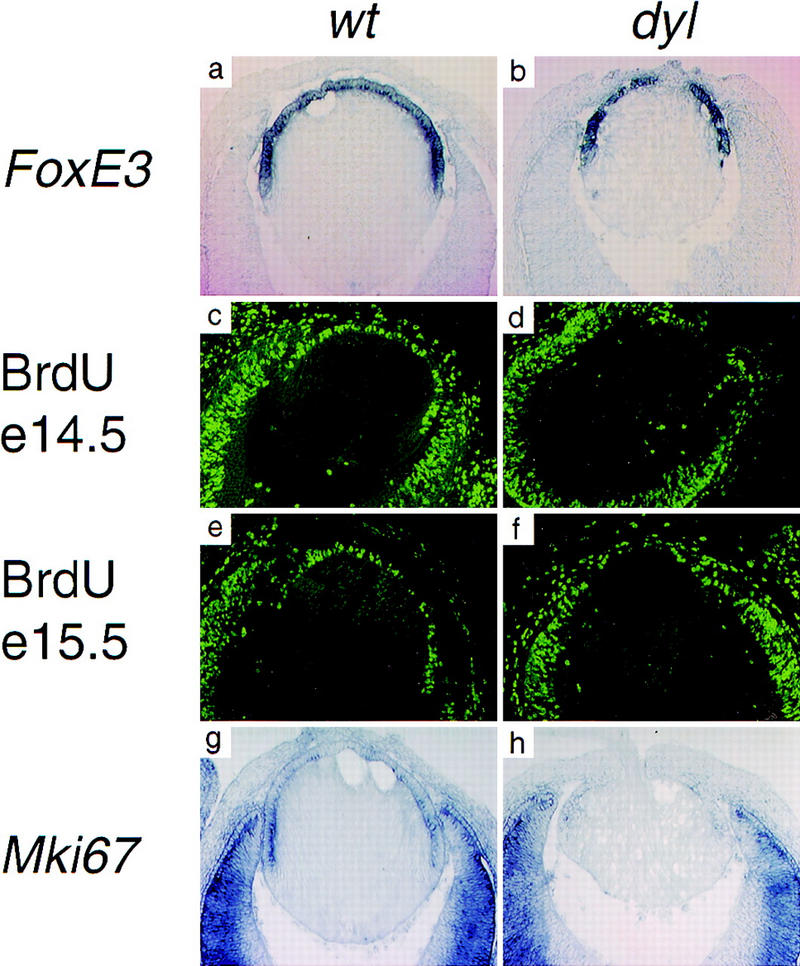
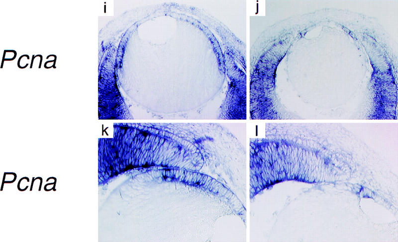
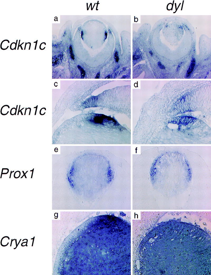
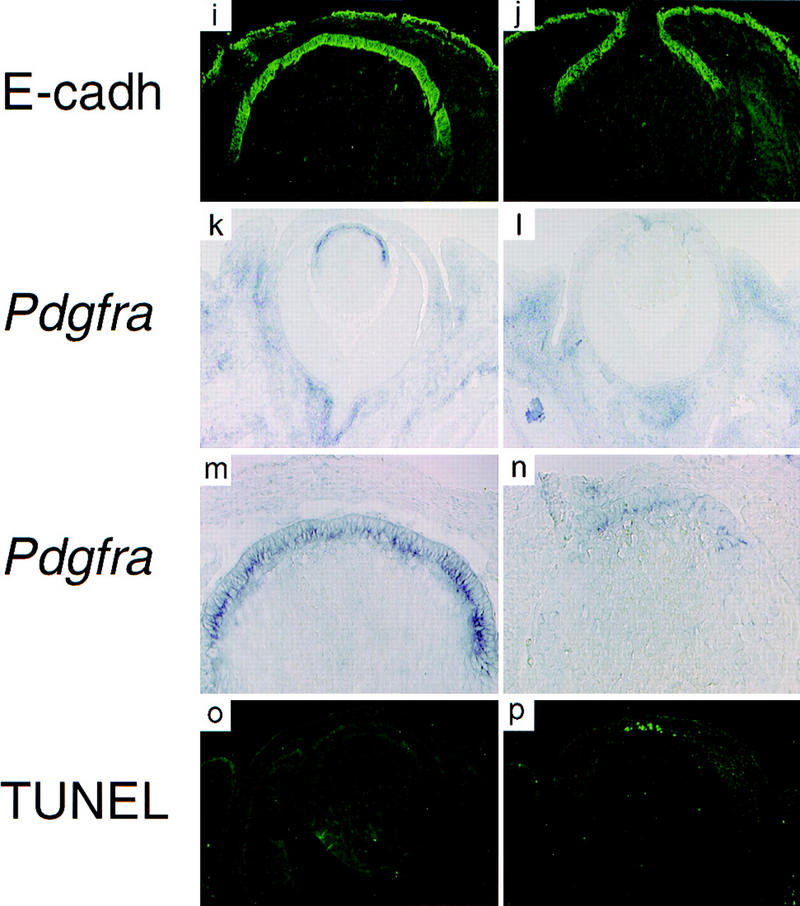
In the mouse mutant dysgenetic lens (dyl) the lens vesicle fails to separate from the ectoderm, causing a fusion between the lens and the cornea. Lack of a proliferating anterior lens epithelium leads to absence of secondary lens fibers and a dysplastic, cataractic lens. We report the cloning of a gene, FoxE3, encoding a forkhead/winged helix transcription factor, which is expressed in the developing lens from the start of lens placode induction and becomes restricted to the anterior proliferating cells when lens fiber differentiation begins. We show that FoxE3 is colocalized with dyl in the mouse genome, that dyl mice have mutations in the part of FoxE3 encoding the DNA-binding domain, and that these mutations cosegregate with the dyl phenotype. During embryonic development, the primordial lens epithelium is formed in an apparently normal way in dyl mutants. However, instead of the proliferation characteristic of a normal lens epithelium, the posterior of these cells fail to divide and show signs of premature differentiation, whereas the most anterior cells are eliminated by apoptosis. This implies that FoxE3 is essential for closure of the lens vesicle and is a factor that promotes survival and proliferation, while preventing differentiation, in the lens epithelium.


Figures

Figure 1
Figure 1
(a) Amino acid sequence of FoxE3 predicted from DNA sequence. The forkhead domain is boxed. (b) The forkhead domain of FoxE3 aligned with other mouse forkhead proteins in the SwissProt database. Phe93 and Phe98 of FoxE3 are indicated with arrows and other amino acids in the methionine-aromatic rosette with solid circles. For conversion between the Fox nomenclature and old names, see http://www.biology.pomona.edu/fox.html.
Figure 1
Figure 1
(a) Amino acid sequence of FoxE3 predicted from DNA sequence. The forkhead domain is boxed. (b) The forkhead domain of FoxE3 aligned with other mouse forkhead proteins in the SwissProt database. Phe93 and Phe98 of FoxE3 are indicated with arrows and other amino acids in the methionine-aromatic rosette with solid circles. For conversion between the Fox nomenclature and old names, see http://www.biology.pomona.edu/fox.html.
Figure 2
Figure 2
FoxE3 expression in embryonic development. (a) Whole mount in situ hybridization of 23-somite mouse embryo showing the first signs of FoxE3 expression on the lateral head ectoderm and on the cephalic neural folds. (b) 25-somite stage embryo (E9.5) with expression in lens placode and caudal forebrain. (c) Side view and (d) frontal view of 30-somite embryo (E10.25) with high expression in lens vesicle and weaker expression in caudal, dorsolateral diencephalon. (e) 35-somite embryo (E10.5) with high expression in lens vesicle and fading brain expression. (f) Dorsal view of clarified 30-somite embryo showing FoxE3 expression in lens vesicles and diencephalon (out of focus). (g) Radioactive in situ hybridization showing lens placode (arrow) expression of FoxE3 (red) in section of E9.5 embryo. Dotted line indicates the border of the optic vesicle (OV). (h) Section through the eye of E12.5 embryo showing FoxE3 expression in the anterior part of the lens vesicle, but turned off in the differentiating primary lens fibers in the posterior part. (i) Section through the eye of E14.5 embryo demonstrating FoxE3 expression confined to the anterior epithelium of the lens.
Figure 2
Figure 2
FoxE3 expression in embryonic development. (a) Whole mount in situ hybridization of 23-somite mouse embryo showing the first signs of FoxE3 expression on the lateral head ectoderm and on the cephalic neural folds. (b) 25-somite stage embryo (E9.5) with expression in lens placode and caudal forebrain. (c) Side view and (d) frontal view of 30-somite embryo (E10.25) with high expression in lens vesicle and weaker expression in caudal, dorsolateral diencephalon. (e) 35-somite embryo (E10.5) with high expression in lens vesicle and fading brain expression. (f) Dorsal view of clarified 30-somite embryo showing FoxE3 expression in lens vesicles and diencephalon (out of focus). (g) Radioactive in situ hybridization showing lens placode (arrow) expression of FoxE3 (red) in section of E9.5 embryo. Dotted line indicates the border of the optic vesicle (OV). (h) Section through the eye of E12.5 embryo showing FoxE3 expression in the anterior part of the lens vesicle, but turned off in the differentiating primary lens fibers in the posterior part. (i) Section through the eye of E14.5 embryo demonstrating FoxE3 expression confined to the anterior epithelium of the lens.
Figure 3
Figure 3
Hematoxylin and eosin-stained sections through the eye of wild-type (a,c,e,g) and dyl (b,d,f,h) mice. (a,b) Eyes from E14.5 embryos. The lens vesicle fails to close in dyl mutants, which in some cases leads to lens material being ejected to the exterior (b). (c–h) Eyes from newborn mice. Note the small, irregularly shaped lens (d) and the persistent connection between lens and cornea (d,f) in dyl mutants. In the equatorial zone of the lens from wild-type animals (g) there is a transition from the well-developed anterior epithelium to posterior elongated lens fibers. In dyl mutants (h) no clear difference is seen between anterior and posterior side and a defined epithelium is missing.
Figure 4
Figure 4
FoxE3 and dyl are colocalized in the mouse genome. (a) Localization of FoxE3 to 4C7 on mouse metaphase chromosomes by fluorescence in situ hybridization (FISH). (b) Comparison between the cytogenetic and genetic maps of mouse chromosome 4. Genes that cytogenetically map proximally or distally of FoxE3 have the same location relative to dyl on the genetic map. Map data, except FoxE3, are taken from the Mouse Genome Database (http://www.informatics.jax.org/). (c) DNA sequence from the forkhead box of FoxE3. dyl mice have two missense mutations that substitute leucine and serine for Phe-93 and Phe-98, respectively.
Figure 4
Figure 4
FoxE3 and dyl are colocalized in the mouse genome. (a) Localization of FoxE3 to 4C7 on mouse metaphase chromosomes by fluorescence in situ hybridization (FISH). (b) Comparison between the cytogenetic and genetic maps of mouse chromosome 4. Genes that cytogenetically map proximally or distally of FoxE3 have the same location relative to dyl on the genetic map. Map data, except FoxE3, are taken from the Mouse Genome Database (http://www.informatics.jax.org/). (c) DNA sequence from the forkhead box of FoxE3. dyl mice have two missense mutations that substitute leucine and serine for Phe-93 and Phe-98, respectively.
Figure 5
Figure 5
The lens epithelium fails to proliferate in dyl mutants. In situ hybridization show similar patterns of FoxE3 expression in wild-type (a) and dyl (b) E14.5 lens. BrdU incorporation reveals intense proliferation of epithelial cells in wild-type lens (c,e), but a sparse distribution of replicating cells in E14.5 dyl lens (d). At E15.5 the difference is even more pronounced with only a few BrdU-positive cells in the mutant (f). The expression of genes encoding proliferation associated antigens Mki67 (g,h) and Pcna (i–l) is down-regulated and displaced anteriorly in dyl mutants. All sections except e and f are from stage E14.5.
Figure 5
Figure 5
The lens epithelium fails to proliferate in dyl mutants. In situ hybridization show similar patterns of FoxE3 expression in wild-type (a) and dyl (b) E14.5 lens. BrdU incorporation reveals intense proliferation of epithelial cells in wild-type lens (c,e), but a sparse distribution of replicating cells in E14.5 dyl lens (d). At E15.5 the difference is even more pronounced with only a few BrdU-positive cells in the mutant (f). The expression of genes encoding proliferation associated antigens Mki67 (g,h) and Pcna (i–l) is down-regulated and displaced anteriorly in dyl mutants. All sections except e and f are from stage E14.5.
Figure 6
Figure 6
The lens epithelial cells in dyl mutants are eliminated by premature differentiation and apoptosis. In situ hybridization (a,b,g,h,k,l,m,n), immunohistochemistry (i,j), and TUNEL assay (o,p). The Cdk inhibitor Cdkn1c is expressed in the equatorial cells of the lens (a,c). In the dyl mutant the expression level of Cdkn1c is reduced and the expression is displaced anteriorly, overlapping with that of FoxE3 (b,d). Prox1 is mainly expressed in the equatorial, nonepithelial cells (e), but in the dyl mutant the highest expression level is seen in the posterior part of the epithelium (f). Crya1 expression is low in the epithelium and highest in the equatorial zone (g). In dyl, Crya1 is evenly expressed throughout the lens, except in the most anterior cells (h). E-cadherin is expressed in the epithelium of wild-type (i) and dyl (j) lens reaching further to the posterior in wild-type. Pdgfra is expressed in the lens epithelium (k,m), but is down-regulated and restricted to the most anterior cells in dyl (l,n). The anterior cells in the lens epithelium undergo apoptosis in dyl mutants (p), whereas no signs of cell death can be detected in wild-type lens (o). All sections are from E14.5 embryos.
Figure 6
Figure 6
The lens epithelial cells in dyl mutants are eliminated by premature differentiation and apoptosis. In situ hybridization (a,b,g,h,k,l,m,n), immunohistochemistry (i,j), and TUNEL assay (o,p). The Cdk inhibitor Cdkn1c is expressed in the equatorial cells of the lens (a,c). In the dyl mutant the expression level of Cdkn1c is reduced and the expression is displaced anteriorly, overlapping with that of FoxE3 (b,d). Prox1 is mainly expressed in the equatorial, nonepithelial cells (e), but in the dyl mutant the highest expression level is seen in the posterior part of the epithelium (f). Crya1 expression is low in the epithelium and highest in the equatorial zone (g). In dyl, Crya1 is evenly expressed throughout the lens, except in the most anterior cells (h). E-cadherin is expressed in the epithelium of wild-type (i) and dyl (j) lens reaching further to the posterior in wild-type. Pdgfra is expressed in the lens epithelium (k,m), but is down-regulated and restricted to the most anterior cells in dyl (l,n). The anterior cells in the lens epithelium undergo apoptosis in dyl mutants (p), whereas no signs of cell death can be detected in wild-type lens (o). All sections are from E14.5 embryos.

References

    1. Brahma SK, Sanyal S. Immunohistochemical studies of lens crystallins in the dysgenetic lens (dyl) mutant mice. Exp Eye Res. 1984;38:305–311. - PubMed
    1. ————— Ontogeny of alpha-crystallin polypeptides during the lens development of a mutant mouse. Curr Eye Res. 1987;6:1291–1297. - PubMed
    1. Brewitt B, Clark JI. Growth and transparency in the lens, an epithelial tissue, stimulated by pulses of PDGF. Science. 1988;242:777–779. - PubMed
    1. Chow RL, Roux GD, Roghani M, Palmer MA, Rifkin DB, Moscatelli DA, Lang RA. FGF suppresses apoptosis and induces differentiation of fibre cells in the mouse lens. Development. 1995;121:4383–4393. - PubMed
    1. Churchill AJ, Booth AP, Anwar R, Markham AF. PAX 6 is normal in most cases of Peters' anomaly. Eye. 1998;12:299–303. - PubMed

Publication types

MeSH terms

Associated data