. 2000 Feb;66(2):638-42.
doi: 10.1128/AEM.66.2.638-642.2000.

A protein disulfide isomerase gene fusion expression system that increases the extracellular productivity of Bacillus brevis

Affiliations

A protein disulfide isomerase gene fusion expression system that increases the extracellular productivity of Bacillus brevis

T Kajino et al. Appl Environ Microbiol. 2000 Feb.

Abstract

We have developed a versatile Bacillus brevis expression and secretion system based on the use of fungal protein disulfide isomerase (PDI) as a gene fusion partner. Fusion with PDI increased the extracellular production of heterologous proteins (light chain of immunoglobulin G, 8-fold; geranylgeranyl pyrophosphate synthase, 12-fold). Linkage to PDI prevented the aggregation of the secreted proteins, resulting in high-level accumulation of fusion proteins in soluble and biologically active forms. We also show that the disulfide isomerase activity of PDI in a fusion protein is responsible for the suppression of the aggregation of the protein with intradisulfide, whereas aggregation of the protein without intradisulfide was prevented even when the protein was fused to a mutant PDI whose two active sites were disrupted, suggesting that another PDI function, such as chaperone-like activity, synergistically prevented the aggregation of heterologous proteins in the PDI fusion expression system.


Figures
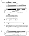
FIG. 1
FIG. 1
Structures of the expression and secretion vectors. (A) Structure of the vector for LC expression. SD, ribosome-binding site of the middle wall protein; SP, signal-peptide-encoding sequence; LC, LC-encoding sequence; Nm, neomycin resistance gene; Pr, promoter of the cell wall protein gene operon in B. brevis; Ori, replication origin. The nucleotide and amino acid sequences of the regions denoted by a, b, c, and d are shown below. ∗, stop codon. (B) Structure of pNH326PDI-GGPS. The abbreviations and symbols are the same as above.
FIG. 2
FIG. 2
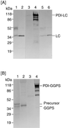
Localization of heterologous proteins expressed by B. brevis. B. brevis 31-OK cells carrying pNH326LC, pNH326LC/PDI, pNH326PDI-LC, pNU212GGPS, or pNH326PDI-GGPS were grown for 6 days at 30°C in YC-P2 medium containing 0.3% Tween 40. The culture supernatant was used as the soluble fraction. The cell pellet was suspended in the same volume as the culture of the sample buffer (62.5 mM Tris-HCl [pH 6.7], 1% SDS, 5% β-mercaptoethanol) and then sonicated. This suspension was incubated in boiling water for 2 min and then centrifuged. The resulting supernatant was used as the insoluble fraction. Ten microliters of each of the insoluble and soluble fractions was subjected to SDS-PAGE, followed by immunoblot analysis as described under Materials and Methods. The gels show the localization of LC (A) and GGPS (B). Lanes 1, 3, and 5, insoluble fraction expressed by itself, expressed as a fusion form with PDI, and coexpressed with PDI, respectively; lanes 2, 4, and 6, soluble fraction expressed by itself, expressed as a fusion form with PDI, and coexpressed with PDI, respectively.
FIG. 3
FIG. 3
The effect of oxidoreductase activity on aggregate formation. B. brevis 31-OK cells carrying the expression vector for LC or GGPS fused to the native or mutant PDI, respectively, were cultured and treated as described in the legend to Fig. 2. Ten microliters of each of the insoluble and soluble fractions of LC (A) and GGPS (B) was analyzed by SDS-PAGE, followed by immunostaining as described under Materials and Methods. Lanes 1 and 3, the accumulated protein fused to the native and mutant PDI, respectively, in the insoluble fraction; lanes 2 and 4, the secreted protein fused to the native and mutant PDI, respectively, in the soluble fraction.
FIG. 4
FIG. 4
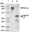
Specific cleavage of the PDI-LC fusion protein by enterokinase. An immunostained SDS-polyacrylamide gel (see Materials and Methods) is shown. Lane 1, marker proteins; lane 2, PDI-LC secreted by B. brevis; lane 3, PDI-LC following cleavage with enterokinase.
FIG. 5
FIG. 5
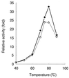
Optimal reaction temperature of PDI-GGPS. Using 50 mM bis-Tris propane (pH 9.5), PDI-GGPS produced by B. brevis was reacted with [14C]isopentenyl diphosphate, dimethylallyl diphosphate, and 5 mM MgCl2 for 30 min at the indicated temperatures. The reaction products were extracted with H2O-saturated butanol, and then the radioactivity of the products was measured with a liquid scintillation counter. The activity at 40°C was defined as 1. Closed and open circles indicate GGPS fused to PDI and native GGPS from the archaea, respectively. The data are the means of at least four independent experiments.

Similar articles

Cited by

References

    1. Burnette W N. “Western blotting”: electrophoretic transfer of proteins from sodium dodecyl sulfate-polyacrylamide gels to unmodified nitrocellulose, and radiographic detection with antibodies and radioiodinated protein A. Anal Biochem. 1981;112:195–203. - PubMed
    1. Ebisu S, Murahashi Y, Takagi H, Kadowaki K, Yamaguchi K, Yamagata H, Udaka S. Nucleotide sequence and replication properties of the Bacillus borstelensis cryptic plasmid pHT926. Appl Environ Microbiol. 1995;61:3154–3157. - PMC - PubMed
    1. Gilbert H F. Protein disulfide isomerase and assisted protein folding. J Biol Chem. 1999;272:29399–29402. - PubMed
    1. Hayano T, Hirose M, Kikuchi M. Protein disulfide isomerase mutant lacking isomerase activity accelerates protein folding in the cell. FEBS Lett. 1995;377:505–511. - PubMed
    1. Hillson D A, Lambert N, Freedman R B. Formation and isomerization of disulfide bonds in proteins: protein disulfide-isomerase. Methods Enzymol. 1984;107:281–294. - PubMed

MeSH terms

LinkOut - more resources