. 2000 Feb;66(2):643-50.
doi: 10.1128/AEM.66.2.643-650.2000.

Endosymbiotic microbiota of the bamboo pseudococcid Antonina crawii (Insecta, Homoptera)

Affiliations

Endosymbiotic microbiota of the bamboo pseudococcid Antonina crawii (Insecta, Homoptera)

T Fukatsu et al. Appl Environ Microbiol. 2000 Feb.

Abstract

We characterized the intracellular symbiotic microbiota of the bamboo pseudococcid Antonina crawii by performing a molecular phylogenetic analysis in combination with in situ hybridization. Almost the entire length of the bacterial 16S rRNA gene was amplified and cloned from A. crawii whole DNA. Restriction fragment length polymorphism analysis revealed that the clones obtained included three distinct types of sequences. Nucleotide sequences of the three types were determined and subjected to a molecular phylogenetic analysis. The first sequence was a member of the gamma subdivision of the division Proteobacteria (gamma-Proteobacteria) to which no sequences in the database were closely related, although the sequences of endosymbionts of other homopterans, such as psyllids and aphids, were distantly related. The second sequence was a beta-Proteobacteria sequence and formed a monophyletic group with the sequences of endosymbionts from other pseudococcids. The third sequence exhibited a high level of similarity to sequences of Spiroplasma spp. from ladybird beetles and a tick. Localization of the endosymbionts was determined by using tissue sections of A. crawii and in situ hybridization with specific oligonucleotide probes. The gamma- and beta-Proteobacteria symbionts were packed in the cytoplasm of the same mycetocytes (or bacteriocytes) and formed a large mycetome (or bacteriome) in the abdomen. The spiroplasma symbionts were also present intracellularly in various tissues at a low density. We observed that the anterior poles of developing eggs in the ovaries were infected by the gamma- and beta-Proteobacteria symbionts in a systematic way, which ensured vertical transmission. Five representative pseudococcids were examined by performing diagnostic PCR experiments with specific primers; the beta-Proteobacteria symbiont was detected in all five pseudococcids, the gamma-Proteobacteria symbiont was found in three, and the spiroplasma symbiont was detected only in A. crawii.


Figures

FIG. 1
FIG. 1
RFLP analysis of bacterial 16S rDNA amplified and cloned from total DNA of A. crawii. Lanes 1 through 10 contained the cloned 16S rDNA fragments digested with either RsaI or HinfI and resolved on a 2% agarose gel. Lanes 1, 3, 4, 7, and 8, type A clones; lanes 2, 5, and 10, type B clones; lanes 6 and 9, type C clones. Lane M contained DNA size markers; the sizes of the markers were (from top to bottom) 2,000, 1,500, 1,000, 700, 500, 400, 300, 200, and 100 bp.
FIG. 2
FIG. 2
Molecular phylogenetic analysis of the endosymbionts of A. crawii based on 16S rDNA sequences. (A) Phylogenetic positions of the γ-symbiont and the β-symbiont in the Proteobacteria. A total of 1,184 unambiguously aligned nucleotide sites were analyzed. (B) Phylogenetic position of the spiroplasma symbiont in the Mycoplasmatales. A total of 1,235 unambiguously aligned nucleotide sites were analyzed. (C) Relationship between the γ-symbiont and the 16S rDNA sequence identified in P. lilacinus that was described in Kantheti et al. (25) but not deposited in DNA databases. A total of 809 unambiguously aligned nucleotide sites were analyzed. Only neighbor-joining phylogenies are shown; maximum-likelihood and maximum-parsimony analyses gave essentially the same results. The bootstrap values obtained with 1,000 resamplings are shown at the nodes. The numbers in brackets are accession numbers.
FIG. 2
FIG. 2
Molecular phylogenetic analysis of the endosymbionts of A. crawii based on 16S rDNA sequences. (A) Phylogenetic positions of the γ-symbiont and the β-symbiont in the Proteobacteria. A total of 1,184 unambiguously aligned nucleotide sites were analyzed. (B) Phylogenetic position of the spiroplasma symbiont in the Mycoplasmatales. A total of 1,235 unambiguously aligned nucleotide sites were analyzed. (C) Relationship between the γ-symbiont and the 16S rDNA sequence identified in P. lilacinus that was described in Kantheti et al. (25) but not deposited in DNA databases. A total of 809 unambiguously aligned nucleotide sites were analyzed. Only neighbor-joining phylogenies are shown; maximum-likelihood and maximum-parsimony analyses gave essentially the same results. The bootstrap values obtained with 1,000 resamplings are shown at the nodes. The numbers in brackets are accession numbers.
FIG. 3
FIG. 3
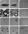
Specific detection of endosymbiotic bacteria in tissue sections of A. crawii by in situ hybridization. Probes DIG-TKSγ, DIG-TKSβ, and DIG-TKSspi targeted the γ-symbionts, the β-symbionts, and the spiroplasma symbionts in panels A, D, and G, panels B, E, and H, and panels C, F, I, J, K, and L, respectively. (A) γ-Symbionts densely populating the mycetome. (B) β-Symbionts localized in the same mycetome. (C) Spiroplasma symbionts not detected in the mycetome. (D) γ-Symbionts visualized as rod-shaped structures in the cytoplasm of mycetocytes. (E) β-Symbionts located in the same cytoplasm of mycetocytes, occupying the spaces between the γ-symbionts. (F) Gut epithelial cells populated by spiroplasma symbionts. (G) Vertical transmission of γ-symbionts through the anterior pole of a developing egg. The junction of the oocyte and nurse cells was specifically infected. (H) Vertical transmission of β-symbionts to a developing egg at the anterior pole of an oocyte, as observed with γ-symbionts. (I) Spiroplasma symbionts detected in various tissues and cells at a low density. (J) Magnified image of spiroplasma symbionts in gut epithelium. (K) Magnified image of spiroplasma symbionts in various types of cells just beneath the cuticle. (L) Magnified image of spiroplasma symbionts in a fat body. Bars = 40 μm. Abbreviations; CU, cuticle; FB, fat body; HC?, hemocyte?; LO, lateral oviduct; MY, mycetome; N, nucleus of mycetocyte; NC, nutritive cord; NR, nurse cell; OO, oocyte.
FIG. 4
FIG. 4
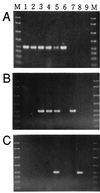
Diagnostic PCR detection of the three types of endosymbionts in representatives of the Pseudococcidae. (A) Detection of the β-symbiont with primers 16SA1 and TKSβsp. (B) Detection of the γ-symbiont with primers 16SA1 and TKSγsp. (C) Detection of the spiroplasma symbiont with primers 16SA1 and TKSSsp. Lane 1, P. citri; lane 2, P. kraunhiae; lane 3, P. citriculus; lane 4, D. wistariae; lane 5, A. crawii; lane 6, plasmid containing 16S rDNA of the β-symbiont; lane 7, plasmid of the γ-symbiont; lane 8, plasmid of the spiroplasma symbiont; lane 9, no-template control. Lanes M contained DNA size markers; the sizes of the markers were (from top to bottom) 2,000, 1,500, 1,000, 700, 500, 400, 300, and 200 bp. Twelve individuals of each species were analyzed to confirm the reproducibility of the results (data not shown).

References

    1. Adachi J, Hasegawa M. MOLPHY version 2.3: programs for molecular phylogenetics based on maximum likelihood. Comput Sci Monogr Inst Stat Math (Tokyo) 1996;28:1–150.
    1. Amann R I, Krumholz L, Stahl D A. Fluorescent-oligonucleotide probing of whole cells for determinative, phylogenetic, and environmental studies in microbiology. J Bacteriol. 1990;172:762–770. - PMC - PubMed
    1. Amann R I, Ludwig W, Schleifer K-H. Phylogenetic identification and in situ detection of individual microbial cells without cultivation. Microbiol Rev. 1995;59:143–169. - PMC - PubMed
    1. Baumann P, Moran N A. Non-cultivable microorganisms from symbiotic associations of insects and other hosts. Antonie Leeuwenhoek. 1997;72:38–48. - PubMed
    1. Baumann P, Baumann L, Lai C-Y, Rouhbakhsh D, Moran N A, Clark M A. Genetics, physiology and evolutionary relationships of the genus Buchnera: intracellular symbionts of aphids. Annu Rev Microbiol. 1995;49:55–94. - PubMed

Publication types

MeSH terms

Associated data

LinkOut - more resources