

Immunolocalization of dinitrogenase reductase produced by Klebsiella pneumoniae in association with Zea mays L
- PMID: 10653751
- PMCID: PMC91896
- DOI: 10.1128/AEM.66.2.783-787.2000
Immunolocalization of dinitrogenase reductase produced by Klebsiella pneumoniae in association with Zea mays L
Abstract
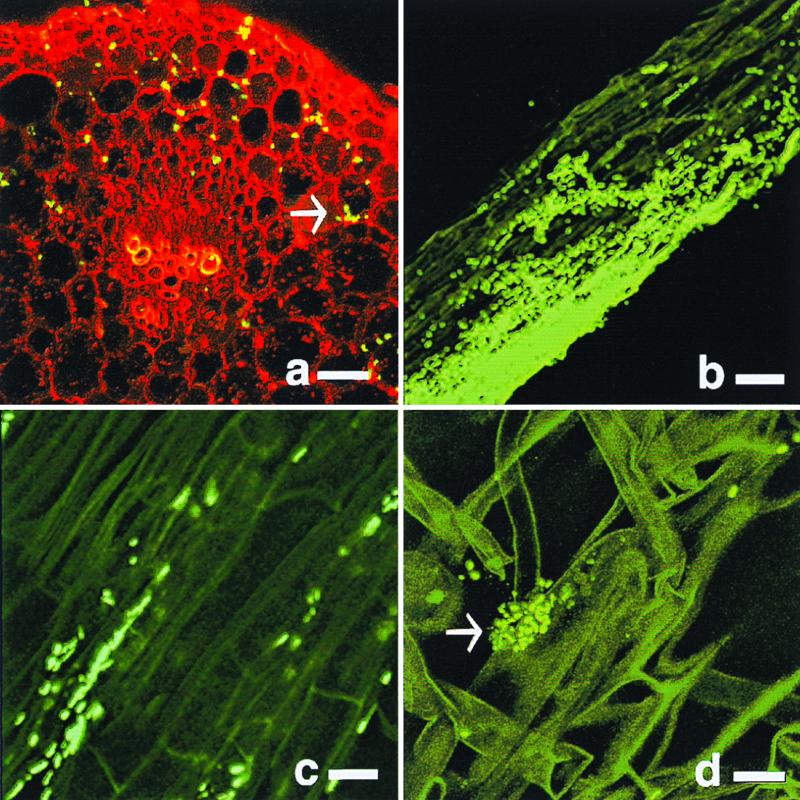
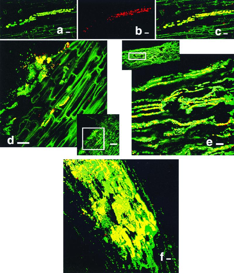
The endophytic lifestyle of Klebsiella pneumoniae is described, including the production of dinitrogenase reductase by bacteria residing in maize root tissue. The green fluorescent protein (GFP) was used to detect the colonization of maize by K. pneumoniae strains 2028 and 342. These strains were found to reside in intercortical layers of the stem and within the region of maturation in the root. The production of dinitrogenase reductase by GFP-tagged bacteria was visualized using immunolocalization. This activity was only apparent when bacteria were supplied with an exogenous carbon source. The results suggest that maize provides a suitable habitat for K. pneumoniae and that this species is capable of producing nitrogenase under the appropriate plant cultivation conditions.
Figures
References
-
- Arsene F, Katupitiya S, Kennedy I R, Elmerich C. Use of lacZ fusions to study the expression of nif genes of Azospirillum brasilense in association with plants. Mol Plant-Microbe Interact. 1994;7:748–757.
-
- Assmus B, Hutzler P, Kirchhof G, Amann R, Lawerence J R, Hartmann A. In situ localization of Azospirillum brasilense in the rhizosphere of wheat with fluorescently labeled, rRNA-targeted oligonucleotide probes and scanning confocal laser microscopy. Appl Environ Microbiol. 1995;61:1013–1019. - PMC - PubMed
-
- Burris R H. Comparative study of the response of Azotobacter vinelandii and Acetobacter diazotrophicus to changes in pH. Protoplasma. 1994;183:62–66.
Publication types
MeSH terms
Substances
LinkOut - more resources
Full Text Sources
Other Literature Sources
