. 2000 Feb 1;19(3):317-23.
doi: 10.1093/emboj/19.3.317.

Crystal structure of a fibrillarin homologue from Methanococcus jannaschii, a hyperthermophile, at 1.6 A resolution

Affiliations

Crystal structure of a fibrillarin homologue from Methanococcus jannaschii, a hyperthermophile, at 1.6 A resolution

H Wang et al. EMBO J. .

Abstract

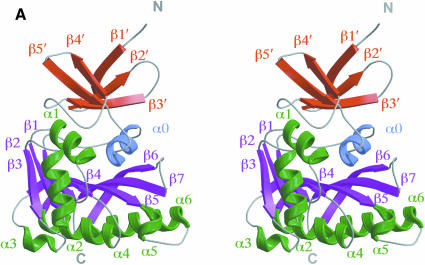
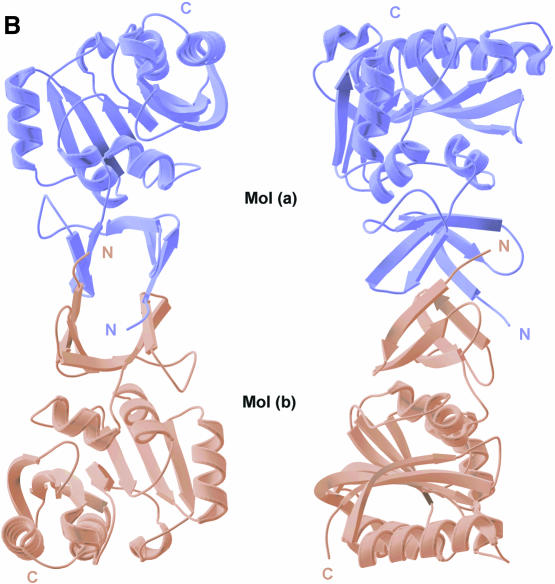
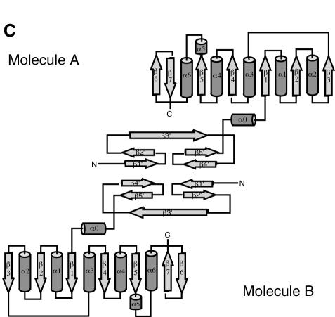
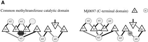
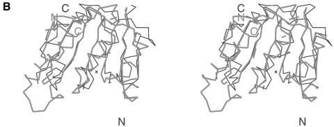
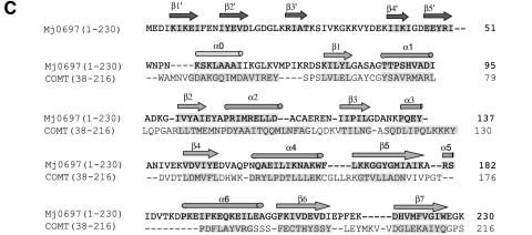
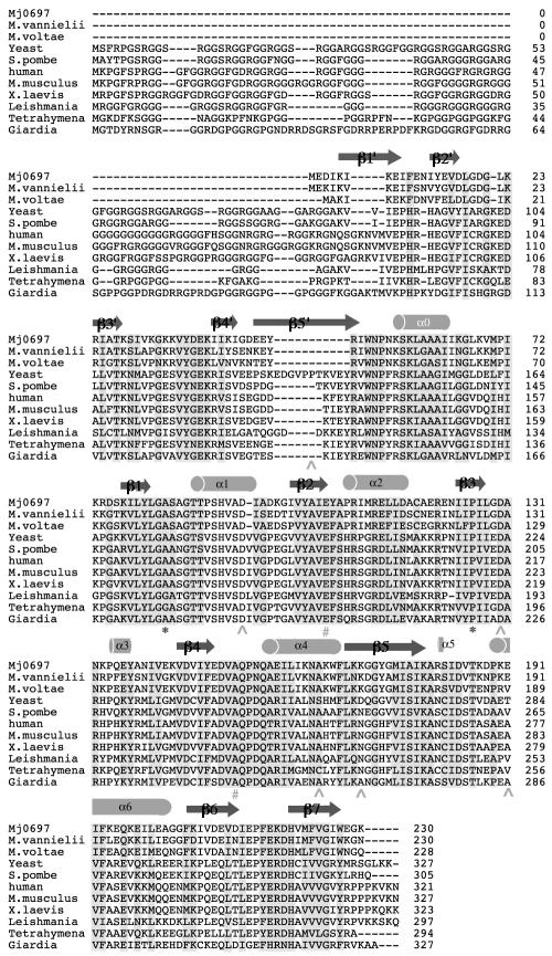
Fibrillarin is a phylogenetically conserved protein essential for efficient processing of pre-rRNA through its association with a class of small nucleolar RNAs during ribosomal biogenesis. The protein is the antigen for the autoimmune disease scleroderma. Here we report the crystal structure of the fibrillarin homologue from Methanococcus jannaschii, a hyperthermophile, at 1.6 A resolution. The structure consists of two domains, with a novel fold in the N-terminal region and a methyltransferase-like domain in the C-terminal region. Mapping temperature-sensitive mutations found in yeast fibrillarin Nop1 to the Methanococcus homologue structure reveals that many of the mutations cluster in the core of the methyltransferase-like domain.


Figures

None
Fig. 1. (A)Ribbon diagram of the Mj0697 monomer. The β-strands in the C-terminal domain (β1–β7) and in the N-terminal domain (β1′–β5′) are shown in magenta and red, respectively, while the α-helices (α1–α6) are shown in green and the connecting helix (α0) in blue. The secondary structure elements were assigned using PROCHECK (Laskowski et al., 1993). The ribbon representation was generated using MOLSCRIPT (Kraulis, 1991). (B)Ribbon diagram of the Mj0697 dimer viewed from two angles, differing by rotations of ∼90° on the vertical axis. (C)Topological diagram of the Mj0697 dimer. Helices are represented as cylinders and β-sheets as arrows.
None
Fig. 2. (A) A comparison of the topological representations of the C-terminal domain of Mj0697 and the consensus catalytic domain derived from other methyltransferases (Vidgren et al., 1994; Schluckebier et al., 1995; Djordjevic and Stock, 1997). Helices are shown as circles, β-strands as triangles. The AdoMet-binding pocket is indicated. The location of the analogous pocket is indicated by an empty circle in the Mj0697 scheme. An extra helix present in the Methanococcus homologue that does not occur in the consensus domain is shaded darker. (B) Superposition of the Cα traces of the C–terminal domain in Mj0697 (black line), and of the methyltransferase catalytic domain in catechol O-methyltransferase, COMT (grey line). The diagram was prepared using INSIGHTII (Molecular Systems Inc.). (C) Structural-based sequence alignment of Mj0697 and COMT.
None
Fig. 3. Amino acid sequence alignment of the fibrillarin homologues. The alignment was performed using CLUSTAL W (Thompson et al., 1994). The secondary structural elements are labelled. Shaded residues are conserved residues, including identical, conserved substitutions and semi-conserved substitutions. The yeast fibrillarin mutants shown include: Nop 1.3 mutants blocking rRNA methylation (*); Nop 1.5 and Nop 1.2 mutants affecting rRNA cleavage (∧); and Nop 1.7 mutants affecting rRNA assembly (#).
None
Fig. 4. Stereo diagram of the electron density map of the α1–β1 loop (82–86) region in the Mj0697 structure. The solvent-flattened experimental electron density map was contoured at 1.5σ, superimposed with the current model. This diagram was generated using the program O (Jones et al., 1991).

Similar articles

Cited by

References

    1. Altschul S.F., Madden, T.L., Schaffer, A.A., Zhang, J., Zhang, Z., Miller, W. and Lipman, D.J. (1997) Gapped BLAST and PSI-BLAST: a new generation of protein database search programs. Nucleic Acids Res., 25, 3389–3402. - PMC - PubMed
    1. Amiri K.A. (1994) Fibrillarin-like proteins occur in the domain Archaea. J. Bacteriol., 176, 2124–2127. - PMC - PubMed
    1. Aris J.P. and Blobel, G. (1991) cDNA cloning and sequencing of human fibrillarin, a conserved nucleolar protein recognized by autoimmune antisera. Proc. Natl Acad. Sci. USA, 88, 931–935. - PMC - PubMed
    1. Brünger A.T. (1992a) X-PLOR Version 3.1. A System for Crystallography and NMR. Yale University Press, New Haven, CT.
    1. Brünger A.T. (1992b) The free R value: a novel statistical quantity for assessing the accuracy of crystal structures. Nature, 335, 472–475. - PubMed

Publication types

MeSH terms

Associated data