. 2000 Feb 1;19(3):445-52.
doi: 10.1093/emboj/19.3.445.

A recurrent RNA-binding domain is appended to eukaryotic aminoacyl-tRNA synthetases

Affiliations

A recurrent RNA-binding domain is appended to eukaryotic aminoacyl-tRNA synthetases

B Cahuzac et al. EMBO J. .

Abstract

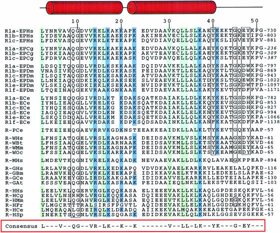
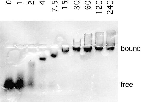
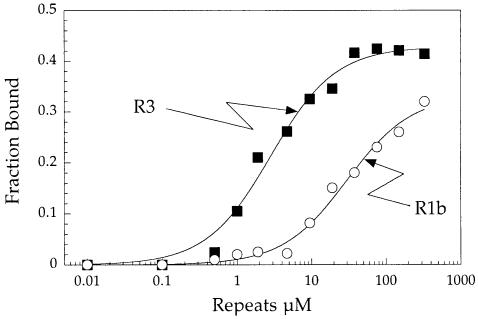
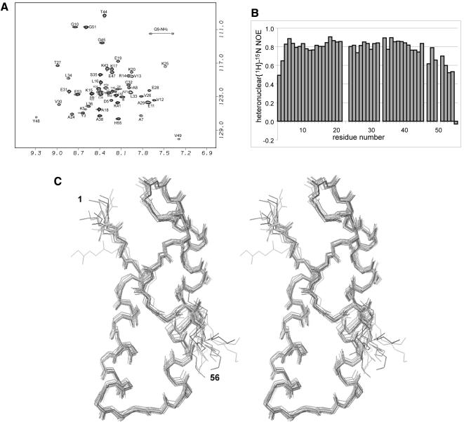
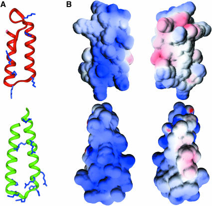
Aminoacyl-tRNA synthetases of higher eukaryotes possess polypeptide extensions in contrast to their prokaryotic counterparts. These extra domains of poorly understood function are believed to be involved in protein-protein or protein-RNA interactions. Here we showed by gel retardation and filter binding experiments that the repeated units that build the linker region of the bifunctional glutamyl-prolyl-tRNA synthetase had a general RNA-binding capacity. The solution structure of one of these repeated motifs was also solved by NMR spectroscopy. One repeat is built around an antiparallel coiled-coil. Strikingly, the conserved lysine and arginine residues form a basic patch on one side of the structure, presenting a suitable docking surface for nucleic acids. Therefore, this repeated motif may represent a novel type of general RNA-binding domain appended to eukaryotic aminoacyl-tRNA synthetases to serve as a cis-acting tRNA-binding cofactor.


Figures

None
Fig. 1. Alignment of the conserved RNA-binding motifs appended to eukaryotic aaRS. The first capital letters of the protein represent the amino acid substrate, while the final two letters are for the species [Hs (Homo sapiens) stands for human, Cg (Cricetulus griseus) for hamster, Dm (Drosophila melanogaster) for fly, Ce (Caenorhabditis elegans) for nematode, Bt (Bos taurus) for cow, Mm (Mus musculus) for mouse, Oc (Oryctolagus cuniculus) for rabbit, Bm (Bombyx mori) for silkworm, At (Arabidopsis thaliana) for cress, Fr (Fugu rubripes) for pufferfish and Sp (Schizosaccharomyces pombe) for yeast]. Residues that match the consensus sequence (defined as residues conserved in 80% of the repeated sequences) are boxed. Conserved hydrophobic residues are in green, basic residues are in blue. The sequence numbers given on the top line relate to the sequence of the R1b motif from hamster used to determine its solution structure. The regions that form helices in R1b are indicated above the sequence alignment.
None
Fig. 2. Gel retardation experiment exemplifying the general RNA-binding capacity of the repeats. RNA homopolymers of guanylate nucleotide were end-labeled with 32P using polynucleotide kinase. R3 (0–240 μM) was incubated with 10 nM [32P]poly(G) in the incubation buffer at 25°C for 15 min. After electrophoresis at 4°C on a native agarose gel, the mobility shift of poly(G) was visualized by autoradiography.
None
Fig. 3. Analysis of the association of poly(G) to the repeats by a filter binding assay. R3 or R1b was incubated at increasing concentrations (0.01–330 μM) with 30 nM [32P]poly(G) as described in the legend to Figure 2. The incubate was applied to nitrocellulose filters (Millipore) to recover radioactivity associated to the proteins. The apparent dissociation constants for poly(G) were determined by non-linear regression of a binding equation to the experimental data. Values of 2.9 and 30 μM were obtained for R3 and R1b, respectively.
None
Fig. 4. (A) 1H–15N HSQC spectrum of R1b. The assignment is labelled with individual amino acid and residue number. (B) Heteronuclear 1H–15N NOE measurement on R1b, showing the relatively high mobility of the Ω–loop and C–terminus. High values of the heteronuclear NOE reflect decreased mobility, typical values for rigid proteins are above 0.7. (C) Stereo view of the 15 calculated structures of R1B (1–56). Backbone atoms are superimposed to the mean structure from residues 3 to 53.
None
Fig. 5. (A) Ribbon diagram of the lowest energy structure of R1B (3–51) (top), compared with another RNA-binding protein, S15 (24–71) (bottom). The conserved basic residues in both families are indicated. (B) Electrostatic surface potential of R1B [in front view, same orientation as in (A)] and S15 [slightly rotated compared with (A)], and 180° rotated view. Positive and negative charges are shown in blue and red, respectively. The figures were generated using GRASP (Nicholls et al., 1991).

References

    1. Alzhanova A.T., Fedorov, A.N., Ovchinnikov, L.P. and Spirin, A.S. (1980) Eukaryotic aminoacyl-tRNA synthetases are RNA binding proteins whereas prokaryotic ones are not. FEBS Lett., 120, 225–229. - PubMed
    1. Arnez J.G. and Cavarelli, J. (1997) Structures of RNA-binding proteins. Q. Rev. Biophys., 30, 195–240. - PubMed
    1. Berglund H., Rak, A., Serganov, A., Garber, M. and Härd, T. (1997) Solution structure of the ribosomal RNA binding protein S15 from Thermus thermophilus. Nature Struct. Biol., 4, 20–23. - PubMed
    1. Biou V., Yaremchuk, A., Tukalo, M. and Cusack, S. (1994) The 2.9 Å crystal structure of T.thermophilus seryl-tRNA synthetase complexed with tRNASer. Science, 263, 1404–1410. - PubMed
    1. Brünger A.T. (1993) X-PLOR Version 3.1: A System for X-ray Crystallography and NMR. Yale University Press, New Haven, CT.

Publication types