. 2000 Feb 1;97(3):1032-7.
doi: 10.1073/pnas.97.3.1032.

Phosphorylation of human progesterone receptors at serine-294 by mitogen-activated protein kinase signals their degradation by the 26S proteasome

Affiliations

Phosphorylation of human progesterone receptors at serine-294 by mitogen-activated protein kinase signals their degradation by the 26S proteasome

C A Lange et al. Proc Natl Acad Sci U S A. .

Abstract

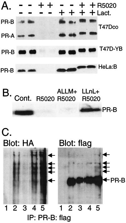
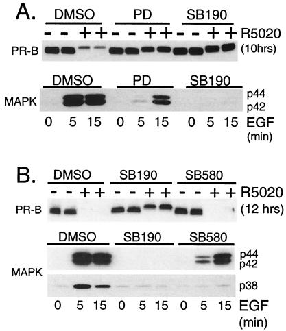
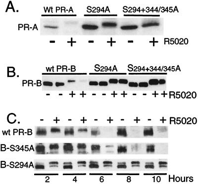
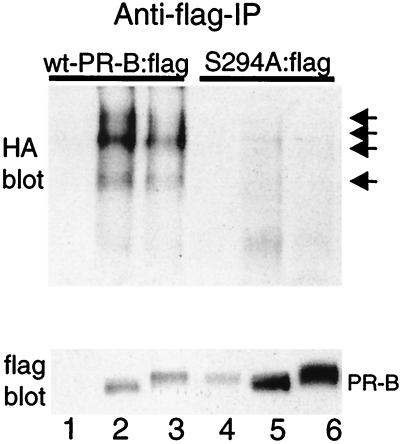
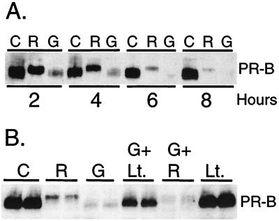
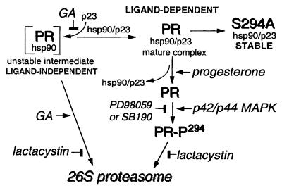
Ligand-dependent down-regulation that leads to rapid and extensive loss of protein is characteristic of several nuclear steroid receptors, including human progesterone receptors (PRs). In breast cancer cells, >95% of PRs are degraded 6 h after the start of progestin treatment. The mechanism for down-regulation is unknown. We examined the role of PR phosphorylation by mitogen-activated protein kinases (MAPKs) in this process. Lactacystin and calpain inhibitor I, specific inhibitors of the 26S proteasome, blocked progestin-induced down-regulation, and ubiquitinated conjugates of PR accumulated in cells. Ligand-dependent PR degradation was also blocked by specific inhibition of p42 and p44 MAPKs. To define the targets of phosphorylation by this kinase, two serine/proline MAPK consensus sites on PR were mutated. We demonstrate that mutation of PR serine-294 to alanine (S294A) specifically and completely prevents ligand-dependent receptor down-regulation. We also find that rapid, ligand-independent degradation of immature PR intermediates occurs by a proteasome-mediated pathway. These results demonstrate that PR destruction, by either of two alternate routes, is mediated by the 26S proteasome. Specifically, down-regulation of mature PRs occurs by a mechanism in which ligand binding activates PR phosphorylation by MAPKs at a unique serine residue, which then targets the receptors for degradation.


Figures

Figure 1
Figure 1
PR down-regulation is mediated by the 26S proteasome. (A) T47Dco breast cancer cells, which express the natural B and A isoforms of PRs constitutively (15), and T47D-YB (16) and HeLa:B cells (17), which stably express the recombinant B isoform of PRs, were treated without or with the progestin R5020 (10 nM) for 12 h in the absence (DMSO solvent) or presence of lactacystin [Lact. (10 μM)], and PR protein (100 μg of total protein per lane) was detected by immunoblotting with PR-specific monoclonal antibodies. (B) Inhibition of PR down-regulation by calpain inhibitor I, but not calpain inhibitor II. T47D-YB cells were treated as in A, except with calpain inhibitor II [ALLM (25 μM)] or calpain inhibitor I [LLnL (25 μM)]. Each compound alone had no effect on PR abundance. (C) PR-ubiquitin conjugates in cells transiently overexpressing ubiquitin and PR-B. HeLa cells were transiently cotransfected with expression vectors encoding HA-tagged ubiquitin and epitope-flag-tagged wild-type PR-B (PR-B:flag) and treated for 4 h without (lane 2) or with R5020 (lane 3; 10 nM) in the absence or presence of lactacystin (lane 4; 10 μM) or LLnL (lane 5; 25 μM). PRs were immunoprecipitated by using anti-flag M2 affinity gel and visualized by immunoblotting with either HA- or flag-specific antibodies. Lane 1, nonspecific antibody and similar affinity gel. High-molecular-weight ubiquitinated forms of PR-B:flag are indicated (arrows).
Figure 2
Figure 2
Activation of MAPK triggers ligand-dependent PR down-regulation. (A) MEK inhibitors block ligand-induced PR down-regulation. T47D-YB cells were pretreated with vehicle (DMSO) or with PD98059 [PD (100 μM)] or SB202190 [SB190 (40 μM)] for 30 min before challenge with R5020 [10 nM (Upper)] or EGF [30 ng/ml (Lower)] for the indicated times. PR-B protein levels (Upper) and MAPK activity (Lower) were measured by immunoblotting with PR- or MAPK-phosphospecific antibodies, respectively. Total MAPK levels remained unchanged (data not shown). (B) Ligand-induced PR down-regulation requires activation of p42 and p44 MAPKs, but not p38 MAPK. T47D-YB cells were pretreated with vehicle (DMSO), SB202190 [SB190 (40 μM)], or SB203580 [SB580 (40 μM)] for 30 min before challenge with R5020 [10 nM (Upper)] or EGF [30 ng/ml (Lower)] for the indicated times. PR-B protein levels (Upper) were measured with specific antibodies, and p42/p44 and p38 MAPK activities (Lower) were measured with phosphospecific antisera for each kinase; 100 μg of protein was loaded per lane.
Figure 3
Figure 3
Mutation of serine-294 to alanine at a MAPK consensus site stabilizes PR in the presence of progestin. (A) Mutant S294A and S294+S344/345A PR-As are resistant to R5020-induced receptor down-regulation. HeLa cells were transiently transfected with cDNA vectors (1 μg) encoding either wild-type PR-A or the S294A or S294+344/345A mutants of PR-A and then treated without or with R5020 (10 nM) for 8 h. PR protein was measured in whole-cell lysates (100 μg) by immunoblotting. (B) Mutant S294A and S294+344/345A PR-Bs are resistant to R5020-induced receptor down-regulation. Duplicate cultures of HeLa cells were transfected with wild-type PR-B or each phosphomutant of PR-B and treated without or with R5020 for 18 h. Protein (100 μg per lane) was loaded, and PR levels were measured by using PR-specific monoclonal antibodies. (C) The S294A PR-Bs stably expressed in T47D-Y cells are resistant to ligand-induced down-regulation. PR-negative breast cell lines stably expressing either an S344/345A (B-S345A) mutant or the S294A (B-S294A) mutant PR-B receptors were produced by transfection of receptor expression vectors containing the neomycin-resistance gene into T47D-Y cells and selected for growth in G418. Neoresistant clonal cell lines were screened for PR expression, and S344/345A or S294A PR-containing cells or wild-type PR-B-containing T47D-YB cells (wt-PR-B) were treated without or with R5020 (10 nM) for 2–10 h; protein levels were measured with PR-specific antibodies. 150–200 μg of protein was loaded per lane.
Figure 4
Figure 4
Mutant S294A PR-Bs fail to undergo ubiquitination. HeLa cells were transiently cotransfected with expression vectors encoding HA-tagged-ubiquitin and either epitope-flag-tagged wild-type PR-B [wt-PR-B:flag (lanes 1–3)] or epitope-flag-tagged S294A mutant PR-B [S294A:flag (lanes 4–6)] and treated for 4 h without (lanes 2 and 5) or with R5020 (lanes 3 and 6). PRs were immunoprecipitated by using an anti-flag M2 affinity gel and were visualized by immunoblotting with either HA- or flag-specific antibodies. Lanes 1 and 3, nonspecific antibody and similar affinity gel. High-molecular-weight ubiquitinated forms in immunoprecipitates from lysates of cells containing wild-type but not S294A mutant PR-Bs are indicated by arrows.
Figure 5
Figure 5
Ligand-independent degradation of immature PRs. (A) Immature PRs are rapidly degraded in the absence of ligand. T47D-YB cells were treated without [C (EtOH vehicle)] or with R5020 alone [R (10 nM)] or with the hsp 90-binding agent GA (G) alone (10 μg/ml) for 2, 4, 6, and 8 h, and PR-B protein in whole-cell lysates was measured by immunoblot analysis. (B) Ligand-independent degradation is mediated by the 26S proteasome. Duplicate cultures of T47D-YB cells were treated as indicated, without [C (EtOH vehicle)] or with R5020 [R (10 nM)] alone, GA [G (10 μg/ml)] alone, or with GA plus lactacystin [Lt. (10 nM; 30-min pretreatment)], GA plus R5020, or lactacystin alone for 6 h. PR-B protein in whole-cell lysates (100 μg of protein per lane) was measured by immunoblot analysis with PR-specific antisera.
Figure 6
Figure 6
Model for regulated loss of PR protein by the 26S proteasome and for ligand-independent and ligand-dependent PR down-regulation. Mature PR complexes bind progesterone, leading to ligand-dependent phosphorylation of serine 294 by p42/p44 MAPKs, which serves as a signal for PR degradation by the 26S proteasome. Inhibition of p42/p44 MAPKs (PD98059 or SB190) or the 26S proteasome (lactacystin) blocks loss of wild-type PR in the presence of progestins. S294A-mutant PRs are resistant to ligand-dependent down-regulation. In the presence of GA, rapid ligand-independent degradation of immature PR complexes also occurs by a 26S proteasome-dependent pathway.

References

    1. Horwitz K B, McGuire W L. Steroids. 1975;25:497–505. - PubMed
    1. Elledge R M, McGuire W L, Osborne C K. Semin Oncol. 1992;19:244–253. - PubMed
    1. Wei L L, Krett N L, Francis M D, Gordon D F, Wood W M, O'Malley B W, Horwitz K B. Mol Endocrinol. 1988;2:62–72. - PubMed
    1. Nardulli A M, Katzenellenbogen B S. Endocrinology. 1988;122:1532–1540. - PubMed
    1. Mimnaugh E G, Chavany C, Neckers L. J Biol Chem. 1996;271:22796–22801. - PubMed

Publication types

MeSH terms

Substances