. 2000 Feb 1;97(3):1125-30.
doi: 10.1073/pnas.97.3.1125.

The sentrin-conjugating enzyme mUbc9 interacts with GLUT4 and GLUT1 glucose transporters and regulates transporter levels in skeletal muscle cells

Affiliations

The sentrin-conjugating enzyme mUbc9 interacts with GLUT4 and GLUT1 glucose transporters and regulates transporter levels in skeletal muscle cells

F Giorgino et al. Proc Natl Acad Sci U S A. .

Abstract

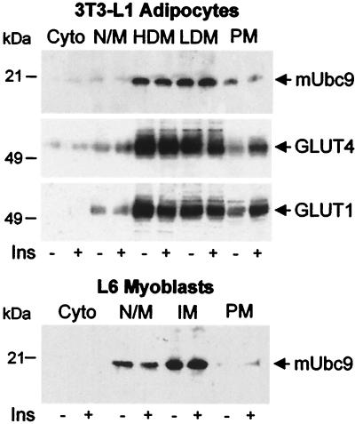
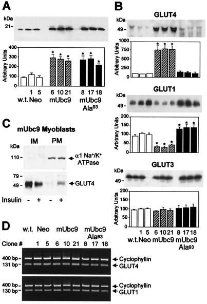
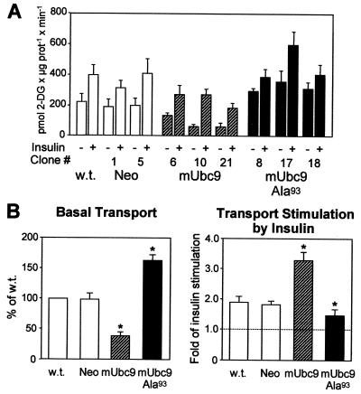
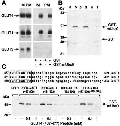
Glucose transport in insulin-regulated tissues is mediated by the GLUT4 and GLUT1 transporters. Using the yeast two-hybrid system, we have cloned the sentrin-conjugating enzyme mUbc9 as a protein that interacts with the GLUT4 COOH-terminal intracellular domain. The mUbc9 enzyme was found to bind directly to GLUT4 and GLUT1 through an 11-aa sequence common to the two transporters and to modify both transporters covalently by conjugation with the mUbc9 substrate, sentrin. Overexpression of mUbc9 in L6 skeletal muscle cells decreased GLUT1 transporter abundance 65%, resulting in decreased basal glucose transport. By contrast, mUbc9 overexpression increased GLUT4 abundance 8-fold, leading to enhanced transport stimulation by insulin. A dominant-negative mUbc9 mutant lacking catalytic activity had effects opposite to those of wild-type mUbc9. The regulation of GLUT4 and GLUT1 was specific, as evidenced by an absence of mUbc9 interaction with or regulation of the GLUT3 transporter isoform in L6 skeletal muscle cells. The mUbc9 sentrin-conjugating enzyme represents a novel regulator of GLUT1 and GLUT4 protein levels with potential importance as a determinant of basal and insulin-stimulated glucose uptake in normal and pathophysiological states.


Figures

Figure 1
Figure 1
Predominant localization of mUbc9 protein in intracellular membranes and nuclei/mitochondria of 3T3-L1 adipocytes and L6 myoblasts. Basal or insulin-stimulated (Ins, 1 μM) cells were processed to obtain the cytosol (Cyto), a fraction enriched in nuclei/mitochondria (N/M), high-density microsomes (HDM), low-density microsomes (LDM), and plasma membranes (PM). Intracellular membranes not fractionated into HDM and LDM (IM) were obtained from L6 cells. Equal amounts of protein (10 μg) were subjected to immunoblotting with anti-mUbc9 antibodies. Subcellular fractions from 3T3-L1 adipocytes (10 μg) also were analyzed by immunoblotting with antibodies to GLUT4 or GLUT1, as indicated.
Figure 2
Figure 2
Regulation of glucose transporters by mUbc9. (A) Overexpression of mUbc9 cDNA in L6 myoblasts. L6 cells were left nontransfected (wild type, w.t.) or stably transfected with plasmids encoding mUbc9 (mUbc9, clones 6, 10, and 21), mUbc9 with mutation of Cys93 to Ala (mUbc9-Ala93, clones 8, 17, and 18), or the G418 resistance gene alone (Neo, clones 1 and 5). Total cell lysates (10 μg) were analyzed by immunoblotting with anti-mUbc9 antibodies (Upper), and the amount of mUbc9 protein was quantified in multiple experiments (Lower, mean ± SE of five experiments). *, P < 0.05 vs. Neo, clones 1 and 5, and w.t. by unpaired Student's t test. (B) Effects of mUbc9 or mUbc9-Ala93 overexpression on GLUT4, GLUT1, and GLUT3 protein levels in L6 myoblasts. Total cellular membranes (10 μg) from wild-type (w.t.), Neo, mUbc9, or mUbc9-Ala93 myoblasts were analyzed by immunoblotting with anti-GLUT4, anti-GLUT1, or anti-GLUT3 antibodies. A representative transporter immunoblot and the quantification of multiple immunoblots (mean ± SE of three experiments) are shown for each. *, P < 0.05 vs. Neo, clones 1 and 5, and wild type by unpaired Student's t tests. (C) GLUT4 transporters in L6 myoblasts overexpressing mUbc9 exhibit insulin-regulatable translocation to the cell surface. IM (10 μg) and PM (10 μg) from basal or insulin-stimulated (1 μM) cells (clone 10) were analyzed by immunoblotting with antibodies to the α1-subunit of Na+/K+ ATPase, as a plasma membrane marker (Upper), or GLUT4 (Lower). IM and PM were depleted or enriched, respectively, in α1 Na+/K+ ATPase. GLUT4 levels in IM and PM fractions from Neo or mUbc9-Ala93 myoblasts were very low (not shown). (D) Unaltered GLUT4 and GLUT1 mRNA levels in nontransfected (w.t.), Neo, mUbc9, and mUbc9-Ala93 myoblasts. Total RNA (10 ng) was subjected to reverse transcription–PCR analysis for determination of GLUT4, GLUT1, and cyclophyllin (coamplified in each reaction as a control for amplification efficiency) mRNA levels.
Figure 3
Figure 3
Modulation of glucose transport by mUbc9. (A) Basal and insulin-stimulated glucose transport rates in wild-type (w.t.), Neo, mUbc9, and mUbc9-Ala93 myoblasts. Cells cultured in 35-mm diameter wells were serum-starved overnight and then incubated in the presence or absence of 1 μM insulin for 30 min. Transport was started by adding 1 μCi/ml 2-[3H]deoxy-d-glucose (NEN) to a concentration of 50 μM for 10 min at 20°C. (B) Basal glucose transport (Left) and the fold stimulation of glucose transport by insulin (Right) in nontransfected (w.t.), Neo, mUbc9, and mUbc9-Ala93 L6 myoblasts. The results represent mean values of four independent experiments performed on individual L6 clones. *, P < 0.05 vs. Neo and wild type by unpaired Student's t tests.
Figure 4
Figure 4
Association of mUbc9 with glucose transporters. (A) Binding of recombinant mUbc9 to GLUT4 and GLUT1, but not GLUT3. GST-mUbc9 or GST (850 nM) was incubated with 75 μg of detergent-solubilized IM and PM from L6 myoblasts overexpressing GLUT4. Transporters bound to the GST fusion proteins were detected by immunoblotting with anti-GLUT4, anti-GLUT1, or anti-GLUT3 antibodies (Right). Solubilized membranes (10 μg) were analyzed by immunoblotting to determine their content of GLUT4, GLUT1, and GLUT3 (Left). (B) Association of GST-mUbc9 with recombinant COOH-terminal fragments of GLUT4 and GLUT1 in vitro. COOH-terminal fragments of GLUT4 or GLUT1 fused to hexahistidine-tagged DHFR (6xHis-DHFR, 850 nM) were incubated with GST-mUbc9 or GST (250 nM). The GST fusion proteins associated with the 6xHis-DHFR fusions were detected by immunoblotting with anti-GST antibodies. Lanes: a, 6xHis-DHFR plus GST; b, 6xHis-DHFR-GLUT1 (residues 451–492) plus GST-mUbc9; c, 6xHis-DHFR-GLUT1 (residues 451–492) plus GST; d, 6xHis-DHFR-GLUT4 (residues 467–509) plus GST-mUbc9; e, 6xHis-DHFR-GLUT4 (residues 467–509) plus GST; f, 6xHis-DHFR plus GST-mUbc9. (C) Transporter domain responsible for mUbc9 binding. (Upper) COOH-terminal sequences of rat GLUT4, rat GLUT1, and rat GLUT3 after the 12th putative transmembrane domain. Amino acid identities in GLUT4 and GLUT1 (uppercase letters) and in GLUT4, GLUT1, and GLUT3 (bold, uppercase letters) are indicated. The boxed amino acid sequences represent highly homologous regions in the transporters. (Lower) Effects of a peptide corresponding to residues 467–477 of rat GLUT4 on the association between GST-mUbc9 and various COOH-terminal fragments of GLUT4 or GLUT1. DHFR-GLUT4 (467–509) 489A 490S indicates a variant of the GLUT4 COOH terminus (residues 467–509) with substitution of Leu489 and Leu490 by Ala and Ser, respectively.
Figure 5
Figure 5
Covalent modification of GLUT4 and GLUT1 by sentrin conjugation. Solubilized membrane fractions from basal or insulin-stimulated (100 nM) 3T3-L1 adipocytes were subjected to immunoprecipitation with monoclonal anti-GLUT4 (A) or polyclonal anti-GLUT1 (B) antibodies. Negative control immunoprecipitations were performed with unrelated antibodies (a, monoclonal antiphosphotyrosine; b, monoclonal antiinsulin receptor; c, polyclonal antiphosphotyrosine; d, polyclonal anti-IRS-1). Immunoprecipitates then were subjected to immunoblotting with polyclonal anti-GLUT4 (A Left), polyclonal anti-sentrin-1 (A Right), polyclonal anti-GLUT1 (B Left), or monoclonal anti-sentrin-1 (B Right) antibodies.

References

    1. Bell G I, Burant C F, Takeda J, Gould G W. J Biol Chem. 1993;268:19161–19164. - PubMed
    1. Pessin J E, Bell G I. Annu Rev Physiol. 1992;54:911–930. - PubMed
    1. Piper R C, Hess L J, James D E. Am J Physiol. 1991;260:C570–C580. - PubMed
    1. Slot J W, Geuze H J, Gigengack S, Lienhard G E, James D E. Proc Natl Acad Sci USA. 1991;88:7815–7819. - PMC - PubMed
    1. Rodnick K J, Slot J W, Studelska D R, Hanpeter D E, Robinson L J, Geuze H J, James D E. J Biol Chem. 1992;267:6278–6285. - PubMed

Publication types

MeSH terms

LinkOut - more resources