. 2000 Feb 1;97(3):1131-6.
doi: 10.1073/pnas.97.3.1131.

Expression of guanylin in "pars tuberalis-specific cells" and gonadotrophs of rat adenohypophysis

Affiliations

Expression of guanylin in "pars tuberalis-specific cells" and gonadotrophs of rat adenohypophysis

L D'Este et al. Proc Natl Acad Sci U S A. .

Abstract

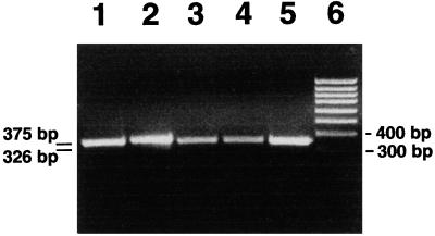
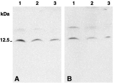
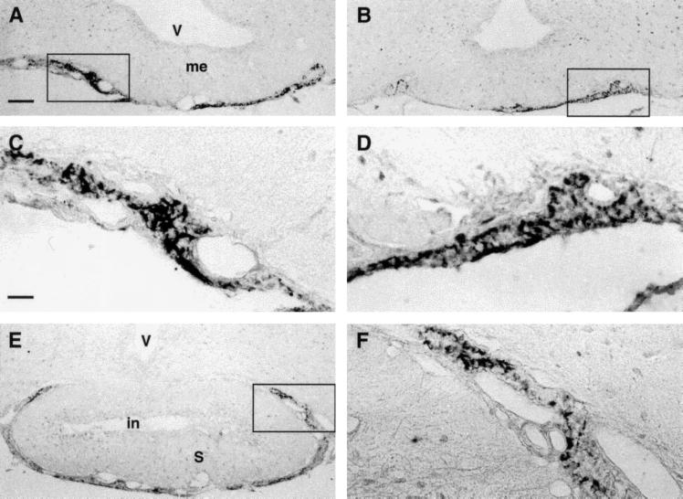
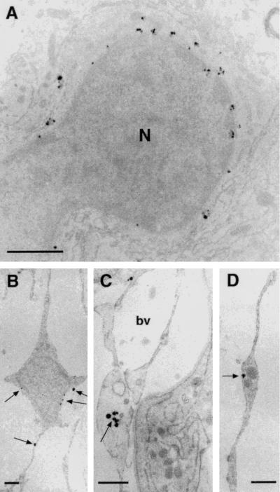
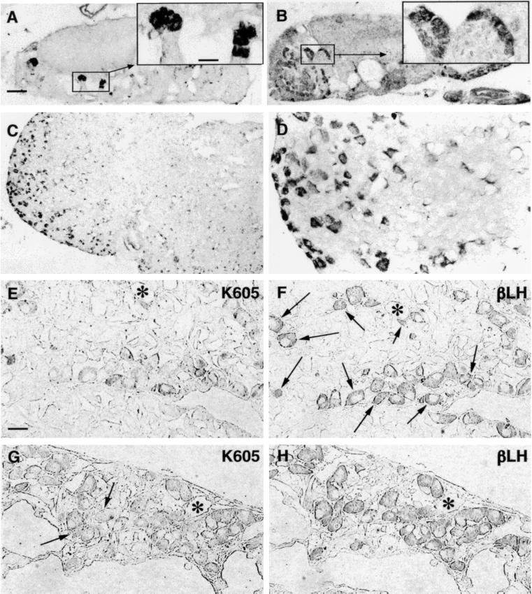
The intestinal peptide guanylin regulates the electrolyte/water transport in the gastrointestinal epithelium by paracrine/luminocrine mechanisms. Because guanylin also circulates in the blood, we investigated the rat hypothalamo-pituitary region for expression and cellular localization of this peptide. Reverse transcriptase-PCR analyses with guanylin-specific primers revealed expression of the peptide in the pars tuberalis and pars distalis of the pituitary. Western blotting analyses in hypophyseal tissue extracts identified the expected 12.5-kDa immunoreactive peptide by using two different region-specific guanylin antisera. Light and electron microscopic immunocytochemistry with the same antisera localized guanylin in "pars tuberalis-specific cells" in the juxtaneural pars tuberalis adjacent to nerve endings and blood vessels of the hypothalamo-pituitary portal system and in gonadotrophic cells within the distal pars tuberalis and ventrolateral part of the pars distalis. The presence and cell-specific localization of guanylin within the hypothalamo-hypophyseal system indicate that this peptide may be specifically involved in paracrine and endocrine regulatory mechanisms.


Figures

Figure 1
Figure 1
RT-PCR analysis with guanylin-specific primers shows amplification products of correct size in rat whole hypophysis, PT of the hypophysis, and duodenum; 15 μl of RT-PCR products were loaded on a 1.8% 89 mM Tris/89 mM boric acid/2 mM EDTA (pH 8.3) agarose gel containing ethidium bromide. (Lane 1) PT, GAPDH; (lane 2) hypophysis, GAPDH; (lane 3) PT, guanylin; (lane 4) hypophysis, guanylin; (lane 5) duodenum, guanylin; (lane 6) 100-bp marker.
Figure 2
Figure 2
Western blot after Tricine-SDS/PAGE of rat tissue extracts immunostained with antiserum K42 (A) and antiserum K605 (B). (Lane 1) duodenum; (lane 2) colon; (lane 3) hypophysis. Note the predominant immunoreactive band of ≈12.5 kDa in the rat hypophysis, duodenum, and colon (positive control). The tissue extraction procedure yielded a faintly immunoreactive band at 17 kDa. The low molecular mass markers from Novex (San Diego, CA) were used for the molecular mass calibration.
Figure 3
Figure 3
(A and B) Panoramic overview of two neighboring sections of juxtaneural PT region to compare the distribution of guanylin immunoreactivities obtained with the antisera K605 (A) and K42 (B). (C and D) Higher magnification of boxed areas in A and B, respectively. The immunoreactive elements showed a stick- or rod-like morphology. (E) Guanylin immunoreactive (K605) elements in PT at the pituitary stalk level. (F) Higher magnification of the boxed area in E. V, third ventricle; me, median eminence; in, infundibular recess; S, pituitary stalk. (A, B, and E, bar = 100 μm; C, D, and F, bar = 25 μm.)
Figure 4
Figure 4
(A–D) Ultrathin sections of juxtaneural PT previously submitted to preembedding gold-silver immunocytochemical procedures with K605 antiserum. Gold-silver-enhanced particles are contained in small vesicles both in the cell body and in the cell elongations (arrows). N, nucleus of a specific cells of PT; bv, blood vessel. (Bars = 1 μm.)
Figure 5
Figure 5
(A and B) Clusters of guanylin immunoreactive cells can be observed in two distal PT sections at different craniocaudal level. (Insets) Higher magnification of boxed areas. (C) Guanylin immunoreactive cells in PD assumed the morphology of the endocrine cells of this region. (D) Higher magnification of an area in C. (E–H) Pair of two consecutive semithin (0.5-μm) sections of PD immunostained for guanylin (K605; E and G) and h-β-LH (βLH; F and H). Almost all guanylin immunoreactive cells show coincident immunoreactivity for h-β-LH, but the number of colocalizing gonadotrophs varies: about one-third in E and F (arrows) and almost all in G and H. Arrows in G indicate the only two guanylin unreactive gonadotrophs. Asterisks label some landmarks to allow the alignment of consecutive sections. (A–C, bar = 100 μm; Insets and E–H, bar = 25 μm.)

References

    1. Currie M G, Fok K F, Kato J, Moore R J, Hamra F K, Duffin K L, Smith C E. Proc Natl Acad Sci USA. 1992;89:947–961. - PMC - PubMed
    1. Gabers D L. Cell. 1992;71:1–4. - PubMed
    1. De Sauvage F, Keshav S, Kuang W J. Proc Natl Acad Sci USA. 1992;89:9089–9093. - PMC - PubMed
    1. Hamra F K, Forte L R, Eber S L, Pidhorodeckyi N V, Krause W J, Freeman R H, Chin D T, Tompkin J A, Fok K F, Smith C E, et al. Proc Natl Acad Sci USA. 1993;90:10464–10468. - PMC - PubMed
    1. Forte L R, Eber S L, Fan X, London R M, Wang Y, Rowland L M, Chin D T, Freeman R H, Krause W J. Endocrinology. 1999;140:1800–1806. - PubMed

Publication types

LinkOut - more resources