. 2000 Feb 1;97(3):1148-53.
doi: 10.1073/pnas.97.3.1148.

Early disruption of centromeric chromatin organization in centromere protein A (Cenpa) null mice

Affiliations

Early disruption of centromeric chromatin organization in centromere protein A (Cenpa) null mice

E V Howman et al. Proc Natl Acad Sci U S A. .

Abstract

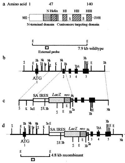
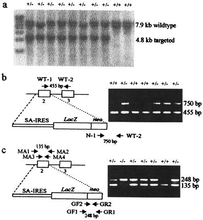
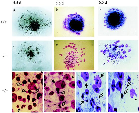
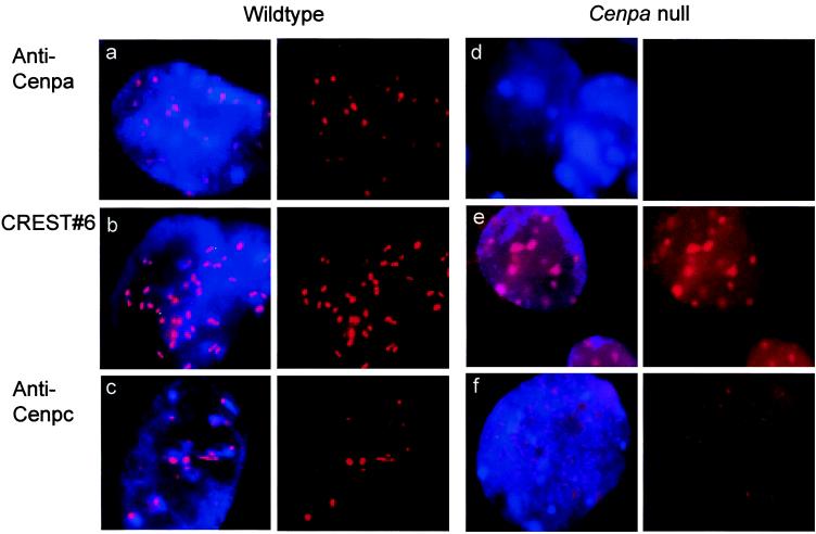
Centromere protein A (Cenpa for mouse, CENP-A for other species) is a histone H3-like protein that is thought to be involved in the nucleosomal packaging of centromeric DNA. Using gene targeting, we have disrupted the mouse Cenpa gene and demonstrated that the gene is essential. Heterozygous mice are healthy and fertile whereas null mutants fail to survive beyond 6.5 days postconception. Affected embryos show severe mitotic problems, including micronuclei and macronuclei formation, nuclear bridging and blebbing, and chromatin fragmentation and hypercondensation. Immunofluorescence analysis of interphase cells at day 5.5 reveals complete Cenpa depletion, diffuse Cenpb foci, absence of discrete Cenpc signal on centromeres, and dispersion of Cenpb and Cenpc throughout the nucleus. These results suggest that Cenpa is essential for kinetochore targeting of Cenpc and plays an early role in organizing centromeric chromatin at interphase. The evidence is consistent with the proposal of a critical epigenetic function for CENP-A in marking a chromosomal region for centromere formation.


Figures

Figure 1
Figure 1
Targeted disruption of the mouse Cenpa gene. (a) The mouse Cenpa protein showing the different subdomains, in particular those at the C terminus that are required for centromere targeting. Our targeting construct (see below) was designed to delete amino acids 29–64 (gray box), which will effectively remove the entire centromere-targeting domain. (b) A restriction map of the Cenpa gene. The exons are denoted by black boxes (23). (c) The gene replacement construct, where the selectable marker cassette consists of a splice-acceptor site (SA), a picornaviral IRES, a lacZ-neomycin-resistance fusion gene, and a simian virus 40 polyadenylation sequence (PA). (d) The Cenpa locus after gene disruption. The positions of external probes used in Southern analysis are shown and the expected size fragments are 7.9-kb wild-type allele and a 4.8-kb targeted allele. ATG and TAA are translation start and stop codons, respectively. Restriction enzymes used were SacI (S), SalI (Sa), EcoRI (E), XbaI (Xb), XhoI (Xh), KpnI (K), NheI (Nh), and SpeI (Sp).
Figure 2
Figure 2
Southern blotting and PCR genotyping of cell line, tail, and embryo DNA. (a) Southern blot analysis of putative targeted ES cell colonies after EcoRI digestion and probed with an external probe (see Fig. 1 b and d). (b) PCR analysis of mouse tail DNA showing a wild-type product of 455 bp detected by WT-1 and WT-2 primers, and a targeted product of 750 bp detected by N-1 and WT-2 primers. SA, splice-acceptor site. (c) Nested PCR of mouse embryos resulting in a 135-bp wild-type product when using primers MA1, MA2, MA3, and MA4, and a 248-bp targeted product when using primers GF1, GR1, GF2, and GR2.
Figure 3
Figure 3
Phase contrast and Giemsa staining images of day-5.5 and -6.5 embryos. Day-5.5 normal embryo photographed by phase (a) or stained with Giemsa (b). (c) Day-6.5 normal embryo stained with Giemsa. Note the compact, dark inner cell mass and the surrounding trophectoderm outgrowth. Day-5.5 −/− embryo photographed by phase (d) or stained with Giemsa (e). (f) Day-6.5 −/− embryo stained with Giemsa. Note the absence of a defined inner cell mass and the incoherent cells in both the day-5.5 and -6.5 −/− embryos. Magnification for af: ×150. (g and h) Close-ups of e and (i and j) close-ups of f, showing micronuclei (empty-triangle arrow), macronuclei (filled-triangle arrow), nuclear bridging (open arrow), nuclear blebbing (filled arrowhead), and highly condensed chromatin bodies (empty arrowhead).
Figure 4
Figure 4
Immunofluorescence analysis of day-5.5 embryos. (ac) Wild-type interphase cells stained with anti-Cenpa, CREST#6 autoimmune serum, and anti-Cenpc, respectively. (df) Cenpa null interphase cells stained with anti-Cenpa, CREST#6, and anti-Cenpc, respectively. Although these pictures represented the results taken at one focal plane of a three-dimensional interphase cell nucleus, direct microscopic analysis through all the planes indicated only variation in the total number of observable signals but not the morphology of the signals (e.g., the more diffuse spots in e, and the higher background signals throughout the nuclei in both e and f compared with their respective controls). (Left) Simultaneous staining of chromatin with 4′,6-diamidino-2-phenylindole (blue) and centromere with anticentromere antibody (red). (Right) Split image of Left showing anticentromere antibody staining (red) only.

References

    1. Choo K H A. The Centromere. Oxford: Oxford Univ. Press; 1997.
    1. Sullivan K, Hechenberger M, Masri K. J Cell Biol. 1994;127:581–592. - PMC - PubMed
    1. Pietras D F, Bennett K L, Siracusa L D, Woodworth-Gutai M, Chapman V M, Gross K W, Kane-Haas C, Hastie N D. Nucleic Acids Res. 1983;11:6965–6983. - PMC - PubMed
    1. Rattner J B. BioEssays. 1991;13:51–56. - PubMed
    1. Hudson D F, Fowler K, Earle E, Saffery R, Kalitsis P, Trowell H, Hill J, Wreford N, de Kretser D, Cancilla M, et al. J Cell Biol. 1998;141:309–319. - PMC - PubMed

Publication types

MeSH terms

LinkOut - more resources