. 2000 Feb 1;97(3):1184-9.
doi: 10.1073/pnas.97.3.1184.

Accelerated development of IgG autoantibodies and autoimmune disease in the absence of secreted IgM

Affiliations

Accelerated development of IgG autoantibodies and autoimmune disease in the absence of secreted IgM

M Boes et al. Proc Natl Acad Sci U S A. .

Abstract

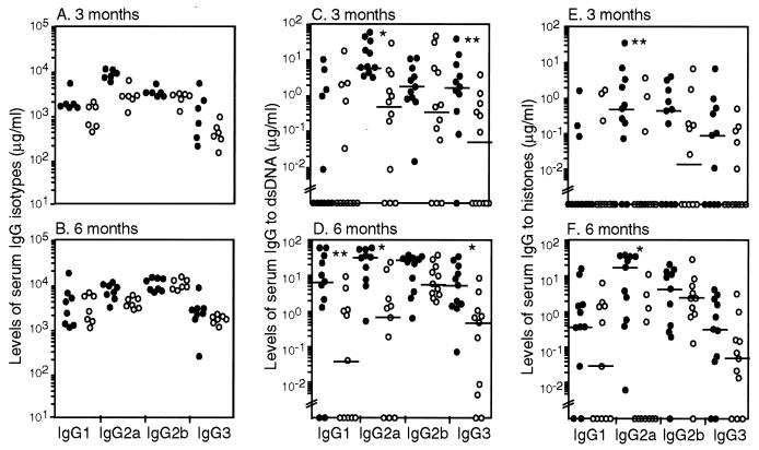
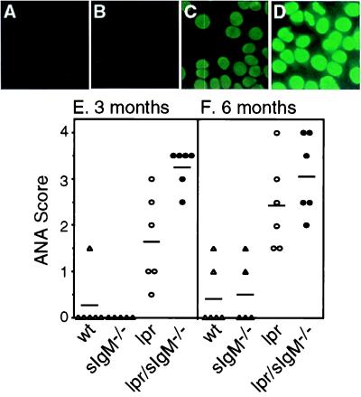
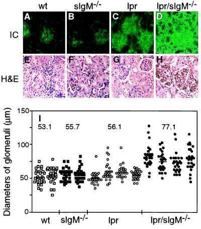
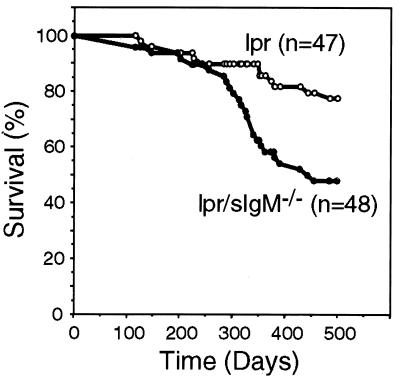
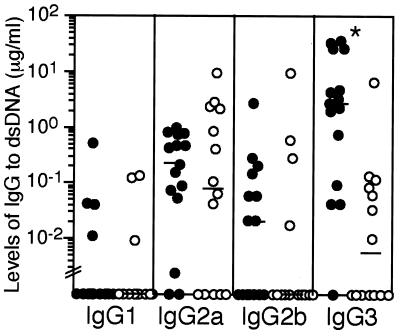
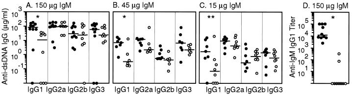
Individuals with systemic lupus erythematosus and rheumatoid arthritis are characterized by the presence of high levels of circulating IgM and IgG autoantibodies. Although IgG autoantibodies often are pathogenic, the role of IgM autoantibodies in autoimmune disease is not clear. Using mice that are unable to secrete IgM but are able to express surface IgM and IgD and to secrete other classes of immunoglobulins, we examined the effect of the absence of secreted IgM in the development of IgG autoantibodies and autoimmune disease in lupus-prone lymphoproliferative (lpr) mice. Compared with regular lpr mice, lpr mice that lack secreted IgM developed elevated levels of IgG autoantibodies to double-stranded DNA and histones and had more abundant deposits of immune complexes in the glomeruli; they also suffered more severe glomerulonephritis and succumbed to the disease at an earlier age. Similarly, the absence of secreted IgM also resulted in an accelerated development of IgG autoantibodies in normal mice. These findings suggest that secreted IgM, including IgM autoantibodies produced naturally or as part of an autoimmune response, may lessen the severity of autoimmune pathology associated with IgG autoantibodies.


Figures

Figure 1
Figure 1
Lpr/sIgM−/− mice developed elevated levels of IgG to dsDNA and histones. Serially diluted sera from 3- and 6-month-old lpr and lpr/sIgM−/− female littermates were assayed by ELISA for the levels of each IgG isotype (A and B) and the levels of IgG specific to dsDNA (C and D) or to histones (E and F). ●, lpr/sIgM−/− mice; ○, lpr mice. Horizontal bars indicate the median levels of IgG antibodies. * and **, P values of <0.005 and <0.05, respectively, between lpr and lpr/sIgM−/− mice. Values below “//” are considered below the detection of the assay.
Figure 2
Figure 2
Comparison of the levels of ANA among various types of mice. (AD) Representative ANA stains of fixed HEp-2 cells with sera (1:150 dilution) from 3-month-old wild-type (wt), sIgM−/−, lpr, and lpr/sIgM−/− females (×40). (E and F) ANA scores of wt, sIgM−/−, lpr, and lpr/sIgM−/− mice at 3 and 6 months of age, respectively.
Figure 3
Figure 3
Lpr/sIgM−/− mice developed more severe glomerulonephritis. (AD) Representative immunofluorescence stains for IgG-containing immune complexes in glomeruli of wild-type (wt), sIgM−/−, lpr, and lpr/sIgM−/− females at 3 months of age. Kidney sections were stained with a FITC-labeled goat anti-mouse IgG Ab (×40). (EH) Representative histological stains of kidney sections of wt, sIgM−/−, lpr, and lpr/sIgM−/− females at 6 months of age. Kidney sections were stained with hematoxylin/eosin (×40). (I) Comparison of the diameters of glomeruli in wt, sIgM−/−, lpr, and lpr/sIgM−/− females at 6 months of age. The diameters of 25 randomly selected glomeruli were measured for two wt mice, two sIgM−/− mice, four lpr mice, and four lpr/sIgM−/− mice. Because glomeruli were not perfectly round, each given diameter was an average of the smallest and largest measurements. Each dot represents one glomerulus. The numbers represent the average diameters of glomeruli of specific genotypes.
Figure 4
Figure 4
Lpr/sIgM−/− mice had a shortened life span. Survival of 47 lpr males and 48 lpr/sIgM−/− males were followed for 500 days and were plotted vs. time.
Figure 5
Figure 5
sIgM−/− mice spontaneously developed elevated levels of IgG3 specific to dsDNA. Sera from 12-month-old wild-type (○) and sIgM−/− (●) females were assayed by ELISA for the levels of IgG specific to dsDNA. *, P value of <0.0006 between wt and sIgM−/− mice. Values below “//” are considered below the detection of the assay.
Figure 6
Figure 6
Reconstitution of lpr/sIgM−/− mice with monoclonal autoreactive IgM specific to DNA. Littermates of lpr/sIgM−/− mice were injected with either 150, 45, or 15 μg monoclonal IgM (●) or saline (○) three times a week for 10 weeks, starting at 2 weeks of age. At the end of the reconstitution (week 12), serum levels of IgG to dsDNA (AC) and to the injected IgM (D) were assayed. The titers of IgG1 specific for IgM represent folds of serum dilutions required to give an OD450 of 1.0. Data shown were from mice injected with 150 μg of either IgM or saline. * and **, P values of < 0.005 and < 0.05, respectively, between IgM and saline-reconstituted mice.

References

    1. Theofilopoulos A N, Dixon F J. Adv Immunol. 1985;37:269–390. - PubMed
    1. Feldmann M, Brennan F M, Maini R N. Cell. 1996;85:307–310. - PubMed
    1. Kotzin B L. Cell. 1996;85:303–306. - PubMed
    1. Hahn B H. N Eng J Med. 1998;338:1359–1368. - PubMed
    1. Ohnishi K, Ebling F M, Mitchell B, Singh R R, Hahn B H, Tsao B P. Int Immunol. 1994;6:817–830. - PubMed

Publication types

MeSH terms