. 2000 Feb 1;97(3):1196-201.
doi: 10.1073/pnas.97.3.1196.

Targeted inhibition of calcineurin prevents agonist-induced cardiomyocyte hypertrophy

Affiliations

Targeted inhibition of calcineurin prevents agonist-induced cardiomyocyte hypertrophy

T Taigen et al. Proc Natl Acad Sci U S A. .

Abstract

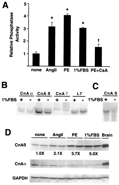
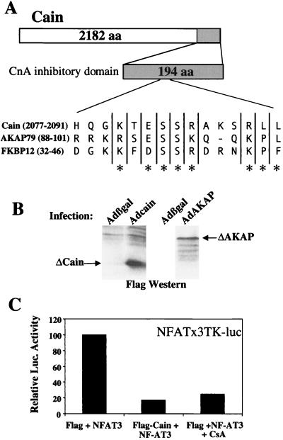
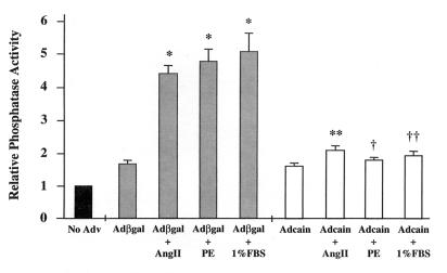
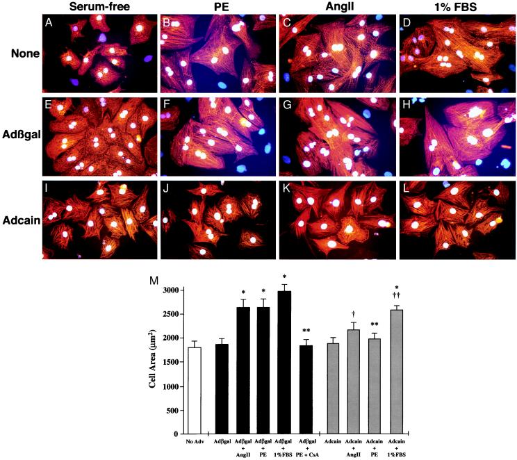
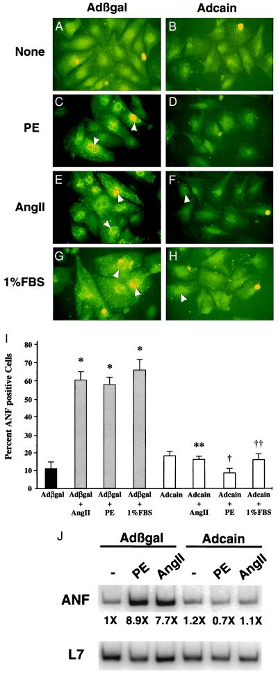
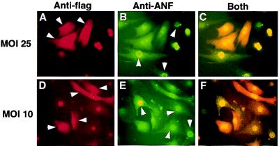
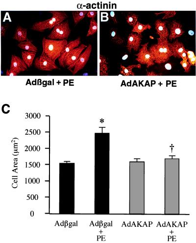
Cardiac hypertrophy is a major predictor of future morbidity and mortality. Recent investigation has centered around identifying the molecular signaling pathways that regulate cardiac myocyte reactivity with the goal of modulating pathologic hypertrophic programs. One potential regulator of cardiomyocyte hypertrophy is the calcium-sensitive phosphatase calcineurin. We show here that calcineurin enzymatic activity, mRNA, and protein levels are increased in cultured neonatal rat cardiomyocytes by hypertrophic agonists such as angiotensin II, phenylephrine, and 1% fetal bovine serum. This induction of calcineurin activity was associated with an increase in calcineurin Abeta (CnAbeta) mRNA and protein, but not in CnAalpha or CnAgamma. Agonist-dependent increases in calcineurin enzymatic activity were specifically inhibited with an adenovirus expressing a noncompetitive peptide inhibitor of calcineurin known as cain [Lai, M. M., Burnett, P. E., Wolosker, H., Blackshaw, S. & Snyder, S. H. (1998) J. Biol. Chem. 273, 18325-18331]. Targeted inhibition of calcineurin with cain or an adenovirus expressing only the calcineurin inhibitory domain of AKAP79 attenuated cardiomyocyte hypertrophy and atrial natriuretic factor expression in response to angiotensin II, phenylephrine, and 1% fetal bovine serum. These data demonstrate that calcineurin is an important regulator of cardiomyocyte hypertrophy in response to certain agonists and suggest that cyclosporin A and FK506 function to attenuate cardiac hypertrophy by specifically inhibiting calcineurin.


Figures

Figure 1
Figure 1
Calcineurin is activated by agonist-stimulated hypertrophy. Cultured cardiomyocytes were stimulated for 48 hr with AngII (100 nM), 1% FBS, or PE (50 μM). (A) Calcineurin phosphatase activity was induced by each agonist whereas cyclosporin A (CsA) significantly attenuated PE-induced calcineurin activity. The data represent two independent experiments, each performed in triplicate. (B) RT-PCR analysis demonstrated a significant induction of CnAβ, but not CnAα, CnAγ, or L7 mRNA in response to 1% FBS in cardiomyocytes. (C) Northern analysis confirms the increase in CnAβ mRNA levels. (D) Western blot analysis demonstrated an increase in CnAβ protein but not CnAα and GAPDH in response to agonist. Rat brain is shown as an enriched source of both CnAα and CnAβ. *, P < 0.0001 vs. control; †, P < 0.001 vs. PE.
Figure 2
Figure 2
Strategy for targeted inhibition of calcineurin. (A) The noncompetitive calcineurin inhibitory domain of cain contains a putative motif that is conserved in AKAP79 and FKBP12. (B) Adenoviral expression of a 194-aa cain peptide (21 kDa) or a 299-aa AKAP79 peptide (33 kDa) in neonatal cardiomyocytes shows stable expression by flag Western blotting. (C) Cotransfection of a NFAT-dependent luciferase reporter plasmid, an expression vector encoding NFAT3, and a flag-tagged cain (194 aa) expression vector or cyclosporin inhibited reporter activation.
Figure 3
Figure 3
Agonist induced increases in calcineurin activity are inhibited by Adcain. Cultured cardiomyocytes were infected with either Adβgal or Adcain, and 24 hr later, stimulated with AngII (100 nM), PE (50 μM), or 1% FBS for 48 hr. Agonist-stimulated cardiomyocytes infected with Adβgal had increased calcineurin phosphatase activity, whereas Adcain infection prevented this increase. The data represent two independent experiments performed in triplicate. *, P < 0.001 vs. Adβgal control; **, P < 0.0001 vs. Adβgal + AngII; †, P < 0.0001 vs. Adβgal + PE; ††, P < 0.0001 vs. Adβgal + 1% FBS.
Figure 4
Figure 4
Adcain infection attenuates agonist induced hypertrophy. Cardiomyocytes were subjected to Adcain infection (IL), Adβgal infection (E--H), or were left uninfected (AD); 24 hr later, cells were either untreated (A, E, and I), treated with 50 μM PE (B, F, and J), 100 nM Ang II (C, G, and K), or 1% FBS (D, H, and L) for 48 hr. Cardiomyocytes were identified with α-actinin antibody (red signal) and nuclei were stained with bis-benzamide (blue/white). (M) Adcain infection or cyclosporin (CsA) treatment prevented the increase in cardiomyocyte cell area induced by AngII and PE, and attenuated the 1% FBS-stimulated increase. The data are means ± SEM from 50–75 random cells measured in each group and were confirmed in two additional experiments. *, P < 0.001 vs. Adβgal control; **, P < 0.01 vs. Adβgal + PE; †, P < 0.05 vs. Adβgal + Ang II; ††, P < 0.05 vs. Adβgal + 1% FBS.
Figure 5
Figure 5
Adcain inhibits ANF expression. The same panels of cardiomyocytes shown in Fig. 4 were colabeled with ANF antibody (green). (A, C, E, and G) Cardiomyocytes infected with Adβgal showed increased ANF protein expression after agonist stimulation, whereas Adcain-infected cells (B, D, F, and H) did not respond to agonist. The white arrowheads indicate positive perinuclear ANF staining. The arrowheads in F and H represent Adcain cardiomyocytes that were barely expressing ANF. (I) Quantitation of percent of ANF expressing cells demonstrated significant expression in Adβgal-infected cultures, whereas Adcain blocked agonist-induced expression. (J) RT-PCR quantitation demonstrated that Adcain blocked ANF mRNA expression in agonist stimulated cardiomyocyte cultures. *, P < 0.0001 vs. Adβgal control; **, P < 0.0001 vs. Adβgal + Ang II; †, P < 0.0001 vs. Adβgal + PE; ††, P < 0.0001 vs. Adβgal + 1% serum.
Figure 6
Figure 6
Concentration independence of Adcain inhibition. Cardiomyocytes were infected with Adcain at a multiplicity of infection of 25 (AC) or 10 (DF) and treated with 50 μM PE for 48 hr. Cardiomyocytes were immunostained with a primary mAb against the flag-epitope (cain) in red and for ANF in green. The data demonstrate that cells expressing cain (arrowheads in A and D) lack ANF expression, and cells expressing ANF (arrowheads in B and E) lack cain expression. (C and F) Overlaid images of both antibodies are shown. The 21-kDa cain-flag peptide fusion was found in both the cytosol and nucleus.
Figure 7
Figure 7
AdAKAP (60–358 amino acids) infection attenuates cardiomyocyte hypertrophy. (A and B) AdAKAP infection, but not Adβgal, attenuated morphologic cardiomyocyte hypertrophy induced by PE. (C) Quantitation of cell surface area demonstrated a significant increase in Adβgal-infected cells treated with PE, which was prevented by AdAKAP infection. *, P < 0.001 vs. Adβgal; †, P < 0.001 vs. Adβgal + PE.

References

    1. Levy D, Garrison R J, Savage D D, Kannel W B, Castelli W P. N Engl J Med. 1990;322:1561–1566. - PubMed
    1. Sadoshima J, Izumo S. Annu Rev Physiol. 1997;59:551–571. - PubMed
    1. Dorn G W, II, Brown J H. Trends Cardiovasc Med. 1999;9:26–34. - PubMed
    1. Sugden P H. Circ Res. 1999;84:633–646. - PubMed
    1. Olson E N, Molkentin J D. Circ Res. 1999;84:623–632. - PubMed

Publication types

MeSH terms