. 2000 Feb 1;97(3):1206-11.
doi: 10.1073/pnas.97.3.1206.

Whole-body optical imaging of green fluorescent protein-expressing tumors and metastases

Affiliations

Whole-body optical imaging of green fluorescent protein-expressing tumors and metastases

M Yang et al. Proc Natl Acad Sci U S A. .

Abstract

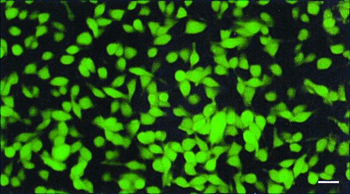
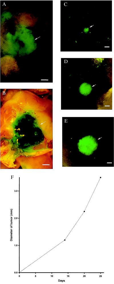
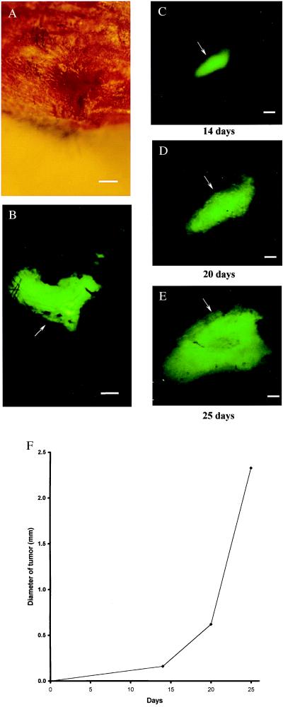
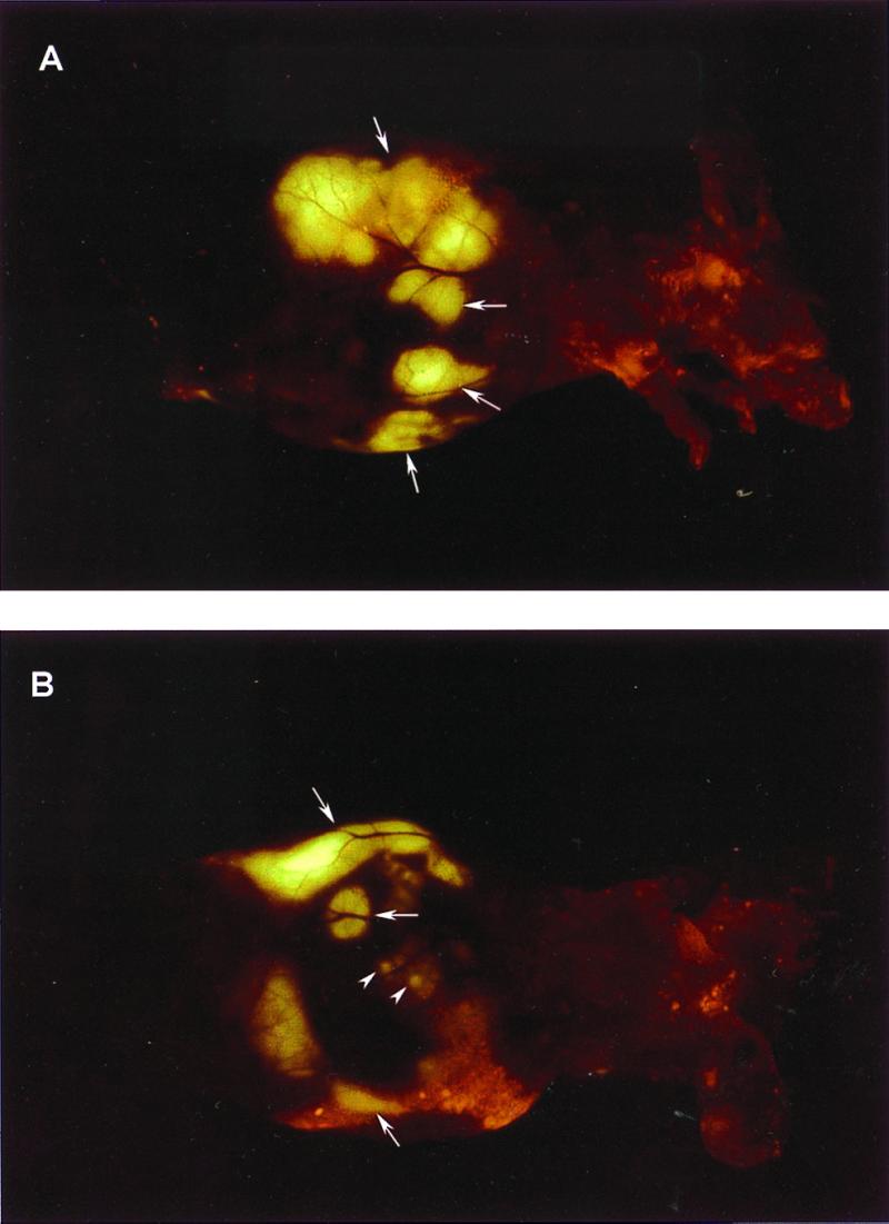
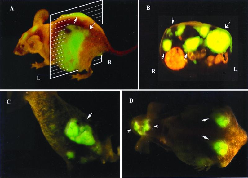
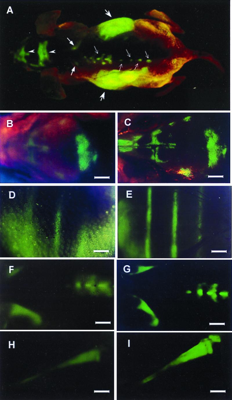
We have imaged, in real time, fluorescent tumors growing and metastasizing in live mice. The whole-body optical imaging system is external and noninvasive. It affords unprecedented continuous visual monitoring of malignant growth and spread within intact animals. We have established new human and rodent tumors that stably express very high levels of the Aequorea victoria green fluorescent protein (GFP) and transplanted these to appropriate animals. B16F0-GFP mouse melanoma cells were injected into the tail vein or portal vein of 6-week-old C57BL/6 and nude mice. Whole-body optical images showed metastatic lesions in the brain, liver, and bone of B16F0-GFP that were used for real time, quantitative measurement of tumor growth in each of these organs. The AC3488-GFP human colon cancer was surgically implanted orthotopically into nude mice. Whole-body optical images showed, in real time, growth of the primary colon tumor and its metastatic lesions in the liver and skeleton. Imaging was with either a trans-illuminated epifluorescence microscope or a fluorescence light box and thermoelectrically cooled color charge-coupled device camera. The depth to which metastasis and micrometastasis could be imaged depended on their size. A 60-microm diameter tumor was detectable at a depth of 0.5 mm whereas a 1, 800-microm tumor could be visualized at 2.2-mm depth. The simple, noninvasive, and highly selective imaging of growing tumors, made possible by strong GFP fluorescence, enables the detailed imaging of tumor growth and metastasis formation. This should facilitate studies of modulators of cancer growth including inhibition by potential chemotherapeutic agents.


Figures

Figure 1
Figure 1
Stable, high-level GFP-expressing B16F0 murine melanoma transductants in vitro. The murine malignant melanoma cell line B16F0 was transduced previously with the RetroXpress vector pLEIN, which expresses EGFP (19) and the neomycin resistance gene on the same bicistronic message (18). Stable, high-expression clones were selected in 800 μg/ml G418 (18). (Bar = 40 μm.)
Figure 2
Figure 2
External images of murine melanoma (B16F0-GFP) metastasis in brain. Murine melanoma metastases in the mouse brain were imaged by GFP expression under fluorescence microscopy. Clear images of metastatic lesions in the brain can be visualized through the scalp and skull. See Materials and Methods for imaging equipment and procedures. (A) External GFP image of brain metastasis through the scalp and scull of an intact mouse 3 weeks after injection of 106 B16F0-GFP cells in the tail vein. (Bar = 1,280 μm.) (B) GFP image of same area as in A, with skull opened. (Bar = 1,280 μm.) (C) External image obtained of the tumor in the brain of the nude mouse on day 14 after GFP tumor cell injection. (Bar = 1,280 μm.) (D) Same as C, day 19 after injection. (Bar = 1,280 μm.) (E) Same as C and D, day 25 after injection. (Bar = 1,280 μm.) (F) Brain tumor growth curve determined by external images (CE).
Figure 3
Figure 3
External images of B16F0-GFP bone metastasis. In the proximal tibia of the left hind leg of C57BL/6 mouse (hair removed). No metastasis can be detected under bright-field microscopy (A). Clear, external images of metastatic lesions of B16F0-GFP in the proximal tibia of the intact mouse were obtained under fluorescence microscopy (B). Time course metastatic growth of B16F0-GFP in the proximal tibia of the intact nude mouse was imaged externally under fluorescence microscopy (CE). (A) Bright-field microscopy of knee joint of hind leg. (Bar = 640 μm.) (B) Same as A; external fluorescent image of knee joint visualizing extensive melanoma metastasis, day 21 after injection. (Bar = 640 μm.) (C) External image obtained in tibia of nude mouse day 14 after tail vein injection. (Bar = 640 μm.) (D) Same as C, day 20. (Bar = 640 μm.) (E) Same as C and D, day 25. (Bar = 640 μm.) (F) Growth curve of tibia metastatic lesion determined by external images (Fig. 2 CE).
Figure 4
Figure 4
External images of B16F0-GFP colonizing the liver. A metastatic lesion of B16F0-GFP in the liver growing at a depth of 0.8 mm after portal vein injection was externally imaged through the abdominal wall of the intact nude mouse. (A) An external image of multilobe liver metastases of the B16F0-GFP cells (large arrows). (B) An external image of small liver metastatic lesions of approximately 1.5 mm in diameter (small arrows) and other larger metastatic lesions (large arrows).
Figure 5
Figure 5
External and internal images of liver lesions of AC3488-GFP. (A) Lateral, whole-body image of metastatic liver lesions of a GFP-expressing human colon cancer in the left (thick arrow) and right lobes (fine arrow) of a live nude mouse at day 21 after surgical orthotopic transplantation. (B) Cross-section of mouse shown in A corresponding to the level of the external image of the tumor in the liver that was acquired (A). Fine arrows show metastatic lesions in the right lobe of liver, and the thick arrow shows the metastatic lesion in the left lobe of liver. (C) Fluorescent whole-body ventral image of primary colon tumor (arrow). (D) Dorsal external image of metastatic tumor in the caudal region of the left and right lobes of the liver (thick arrows) and skull metastasis (arrowheads).
Figure 6
Figure 6
External and internal images of bone metastasis of AC3488 GFP. External fluorescent whole-body images compared with direct images of skeletal metastases. (A) External images of tumors in the skeletal system including the skull (arrow heads), scapula (thick arrows), spine (fine arrows), and liver metastasis (largest arrows) in a dorsal view of live, intact nude mouse. (BI) Series of external fluorescence images of metastatic lesions in the skull, ribs, spine, and tibia, (B, D, F, and H) compared with corresponding images of the exposed skeletal metastases (C, E, G, and I) (Bars = 1280 μm).

References

    1. Tearney G J, Brezinski M E, Bouma B E, Boppart S A, Pitris C, Southern J F, Fujimoto J G. Science. 1997;276:2037–2039. - PubMed
    1. Taubes G. Science. 1997;276:1991–1993. - PubMed
    1. Baum P R, Brummendorf T H. Q J Nucleic Med. 1998;42:33–42. - PubMed
    1. Teates C D, Parekh J S. Curr Probl Diagn Radiol. 1993;22:229–226. - PubMed
    1. Dessureault S. Breast Cancer Res Treat. 1997;45:29–37. - PubMed

MeSH terms

Substances