

doi: 10.1038/72360.
Structure and assembly of large lipid-containing dsDNA viruses
Affiliations
- PMID: 10655609
- PMCID: PMC4167659
- DOI: 10.1038/72360
Item in Clipboard
Structure and assembly of large lipid-containing dsDNA viruses
Nat Struct Biol.
2000 Feb.
No abstract available
Figures
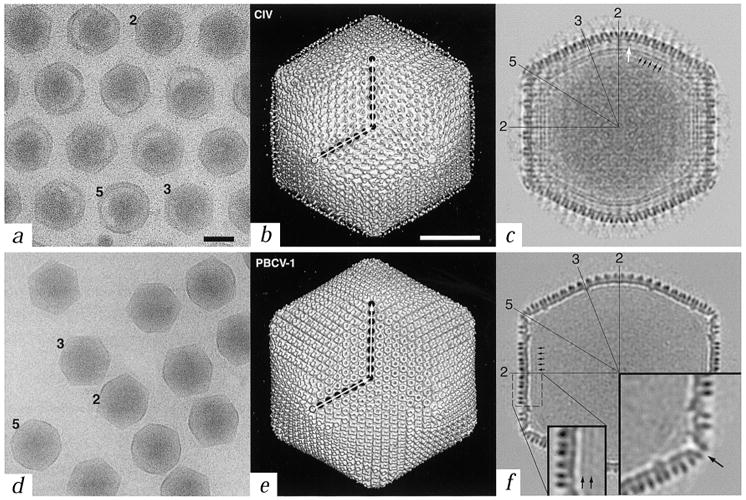
Comparison of CIV and PBCV-1 virions. a, d, Micrographs of vitrified samples of CIV and PBCV-1 virions (black bar = 1000 Å). Representative particles oriented with their two-, three-, or five-fold axes nearly parallel to the view direction are identified with numbers. Long, thin fibers emanate from the surface of CIV virions and maintain a regular intervirion spacing (a). b, e, Shaded-surface representations of the three-dimensional reconstructions of CIV and PBCV-1, each viewed down an icosahedral three-fold axis (white bar = 500 Å). Each capsid consists of twelve pentavalent and either 1,460 (CIV) or 1,680 (PBCV-1) hexavalent ‘capsomers’, arranged with T = 147 (h = 7, k = 7 in CIV) and T = 169d (h = 7, k = 8 in PBCV-1) icosahedral, quasi-equivalent lattices. The T (triangulation) number, which describes the geometrical arrangement of capsomers on a two-dimensional lattice, is given by the relationship T = h2+hk+k2, where h and k are integers that define lattice points. The d (for dextro) in T = 169d indicates that the PBCV-1 capsid belongs to a right-handed, skew class of T lattice. Filled circles mark the positions of capsomers along h and k directions (open circle is the origin of h, k lattice). Only the proximal end of each fiber in CIV is visible, presumably because the fibers are inherently flexible resulting in the distal portions smearing out in the reconstruction process. c, f, Central sections of the reconstruction density maps as viewed along a two-fold axis (same magnification as panels (b) and (e)). A lipid bilayer exists beneath the capsid shell in both virions (black arrows in (c) and (f) and in inset in (f)), although it is less prominent in PBCV-1. The PBCV-1 bilayer is more easily detected in images of disrupted virions, prepared by vitrification or negative staining (data not shown). Numerous connections, which constitutes an additional shell, occur between the outer shell and the bilayer in CIV (white arrow in (c)). Several icosahedral two-, three-, and five-fold symmetry axes are identified in both sections. Large inset in (f) is a view of the region near a pentavalent capsomer and reveals an axial channel (arrow). Both insets in (f) are at 2× magnification compared to (b) (c) (e), and (f).
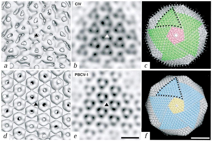
Close-up views of the trimers of CIV and PBCV-1. a, d, Shaded-surface representations of the reconstructions viewed down an icosahedral three-fold axis (triangles). Fibers in CIV are centered over the trimers whereas the trimers in PBCV-1 have a central, concave depression and axial channel. b, e, Planar sections through the reconstructed density maps viewed along an icosahedral three-fold axis (triangles). (Bar, 100 Å for panels (a) (b) (d), and (e)). c, f, Comparison of the symmetron facets of CIV (c) and PBCV-1 (f). Five trisymmetrons are highlighted in each reconstruction (green in CIV and blue in PBCV-1) and a single pentasymmetron is colored pink in CIV and yellow in PBCV-1. A pentavalent capsomer (uncolored) lies at the center of each pentasymmetron. Ten capsomers in CIV and eleven in PBCV-1 form the edge of each trisymmetron (black dots). Bar, 500 Å.
References
Publication types
MeSH terms
Substances
Grants and funding
LinkOut - more resources
Full Text Sources