

Defensive extrusive ectosymbionts of Euplotidium (Ciliophora) that contain microtubule-like structures are bacteria related to Verrucomicrobia
- PMID: 10660683
- PMCID: PMC26518
- DOI: 10.1073/pnas.030438197
Defensive extrusive ectosymbionts of Euplotidium (Ciliophora) that contain microtubule-like structures are bacteria related to Verrucomicrobia
Abstract
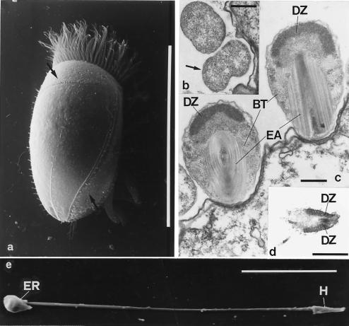
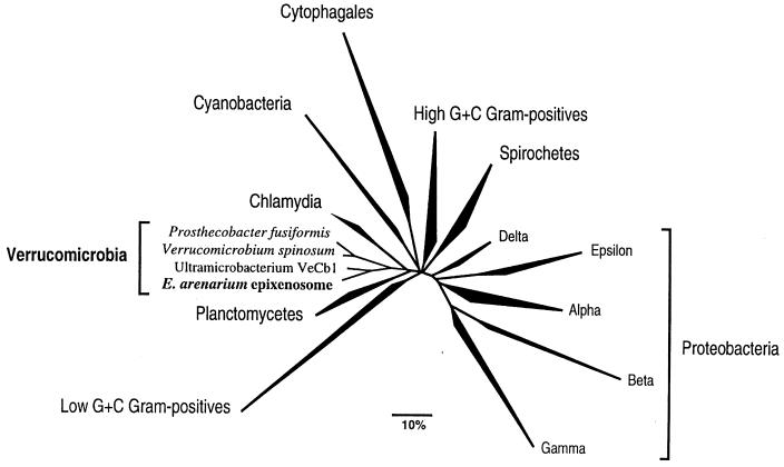
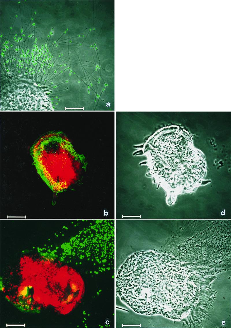
Epixenosomes, ectosymbionts on hypotrich ciliates (genus Euplotidium) defend their host against the ciliate predator Litonotus lamella. Although here only Euplotidium itoi and Euplotidium arenarium from tide pools along a rocky shore near Leghorn (Ligurian sea) were studied in detail, these epibionts are certainly present on specimens of E. itoi and on other Euplotidium species in similar north coastal habitats. The complex life history of epixenosomes has two main stages. In stage I, cells with typical prokaryotic structure divide by binary fission. Stage II cells show complex organization with different cytoplasmic compartments where an extrusive apparatus within a proteinaceous matrix, although not membrane-bounded, differs from the remaining cytoplasm. The ejection process is involved in defense; extrusive apparatus is surrounded by a basket consisting of bundles of tubules. These tubules, 22 +/- 3 nm in diameter, delimited by a wall made up of globular structures, are sensitive to inhibitor of tubulin polymerization (nocodazole/4 degrees C temperature) and react positively with different antitubulin antibodies, two of which are monoclonal. The prokaryotic vs. eukaryotic nature of epixenosomes was resolved by comparative sequence analysis of amplified small subunit rRNA genes and in situ hybridization with fluorescently labeled rRNA-targeted polynucleotide probes. These unique ectosymbionts are phylogenetically related to Verrucomicrobia. Epixenosomes represent marine symbionts in this recently discovered division of the Bacteria.
Figures
References
-
- Corliss J O. J Protozool. 1985;32:373–376.
-
- Verni F, Rosati G. J Protozool. 1990;37:337–343.
-
- Rosati G. Symbiosis. 1999;26:1–23.
-
- Rosati G, Verni F, Lenzi P. Eur J Protistol. 1993;29:238–245. - PubMed
-
- Margulis L. Symbiosis in Cell Evolution. 2nd Ed. San Francisco: Freeman; 1993. pp. 217–261.
Publication types
MeSH terms
Substances
Associated data
- Actions
- Actions
LinkOut - more resources
Full Text Sources
Molecular Biology Databases
