

Comment
doi: 10.1084/jem.191.3.411.
The induction of tolerance by dendritic cells that have captured apoptotic cells
Affiliations
- PMID: 10662786
- PMCID: PMC2195815
- DOI: 10.1084/jem.191.3.411
Item in Clipboard
Comment
The induction of tolerance by dendritic cells that have captured apoptotic cells
J Exp Med.
.
No abstract available
Figures
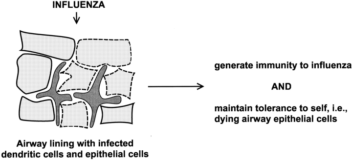
An illustration of the self-nonself problem from the perspective of DCs. During influenza infection of the airway, a site rich in DCs (reference 59), there is extensive apoptotic death of infected airway epithelial cells, which the DCs would likely process simultaneously with the influenza virus (references 29, 30).
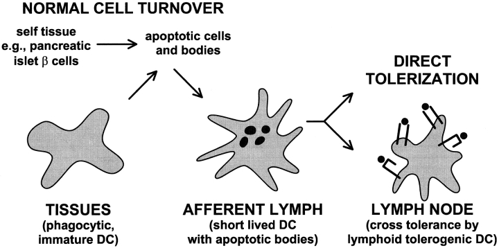
Pathways whereby DCs might induce peripheral tolerance to antigens within tissue cells undergoing normal cell turnover by apoptosis. DC precursors traffic through tissues, phagocytose dying cells or apoptotic bodies, and then enter the lymph. Upon reaching the lymph node, the DCs tolerize naive, self-reactive T cells either directly, or indirectly (as diagrammed here) after reprocessing by different regulatory or tolerogenic DCs in the lymph node.
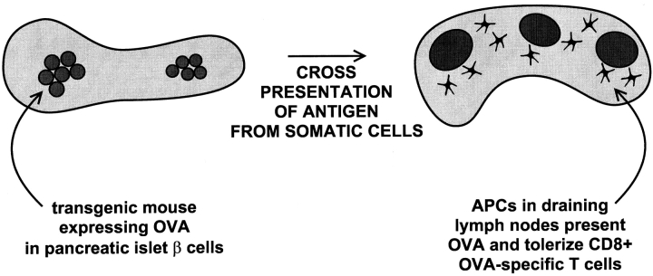
Diagram of the model of Kurts et al. (references 34, 35). Self-peptides in tissue cells, here the insulin-producing β cells of the pancreatic islets, are presented in a tolerogenic way by bone marrow–derived cells in the draining pancreatic lymph nodes.
Comment on
-
Consequences of cell death: exposure to necrotic tumor cells, but not primary tissue cells or apoptotic cells, induces the maturation of immunostimulatory dendritic cells.J Exp Med. 2000 Feb 7;191(3):423-34. doi: 10.1084/jem.191.3.423. J Exp Med. 2000. PMID: 10662788 Free PMC article.
-
A discrete subpopulation of dendritic cells transports apoptotic intestinal epithelial cells to T cell areas of mesenteric lymph nodes.J Exp Med. 2000 Feb 7;191(3):435-44. doi: 10.1084/jem.191.3.435. J Exp Med. 2000. PMID: 10662789 Free PMC article.
References
-
- Romani N., Koide S., Crowley M., Witmer-Pack M., Livingstone A.M., Fathman C.G., Inaba K., Steinman R.M. Presentation of exogenous protein antigens by dendritic cells to T cell clonesintact protein is presented best by immature, epidermal Langerhans cells. J. Exp. Med. 1989;169:1169–1178. - PMC - PubMed
-
- Stumbles P.A., Thomas J.A., Pimm C.L., Lee P.T., Venaille T.J., Proksch S., Holt P.G. Resting respiratory tract dendritic cells preferentially stimulate T helper cell type 2 (Th2) responses and require obligatory cytokine signals for induction of Th1 immunity. J. Exp. Med. 1998;188:2019–2031. - PMC - PubMed
Publication types
MeSH terms
Substances
LinkOut - more resources
Full Text Sources
Other Literature Sources
