. 2000 Feb 15;20(4):1386-92.
doi: 10.1523/JNEUROSCI.20-04-01386.2000.

Caspase-2 mediates neuronal cell death induced by beta-amyloid

Affiliations

Caspase-2 mediates neuronal cell death induced by beta-amyloid

C M Troy et al. J Neurosci. .

Abstract

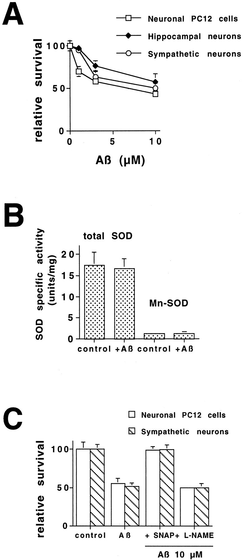
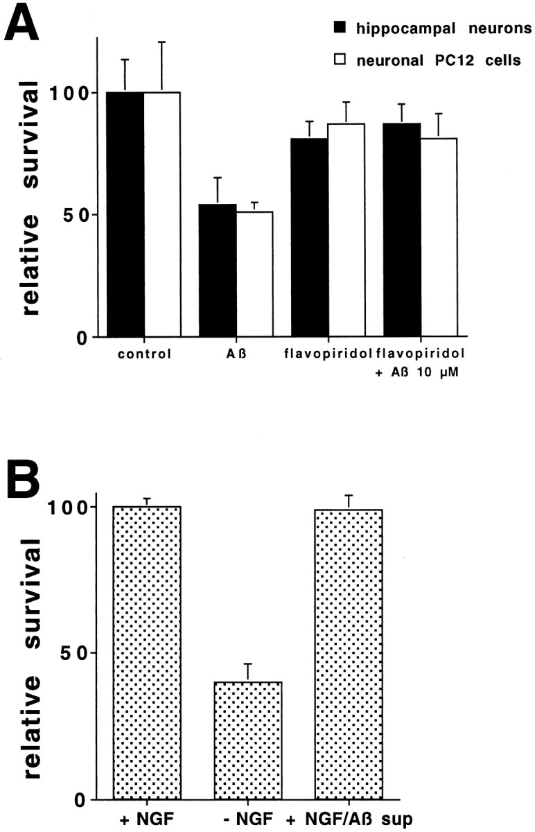
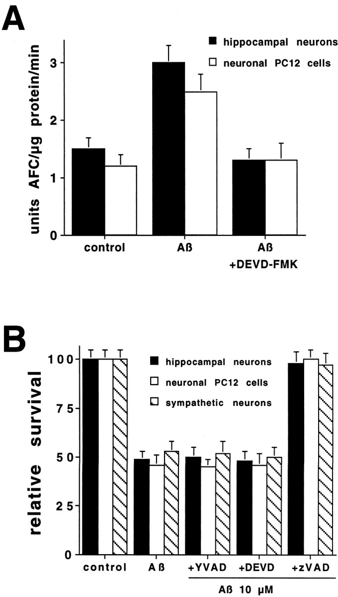
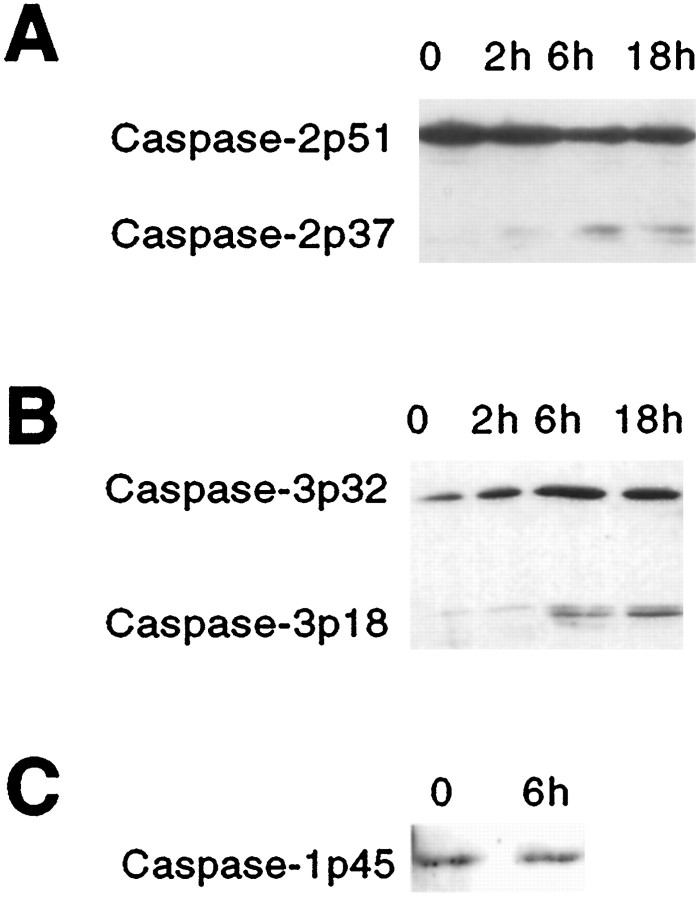
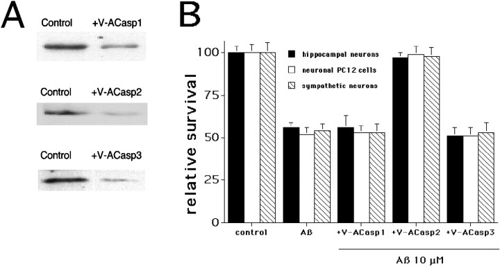
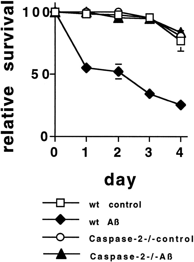
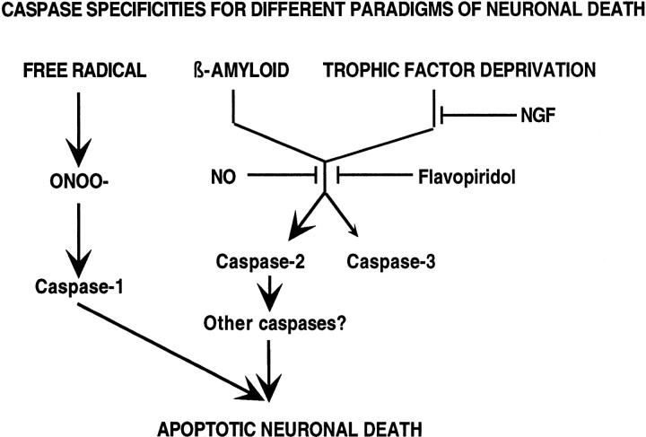
beta-amyloid (Abeta) has been proposed to play a role in the pathogenesis of Alzheimer's disease (AD). Deposits of insoluble Abeta are found in the brains of patients with AD and are one of the pathological hallmarks of the disease. It has been proposed that Abeta induces death by oxidative stress, possibly through the generation of peroxynitrite from superoxide and nitric oxide. In our current study, treatment with nitric oxide generators protected against Abeta-induced death, whereas inhibition of nitric oxide synthase afforded no protection, suggesting that formation of peroxynitrite is not critical for Abeta-mediated death. Previous studies have shown that aggregated Abeta can induce caspase-dependent apoptosis in cultured neurons. In all of the neuronal populations studied here (hippocampal neurons, sympathetic neurons, and PC12 cells), cell death was blocked by the broad spectrum caspase inhibitor N-benzyloxycarbonyl-val-ala-asp-fluoromethyl ketone and more specifically by the downregulation of caspase-2 with antisense oligonucleotides. In contrast, downregulation of caspase-1 or caspase-3 did not block Abeta(1-42)-induced death. Neurons from caspase-2 null mice were totally resistant to Abeta(1-42) toxicity, confirming the importance of this caspase in Abeta-induced death. The results indicate that caspase-2 is necessary for Abeta(1-42)-induced apoptosis in vitro.


Figures

Fig. 1.
Fig. 1.
Nitric oxide protects against β-amyloid-induced death in neuronal cells. A, Aβ1–42induces dose-dependent death in three different neuronal cell types. E18 hippocampal neurons were grown in culture for 3 d and then exposed to increasing concentrations of Aβ1–42. Survival was assessed after 1 d by counting nuclei in cell lysates (n = 3). Survival is reported relative to untreated cultures and is given as mean ± SEM. Sympathetic neurons were grown in culture for 5 d and then exposed to increasing concentrations of Aβ1–42. Survival was assessed after 1 d by counting cells in the living cultures. Survival is reported relative to that in the same cultures before Aβ1–42treatment and is given as mean ± SEM (n = 3). PC12 cells were exposed to increasing concentrations of Aβ1–42. Survival was assessed after 1 d by counting nuclei in cell lysates (n = 3). Survival is reported relative to untreated cultures and is given as mean ± SEM. These are representative experiments. Comparable results were obtained in six additional independent experiments with hippocampal neurons, in three additional experiments with sympathetic neurons, and in five additional experiments with PC12 cells. B, Mn-SOD is not induced by Aβ1–42 treatment. PC12 cells were treated with or without Aβ1–42 (10 μm) for 6 hr (n = 3). Cells were extracted with 0.5% NP-40, and protein was measured by the Bradford method. Total SOD and Mn-SOD levels were determined by the xanthine–xanthine oxidase system, with measurement of the reduction of nitroblue tetrazolium at 560 nm in the presence and absence of KCN. Mn-SOD activity was determined from an SOD standard curve and is reported as the KCN-insensitive activity ± SEM. C, Increasing NO protects from Aβ1–42-induced neuronal cell death. PC12 cells and sympathetic neurons were exposed to Aβ1–42 (10 μm) in the presence or absence of SNAP (100 μm) or l-NAME (10 μm). Survival was assessed after 1 d as described above (n = 3). This is a representative experiment; comparable results were obtained in three additional independent experiments. Survival is reported relative to untreated cultures and is given as mean ± SEM. Similar results were obtained with cultured sympathetic neurons.
Fig. 2.
Fig. 2.
A, The cell cycle inhibitor flavopiridol protects hippocampal neurons and neuronal PC12 cells from Aβ1–42 toxicity. Hippocampal cultures and neuronal PC12 cells were treated with Aβ1–42 in the presence or absence of flavopiridol (1 μm) (n = 3). Survival was assessed after 1 d as described in Figure 1, is reported relative to untreated cultures, and is given as mean ± SEM. This is a representative experiment; comparable results were obtained in six additional independent experiments for hippocampal cultures and three additional experiments for PC12 cells.B, Aβ1–42 does not inhibit NGF activity. RPMI with NGF was incubated with or without Aβ1–42 (10 μm) for 30 min at 37°C, and Aβ1–42 was removed by centrifugation. The various media were added to PC12 cells, which had been subjected to trophic factor deprivation. Survival was quantified at 1 d and is given as mean ± SEM (n = 3).
Fig. 3.
Fig. 3.
A, Aβ1–42 induces caspase activity in hippocampal neurons and PC12 cells. Hippocampal neurons and neuronal PC12 cells were treated with Aβ1–42(10 μm) with or without DEVD-FMK (10 μm) for 6 hr. Cells were lysed, and 25 μg of protein of each treatment was incubated with the fluorogenic substrate DEVD-AFC (15 μm). The release of AFC was quantified in an LS50B fluorometer. This is a representative experiment; comparable results were obtained in three additional independent experiments.B, Differential protection by caspase inhibitors from Aβ1–42-induced death. Cultures of hippocampal neurons, PC12 cells, and sympathetic neurons were exposed to Aβ1–42 (10 μm) in the presence or absence of the indicated inhibitors (n = 3): YVAD-FMK at 100 μm, DEVD-FMK at 10 μm, and zVAD-FMK at 50 μm. Cells were counted after 1 d as described in Figure 1. Survival is reported relative to untreated cultures and is given as mean ± SEM.
Fig. 4.
Fig. 4.
Caspase-2 and caspase-3 are activated in hippocampal neurons after Aβ1–42 treatment. Hippocampal cultures were incubated with or without Aβ1–42 for the indicated times. Cell lysates (equal amounts of protein, determined by the Bradford method) were subjected to Western blotting using the indicated antisera. Ponceau staining confirmed equal loading. These are representative blots; comparable results were obtained in three independent experiments. A, Caspase-2. B, Caspase-3. C, Caspase-1.
Fig. 5.
Fig. 5.
Caspase-2 is necessary for Aβ1–42-induced neuronal cell death. A, Specific downregulation of caspase-1, -2, or -3. PC12 cells were treated with the indicated antisense oligonucleotides (240 nm) for 6 hr. Cells lysates were subjected to Western blotting using the appropriate antisera, i.e., anti-caspase-1 for V-ACasp1-treated cells. B, Only downregulation of caspase-2 protects against Aβ1–42-induced neuronal cell death. Cultures of hippocampal neurons, PC12 cells, and sympathetic neurons were treated with 10 μm1–42 in the presence or absence of the indicated antisense oligonucleotides, each at a concentration of 240 nm (n = 3). Survival was quantified after 1 d, is reported relative to untreated cultures, and is given as mean ± SEM. This is a representative experiment; comparable results were obtained in three additional independent experiments with hippocampal cultures, as well as three additional experiments each with PC12 cells and sympathetic neurons.
Fig. 6.
Fig. 6.
Sympathetic neurons from caspase-2 null mice are resistant to Aβ1–42 toxicity. Sympathetic neuron cultures from 1-d-old wild-type or caspase-2 null pups were treated with Aβ1–42 (n = 3). Survival was quantified daily, as described in Figure 1. Survival is reported relative to that in the same cultures before Aβ1–42treatment and is given as mean ± SEM (n = 3). This is a representative experiment; comparable results were obtained in four additional independent experiments.
Fig. 7.
Fig. 7.
Caspase specificities in different paradigms of cell death. Schematic illustration of the pathways to cell death for β-amyloid, trophic factor deprivation, and free radical-mediated oxidative stress.

References

    1. Ahmad M, Srinivasula SM, Hegde R, Mukattash R, Fernandes-Alnemri T, Alnemri ES. Identification and characterization of murine caspase-14, a new member of the caspase family. Cancer Res. 1998;58:5201–5205. - PubMed
    1. Bergeron L, Perez GI, Macdonald G, Shi L, Sun Y, Jurisicova A, Varmuza S, Latham KE, Flaws JA, Salter JC, Hara H, Moskowitz MA, Li E, Greenberg A, Tilly JL, Yuan J. Defects in regulation of apoptosis in caspase-2-deficient mice. Genes Dev. 1998;12:1304–1314. - PMC - PubMed
    1. Chan SL, Griffin WS, Mattson MP. Evidence for caspase-mediated cleavage of AMPA receptor subunits in neuronal apoptosis and Alzheimer's disease. J Neurosci Res. 1999;57:315–323. - PubMed
    1. Cotman CW, Su JH. Mechanisms of neuronal death in Alzheimer's disease. Brain Pathol. 1996;6:493–506. - PubMed
    1. Cotman CW, Whittemore ER, Watt JA, Anderson AJ, Loo DT. Possible role of apoptosis in Alzheimer's disease. Ann NY Acad Sci. 1994;747:36–49. - PubMed

Publication types

MeSH terms