. 2000 Feb 15;20(4):1393-403.
doi: 10.1523/JNEUROSCI.20-04-01393.2000.

Interstitial cells of Cajal mediate cholinergic neurotransmission from enteric motor neurons

Affiliations

Interstitial cells of Cajal mediate cholinergic neurotransmission from enteric motor neurons

S M Ward et al. J Neurosci. .

Abstract

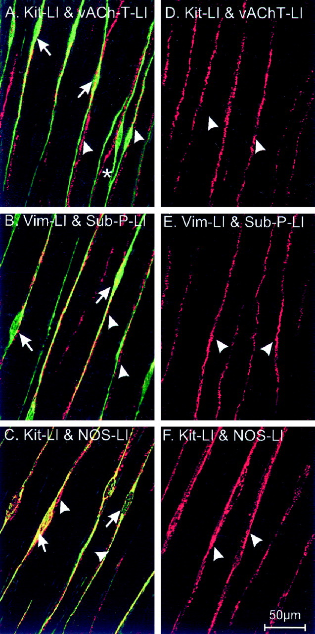
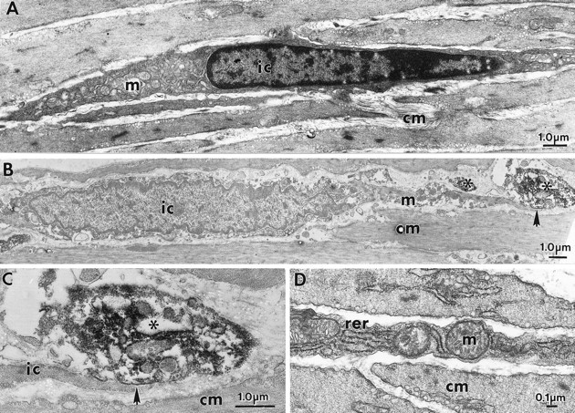
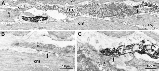
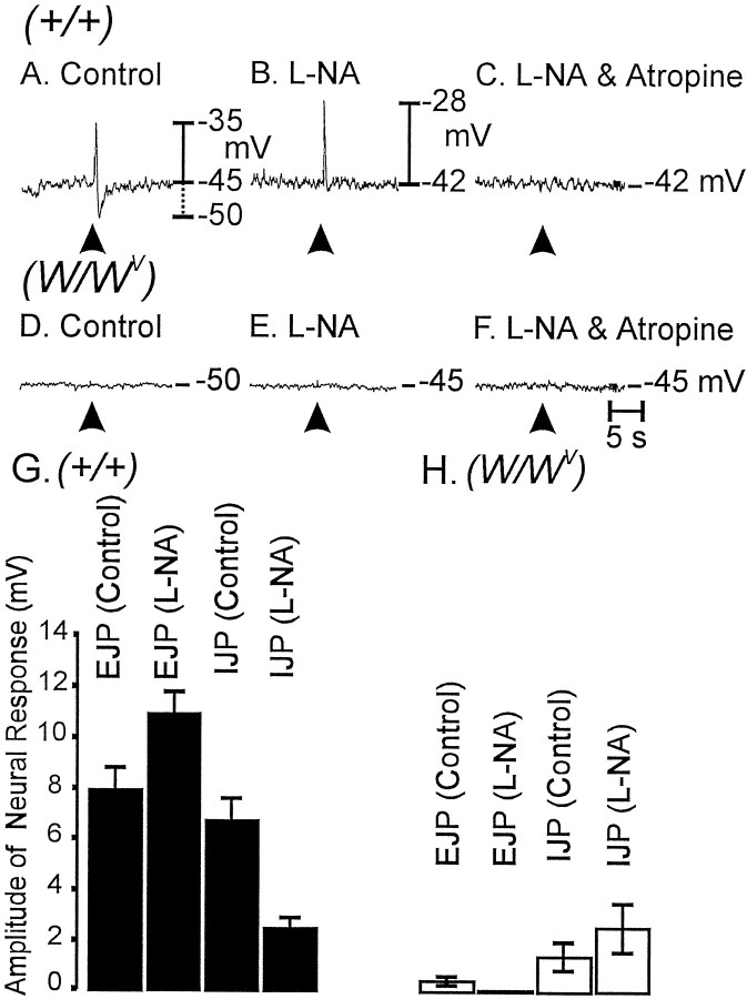
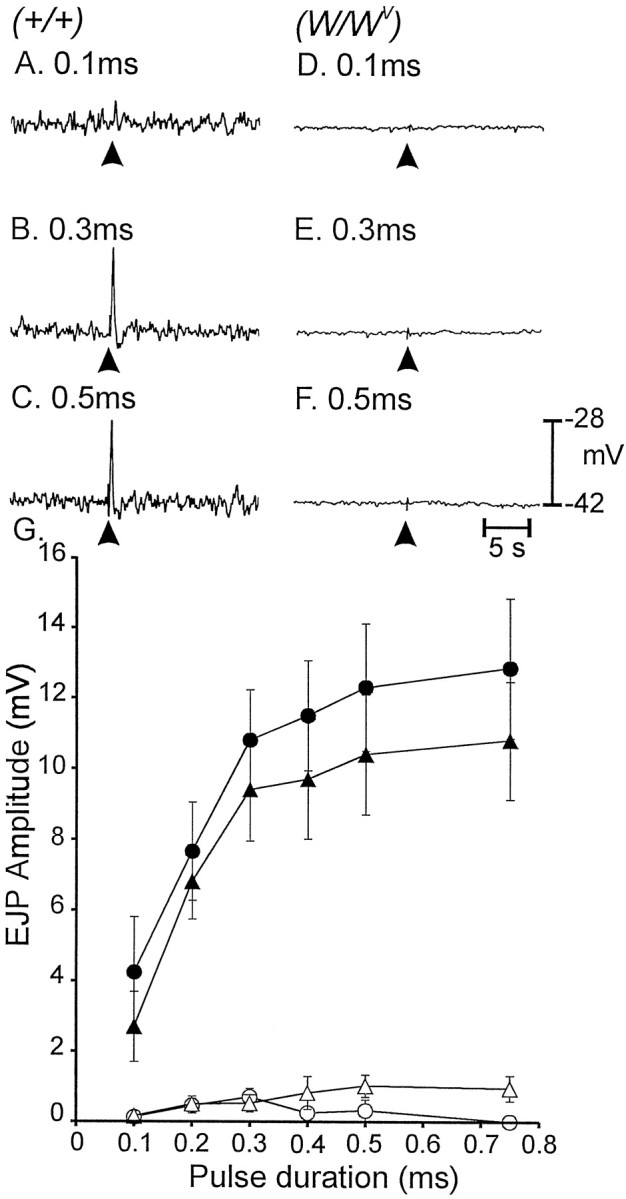
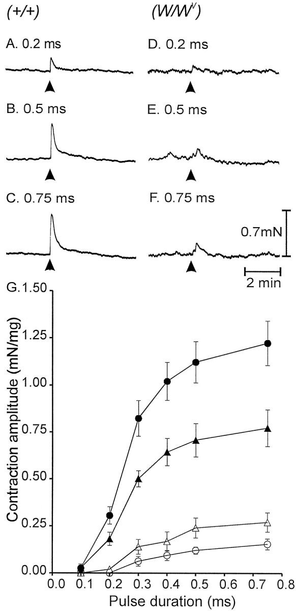
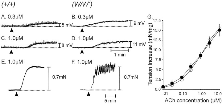
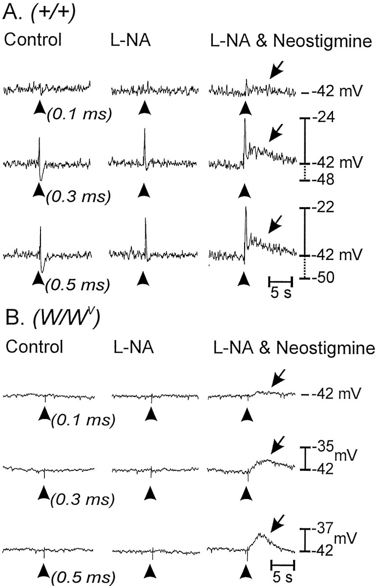
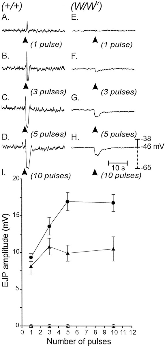
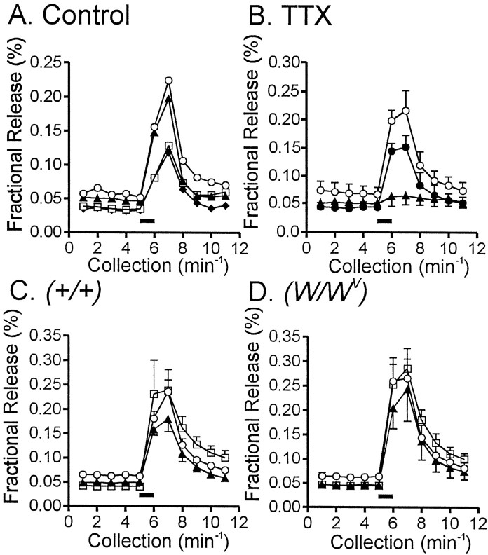
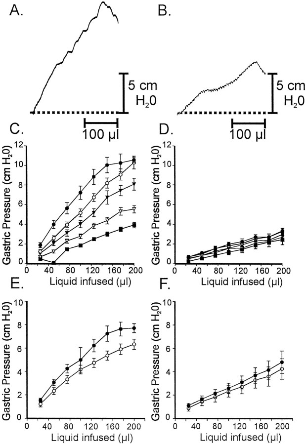
Interstitial cells of Cajal (ICC) are interposed between enteric neurons and smooth muscle cells in gastrointestinal muscles. The role of intramuscular ICC (IC-IM) in mediating enteric excitatory neural inputs was studied using gastric fundus muscles of wild-type animals and W/W(v) mutant mice, which lack IC-IM. Excitatory motor neurons, labeled with antibodies to vesicular acetylcholine transporter or substance-P, were closely associated with IC-IM. Immunocytochemistry showed close contacts between enteric neurons and IC-IM. IC-IM also formed gap junctions with smooth muscle cells. Electrical field stimulation yielded fast excitatory junction potentials in the smooth muscle that were blocked by atropine. Neural responses were greatly reduced in muscles of W/W(v) animals. Loss of cholinergic responses in W/W(v) muscles seemed to be caused by the loss of close synaptic contacts between motor neurons and IC-IM, because these muscles were not less responsive to exogenous acetylcholine than were wild-type muscles. W/W(v) muscles also responded to excitatory nerve stimulation when the breakdown of acetylcholine was blocked by neostigmine. The density of cholinergic nerve bundles within the muscles was not significantly different in wild-type and W/W(v) muscles, and similar amounts of (14)[C]choline were released from preloaded wild-type and W/W(v) muscles in response to nerve stimulation. The impact of losing IC-IM on gastric compliance was also evaluated in intact stomachs. Pressure increased as a function of fluid volume and infusion rate in wild-type animals, but W/W(v) animals showed little basal tone and minimal increases in pressure with fluid infusions. These data suggest that IC-IM play a major role in receiving cholinergic excitatory inputs from the enteric nervous system in the murine fundus.


Figures

Fig. 1.
Fig. 1.
Distribution of ICC and enteric nerves in the murine gastric fundus of wild type (A–C) and W/Wv mutants (D–F). Kit-like immunoreactivity (Kit-LI; A,C;green) and vimentin-like immunoreactivity (Vim-LI; B; green) reveal IC-IM (arrows) within the circular muscle layer (single optical section 0.6 μm thick in z-axis). Double-labeling immunohistochemical experiments using antibodies for the vesicular acetylcholine transporter (vAChT-LI;A; red) and substance-P (Sub-P-LI; B; red) identify processes within the circular muscle (arrowheads) of enteric excitatory neurons. Nitric oxide synthase-like immunoreactivity (NOS-LI;C; red) identifies processes of inhibitory motor neurons within the circular muscle layer. Note the close apposition of IC-IM with varicose terminals of excitatory and inhibitory neurons. Individual nerve processes are associated with multiple IC-IM (asterisk). Excitatory and inhibitory neurons (arrowheads) within the gastric fundus ofW/Wv mutants appeared normal (as labeled in D–F; red), but these tissues lacked IC-IM (note absence of green-labeled cells). Scale bar in F applies to allpanels.
Fig. 2.
Fig. 2.
A, D, IC-IM and smooth muscle cells are of similar spindle-like shape in the circular muscle layer of the fundus. These cells have distinct ultrastructural features. IC-IM (ic) contain numerous mitochondria (m), smooth and rough endoplasmic reticulum, dense heterochromatic nuclei, and few myofilaments and dense bodies. Smooth muscle with typical ultrastructural features surrounds the IC-IM. IC-IM and enteric neurons with vAChT-LI form intimate contacts as revealed by immunoelectron microscopy. B, An IC-IM within the circular muscle layer (cm) is shown. Areas of close, synaptic-like structures (arrow) exist between vAChT-containing nerve terminals (asterisks) and IC-IM. C, The region denoted by the arrow in B is shown in higher power.
Fig. 3.
Fig. 3.
IC-IM and enteric neurons with NOS-LI form intimate contacts as revealed by immunoelectron microscopy.A, An IC-IM (ic) within the circular muscle layer (cm) is shown. IC-IM were readily identified by the large number of mitochondria (m), rough endoplasmic reticulum, and free ribosomes. NOS-LI in enteric nerve terminals is observed as a dense DAB reaction product (asterisk). A, C, A close, synaptic-like region of membrane exists between a NOS-LI nerve terminal and IC-IM (shortarrow; at higher magnification in C). A, B, The IC-IM in A also forms a gap junction with a neighboring smooth muscle cell (longarrow; at higher power in B).
Fig. 4.
Fig. 4.
Differences in responses to EFS in wild-type andW/Wv mutant animals.A, EFS (arrowhead; 0.5 msec pulse supramaximal voltage) produced a biphasic electrical response in wild-type muscles, characterized by a rapid EJP. The EJP was followed by an IJP. B, l-NA (100 μm) reduced the IJPs and increased the amplitudes of the EJPs.C, After l-NA, atropine (1 μm) completely blocked the EJPs, suggesting that muscarinic receptors mediated the EJP responses. D, InW/Wv mutant animals, EJPs and IJPs were greatly attenuated. E, F, l-NA had little or no effect on responses to EFS (E), and after l-NA, atropine had no effect (F). G, H, The effects ofl-NA on EJPs and IJPs from experiments on muscles of wild type (n = 17; filledverticalbars) and ofW/Wv mutants (n = 13; openverticalbars) are summarized in G and H,respectively.
Fig. 5.
Fig. 5.
Pulses of EFS of different durations (0.1–0.75 msec) altered responses of gastric fundus muscles. A–C, Increasing the duration of pulses (arrowheads) potentiated the amplitude of EJPs in wild-type tissues (A–C for pulse durations of 0.1, 0.3, and 0.5 msec, respectively). D–F, EFS of the same pulse duration had no effect in W/Wv muscles (D–F for pulses of 0.1, 0.3, and 0.5 msec, respectively). G, The electrical responses to EFS as a function of pulse duration in wild type before (filledtriangles) and after (filledcircles) addition ofl-NA (100 μm) are summarized. Responses were greatly diminished in W/Wv muscles using the same stimulus parameters before (opentriangles) and after (opencircles) l-NA.
Fig. 6.
Fig. 6.
Mechanical responses to EFS in wild-type and mutant gastric tissues. A–C, Isometric contractions to single pulses of EFS (arrowheads; 0.1, 0.5, and 0.75 msec, respectively) are shown. D–F, Mechanical responses of a W/Wv muscle using the same stimulus parameters are shown. G, Mechanical responses to EFS of wild-type muscles before (filledtriangles) and after (filledcircles) l-NA (100 μm) are summarized. Responses ofW/Wv muscles were greatly attenuated using EFS with the same stimulus parameters. A summary of responses ofW/Wv muscles to EFS before (opentriangles) and after (opencircles) l-NA are shown.
Fig. 7.
Fig. 7.
Electrical and mechanical responses of wild-type and W/Wv muscles to exogenous ACh. Application of ACh (arrowheads) tonically depolarized muscles in a concentration-dependent manner. A,C, Responses of wild-type muscles to 0.3 and 1.0 μm, respectively. B,D, Responses of W/Wvmuscles to the same doses. Acetylcholine caused contractions of wild-type and W/Wv muscles.E, The mechanical response of a wild-type muscle to 1.0 μm ACh. F, The response of aW/Wv muscle to the same concentration of ACh. Many W/Wv muscles displayed oscillations in tone superimposed on the tonic contraction. This type of response also occurred, but more rarely in wild-type muscles.G, A summary of the responses of wild-type (filledcircles;n = 5) and W/Wv(opencircles; n = 5) muscles to ACh (0.01–10 μm).
Fig. 8.
Fig. 8.
Inhibition of endogenous cholinesterase with neostigmine revealed a slowly developing membrane depolarization in wild-type and W/Wv mutant tissues.A, The electrical responses of wild-type muscles to EFS (arrowheads; single pulses of 0.1, 0.3, and 0.5 msec duration) before (left) and after (middle) l-NA (100 μm) and after l-NA with neostigmine (0.5 μm;right) are shown. l-NA blocked IJPs and potentiated EJPs. In the presence of l-NA, neostigmine increased the amplitude of the fast EJP and revealed a slowly developing, second depolarization (arrows).B, Responses of W/Wvmuscles to EFS were negligible under control conditions (left) and after l-NA (middle). However, neostigmine added afterl-NA revealed a slowly developing membrane depolarization in response to EFS (right) that was similar in detail and time course to the second depolarization response of wild-type muscles (arrows).
Fig. 9.
Fig. 9.
Electrical responses of wild-type andW/Wv muscles to EFS.A–D, Multiple pulses of EFS (arrowheads; 1–10 pulses delivered within 100 msec) caused pronounced EJPs and IJPs in wild-type muscles (1–10 pulses as labeled). E–H, EJPs were not observed inW/Wv muscles; however 3–10 pulses (delivered within 100 msec) caused slowly developing hyperpolarization responses in these tissues. l-NA had no effect on the IJPs elicited in W/Wv muscles with 3 or more pulses. I, Data for the effect of l-NA on control and W/Wv mutant tissues are summarized. EJPs elicited in wild-type muscles (filledtriangles) were potentiated by l-NA (100 μm;filledcircles), whereas EJPs were absent in W/Wv muscles (opentriangles) and not revealed by l-NA (opencircles).
Fig. 10.
Fig. 10.
[14C]Choline release from wild-type and W/Wv muscles in response to EFS. A, The typical [14C]choline overflow from a wild-type fundus muscle in response to EFS is shown. Tissues were stimulated at 40 min intervals for 1 min (5 Hz; 0.1-msec-duration pulses;filledhorizontalbar), and [14C]choline overflow was assayed from superfusion samples. [14C]Choline overflow is plotted as a percentage of fractional release with time of collection. Overflow decreased with subsequent stimulations (S1, discarded; S2,opencircles; S3,filledtriangles; S4,opensquares; S5,filleddiamonds).B, The overflow of [14C]choline in response to EFS (opencircles) was inhibited by TTX (1 μm; n = 9 tissues of 3 animals; filledtriangles), and this was reversible after washout (filledcircles). C, D, EFS caused release of similar amounts of [14C]choline from wild-type (C) orW/Wv (D) muscles (p > 0.05). [14C]Choline release under control conditions (opencircles, S2) was not affected by l-NA (100 μm;filledtriangles, S3) but was slightly increased by neostigmine (0.5 μm;opensquares, S4).
Fig. 11.
Fig. 11.
Gastric compliance measurements from ex vivo stomachs of wild-type andW/Wv animals. Gastric pressure (centimeters of H2O) was recorded as a function of constant volume infusions at rates varying from 1.01 to 20.28 μl sec−1. Three infusions were performed at each rate, and the data were averaged (30-d-old age-matched animals;n = 5 for each genotype). A, Gastric pressure increased as fluid was infused in the stomachs of wild-type animals. The gastric pressure response increased as a function of infusion rate. B, Gastric pressure responses to fluid infusion were greatly diminished in the stomachs ofW/Wv mutants (p < 0.05 when compared with wild-type stomachs). C, D, Summary data from five wild-type animals (C) and fiveW/Wv mutants (D) are shown (1.01 μl sec−1, filledsquares; 2.02 μl sec−1,openinvertedtriangles; 4.09 μl sec−1, filledinvertedtriangles; 8.21 μl sec−1, opencircles; 20.28 μl sec−1, filledcircles). E, F, Atropine (1 μm) in the presence of l-NA (100 μm) increased gastric compliance of wild-type stomachs (E) but had little or no effect on the stomachs of W/Wv animals (F;l-NA, filledcircles;l-NA and atropine, opencircles for both E,F).

References

    1. Alberts P, Bartfai T, Stjarne L. The effects of atropine on [3H]acetylcholine secretion from guinea-pig myenteric plexus evoked electrically or by high potassium. J Physiol (Lond) 1982;329:93–112. - PMC - PubMed
    1. Burns AJ, Lomax AE, Torihashi S, Sanders KM, Ward SM. Interstitial cells of Cajal mediate inhibitory neurotransmission in the stomach. Proc Natl Acad Sci USA. 1996;93:12008–12013. - PMC - PubMed
    1. Burnstock G. Review lecture. Neurotransmitters and trophic factors in the autonomic nervous system. J Physiol (Lond) 1981;313:1–35. - PMC - PubMed
    1. Daniel EE, Posey-Daniel V. Neuromuscular structures in opossum esophagus: role of interstitial cells of Cajal. Am J Physiol. 1984;246:G305–G315. - PubMed
    1. Faussone-Pellegrini MS, Cortesini C. The muscle coat of the lower esophageal sphincter in patients with achalasia and hypertensive sphincter. An electron microscopic study. J Submicrosc Cytol. 1985;17:673–685. - PubMed

Publication types

MeSH terms

LinkOut - more resources