. 2000 Feb 15;20(4):1446-57.
doi: 10.1523/JNEUROSCI.20-04-01446.2000.

Consequences of neural cell adhesion molecule deficiency on cell migration in the rostral migratory stream of the mouse

Affiliations

Consequences of neural cell adhesion molecule deficiency on cell migration in the rostral migratory stream of the mouse

G Chazal et al. J Neurosci. .

Abstract

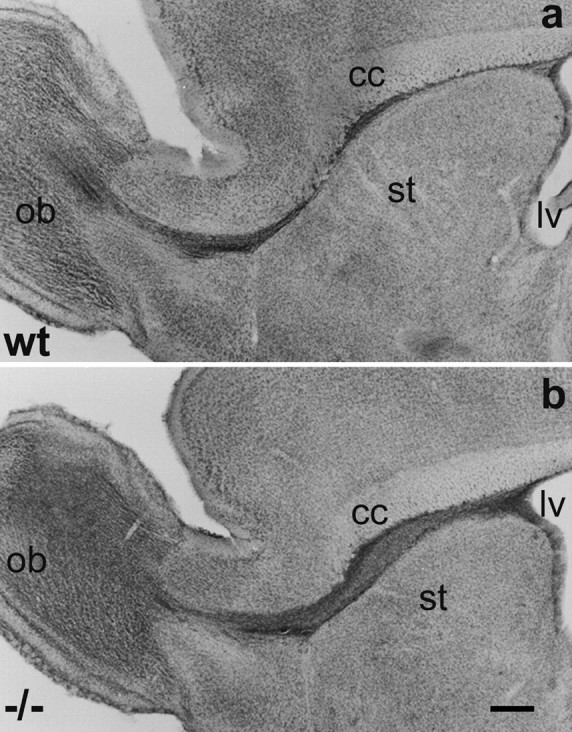
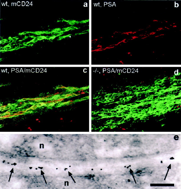
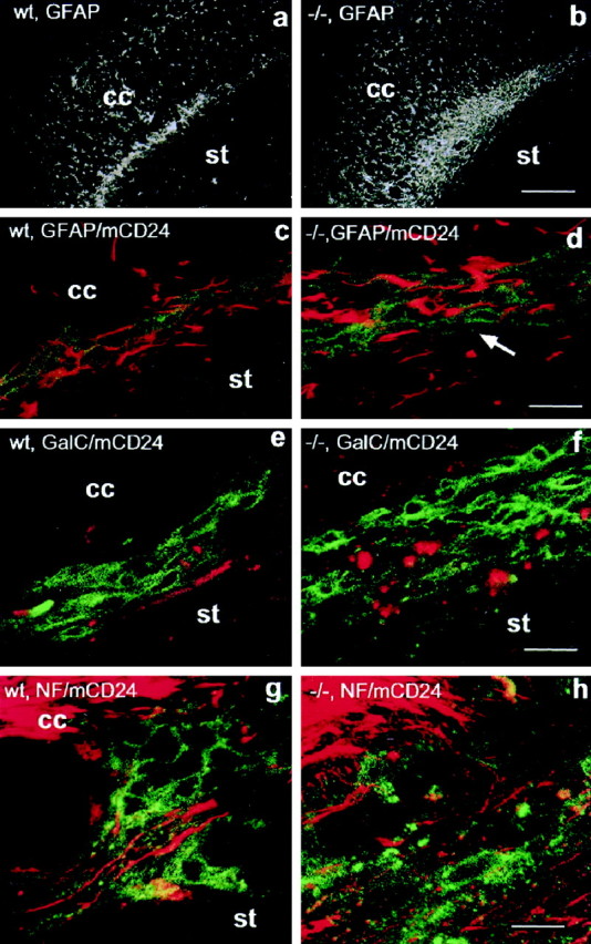
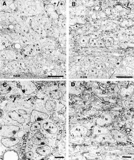
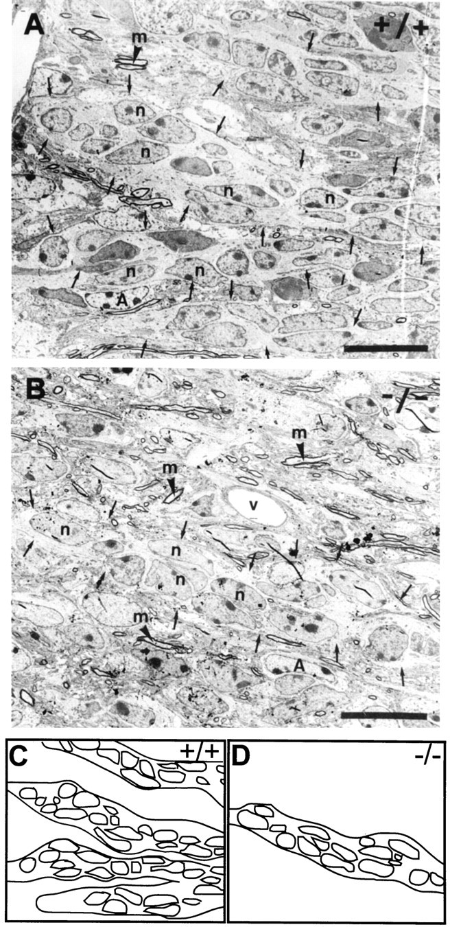
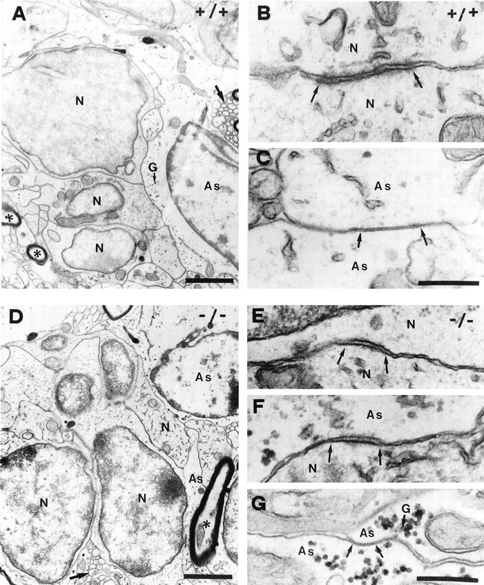
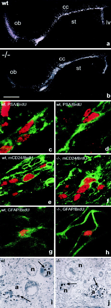
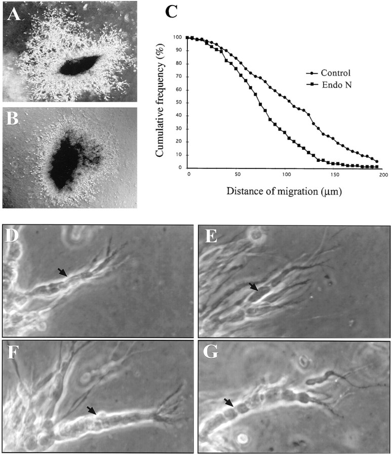
In vertebrates, interneurons of the olfactory bulb (OB) are generated postnatally and throughout life at the subventricular zone of the forebrain. The neuronal precursors migrate tangentially through the forebrain using a well defined pathway, the rostral migratory stream (RMS), and a particular mode of migration in a chain-like organization. A severe size reduction of the OB represents the most striking morphological phenotype in neural cell adhesion molecule (NCAM)-deficient mice. This defect has been traced back to a migration deficit of the precursors in the RMS and linked to the lack of the polysialylated form of NCAM. In this study we investigate the morphological alterations and functional properties of the RMS in mice totally devoid of all isoforms of NCAM and polysialic acid (PSA). We show that a morphologically altered, but defined and continuous pathway exists in mutants, and we present in vivo and in vitro evidence that PSA-NCAM in the RMS is not essential for the formation and migration of chains. Instead, we find a massive gliosis associated with the formation of membrane specializations in a heterotypic manner, linking precursors to astrocytes. This finding and the over-representation and defasciculation of axons in the pathway suggest that important interactions between migrating cells and their stationary environment are perturbed in the mutants. Finally, we used transplantation experiments to demonstrate that lack of PSA-NCAM leads to a decrease but not a total blockade of migration and demonstrate that the mutant RMS is functional in transporting normal neuronal precursors to the OB.


Figures

Fig. 1.
Fig. 1.
Localization of the RMS in adult wild-type (a, wt) and NCAM-deficient (b,−/−) mice. Light micrographs of Nissl-stained sagittal sections through the forebrain reveal in both animals a continuous pathway connecting the anterior horn of the lateral ventricle (lv) to the center of the olfactory bulb (ob). In the wild-type, the pathway is of small diameter over its entire length. Only at its most ventral part it appears slightly dispersed. In the mutant, the pathway is larger in diameter, especially in its caudal portion between the corpus callosum (cc) and the striatum (st), but represents an unbroken and anatomically correct localized structure. Scale bar, 0.5 mm.
Fig. 2.
Fig. 2.
Expression of PSA–NCAM and mCD24 in the wild-type (a–c, e, wt) and NCAM-deficient (d, −/−) RMS. In the wild type, confocal microscopy reveals that staining for mCD24 (a) and PSA–NCAM (b) are largely overlapping (c) and shows the chain-like arrangement of neural precursors migrating in the pathway. In mutants (d), mCD24 labeling demonstrates the massive accumulation of cells in the RMS. Nevertheless, the chain-like organization is still obvious, whereas PSA–NCAM staining is totally absent, as expected. e, Immunoelectron microscopic image of the contact area of two neural precursors (n) in the wild-type RMS at 20,000× magnification. PSA-associated gold particles are found in clusters (arrows) distributed over the membranes. Scale bar: a–d, 50 μm; e, 300 nm.
Fig. 3.
Fig. 3.
Arrangement of glia and axons in the wild-type (a, c, g, wt) and NCAM-deficient (b, d, h, −/−) RMS. Glial fibrillary acidic protein (GFAP) staining reveals the presence of astrocytes over the entire length of the pathway in wild-type (a) and mutant animals (b). In mutants, there is a massive accumulation of GFAP immunoreactivity distributed over a wider area. Double staining for GFAP (red) and mCD24 (green) demonstrates that in the wild-type (c) glial processes, which are of small diameter and oriented in the direction of migration, ensheath and cover the entire free surface of chains. In the mutant (d), GFAP immunoreactivity is strongly increased; the processes appear thicker in diameter and less organized in the rostrocaudal direction. In addition, the covering of precursors appears discontinuous, leaving them exposed to the environment (arrow). Labeling for the oligodendrocyte marker GalC (e, f, red; mCD24, green) reveals the presence of this cell type in the control (e) as well as the mutant (f) RMS. As for GFAP, GalC appears to be more expressed in the mutant pathway. Double staining for the axon marker neurofilament (NF, red) and mCD24 (green) demonstrates the presence of axons in the RMS. In the wild-type (g), neurofilament positive structures are always well defined and oriented in the direction of migration, allowing the tracing of axon fascicles over considerable distances. In the mutant (h), neurofilament immunoreactivity was much more abundant but dispersed, not showing the high degree of organization and orientation found in the wild-type. The punctuate aspect of the NF immunoreactivity indicates axons leaving the plane of the section. cc,Corpus callosum; st, striatum. Scale bars:a, b, 400 μm; c–h, 30 μm.
Fig. 4.
Fig. 4.
Ultrastructural organization of the wild-type (A,C) and PSA–NCAM-deficient (B,D) RMS. In frontal section, the wild-type RMS (A) appears as a highly organized structure containing groups of neural precursors (n) accompanied by astrocytes (a). Individual oligodendrocytes (O), axon profiles, and blood vessels were visible. The mutant pathway (B) shows a lower degree of cell grouping and a massive invasion of axonal structures. In higher magnification (C, D), it becomes obvious that in the wild-type (C) groups of neural precursor (N), representing chains (small arrows), are accompanied by astrocytes (As), together forming dense groups. Individual myelinated (M) and bundles of nonmyelinated axons (large arrow) appear in proximity but are spatially separated from precursors. The equivalent diameter of these fibers shows the strictly parallel orientation, indicative of fasciculation. In contrast, the mutant pathway (D) shows striking disorganization: a lower, but still considerable, degree of cell grouping (small arrows), astrocytes (As) separated from grouped precursors by intercalating axon profiles, and a massive invasion of these axonal structures. Note that these axons show no obvious signs of orientation or bundling.noa, Nucleus olfactorius anterior; Scale bars:A, B, 10 μm; C,D, 3 μm.
Fig. 5.
Fig. 5.
Organization of the RMS in sagittal section. In the wild-type (A), the neural precursors (n) show an elongated shape and are organized in continuous chains (small arrows). In the mutant (B) neural precursors (n) appear more variable in shape, but the majority is still organized in rostrocaudally oriented chain-like arrangements, which can be traced over long distances (C, D, schematic representations). Nevertheless, chains are less frequent and more dispersed. The entire pathway in mutants appears highly disorganized because of the presence of many nonmyelinated and myelinated (m) axons, which show no preferential orientation or organization. A, Astrocyte;v, blood vessel. Scale bar, 10 μm.
Fig. 6.
Fig. 6.
Cell contacts in the wild-type (A–C) and mutant (D–G) RMS. In the wild-type (A), groups of neural precursor (N) are closely apposed and separated from axons (asterisks) and astrocytes (As), containing clusters of glycogen granules (G). Contacts between precursor (B) are of thezona adherens type with flocculent material in the intercellular cleft and stained material on the cytoplasmic side. Contacts between glial cells were characterized as typical gap junctions (C). Heterotypic contacts between precursors and glia were never seen. In the mutant (D), precursors can still be found as groups but appear less organized. Astrocytic processes (As) enwrap precursors only partially, leaving them exposed to surrounding parenchyma. Homotypic contacts (N/N; As/As) in mutants were comparable to the wild-type (E, G), but in addition many heterotypic precursor astrocyte membrane specializations of thezona adherens type are found (F). Scale bars: A, D, 1 μm;B, C, E, F,G, 200 nm.
Fig. 7.
Fig. 7.
Proliferation in the RMS. BrdU immunohistochemistry on sagittal sections revealed the presence of dividing cells over the entire length of the wild-type (a) and mutant (b) RMS when BrdU was administered over 48 hr. Double labeling and confocal microscopy in the wild-type demonstrated the presence of PSA (green)/BrdU (red) double-positive cells integrated into chains 1 hr after BrdU injection (c,d). In the wild-type and mutant, mCD24 (green)/BrdU (red)-labeled cells were comparably located within chain-like structures (e, f). The RMS of both groups contained also individual GFAP (green)/BrdU (red)-stained cells, demonstrating the production of new astrocytes in the pathway. Both cell types, dividing neural precursors (n) and astrocytes (a) were also identified in the wild-type (i) and mutants (j) using immunogold labeling for BrdU and electron microscopy. Arrows indicate BrdU-associated gold particles overlying nuclei of dividing cells. Scale bar:a, b, 1 mm; c–h, 10 μm;i, j, 4 μm.
Fig. 8.
Fig. 8.
Cell migration from SVZ explants cultured in Matrigel. SVZ tissue was cultured in the absence (A, D, F) or presence (B, E, G) of EndoN. Phase-contrast image of typical explants cultured in absence (A) or presence (B) of EndoN as has been used for the migration distance analyses. Note at this low magnification the more pronounced development of chains in the control (A) compared to the EndoN-treated culture (B). C, Cumulative frequency distribution plot of the distance of SVZ cell migration in the absence (circles) or presence (squares) of EndoN. The values represent pooled data from three independent experiments. Aspect of migrating cells in absence of EndoN after 24 (D) or 48 (F) hr of culture. Note the compacted appearance of the cells integrated in chains at both time points. Borders between individual cells were not prominent (D, F, black arrows). In the presence of EndoN for 24 hr, fully developed chains were rare (compare A, B), but clearly distinguishable when they appeared (E). In these aggregates individual cells (arrow) were easily distinguishable, contrary to the wild-type. After 48 hr (G) well-defined aggregates became visible in the EndoN-treated cultures. Nevertheless, as at 24 hr, borders between cells were more prominent, allowing the identification of individual cells (F, G, compareblack arrows).
Fig. 9.
Fig. 9.
Functional properties of the PSA–NCAM-deficient RMS. PSA–NCAM-expressing cells, isolated from the SVZ of LacZ-expressing adult mice, were transplanted into the anterior horn of the lateral ventricle of wild-type and mutant mice. After a survival time of 30 d Xgal-positive cells were found integrated in the SVZ (a, b), the RMS (c, d), and the OB (e, f) in both groups of animals. Scale bar, 30 μm.

References

    1. Alvarez-Buylla A. Mechanism of migration of olfactory bulb interneurons. Cell Dev Biol. 1997;8:207–213. - PubMed
    1. Altman J. Autoradiographic and histological studies of postnatal neurogenesis. IV. Cell proliferation and migration in the anterior forebrain with special reference to persisting neurogenesis in the olfactory bulb. J Comp Neurol. 1969;137:433–458. - PubMed
    1. Berryman MA, Rodewald RD. An enhanced method for post-embedding immunocytochemical staining which preserves cell membranes. J Histochem Cytochem. 1990;38:159–170. - PubMed
    1. Calaora V, Chazal G, Nielsen PJ, Rougon G, Moreau H. mCD24 expression in the developing mouse brain and in zones of secondary neurogenesis in the adult. Neuroscience. 1996;73:581–594. - PubMed
    1. Cohen-Tannoudji M, Morello D, Babinet C. Unexpected position-dependence of H-2 and β2 microglobulin/lacZ transgenes. Mol Reprod Dev. 1992;33:149–159. - PubMed

Publication types

MeSH terms