. 2000 Feb 15;20(4):1495-504.
doi: 10.1523/JNEUROSCI.20-04-01495.2000.

Developmental changes in the transmitter properties of sympathetic neurons that innervate the periosteum

Affiliations

Developmental changes in the transmitter properties of sympathetic neurons that innervate the periosteum

S E Asmus et al. J Neurosci. .

Abstract

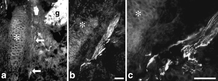
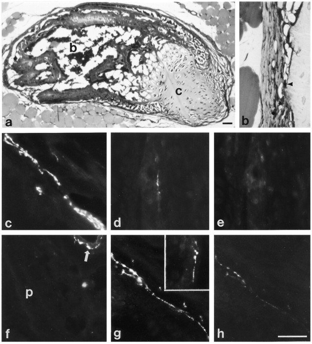
During the development of sweat gland innervation, interactions with the target tissue induce a change from noradrenergic to cholinergic and peptidergic properties. To determine whether the change in neurotransmitter properties that occurs in the sweat gland innervation occurs more generally in sympathetic neurons, we identified a new target of cholinergic sympathetic neurons in rat, the periosteum, which is the connective tissue covering of bone, and characterized the development of periosteal innervation of the sternum. During development, sympathetic axons grow from thoracic sympathetic ganglia along rib periosteum to reach the sternum. All sympathetic axons displayed catecholaminergic properties when they reached the sternum, but these properties subsequently disappeared. Many axons lacked detectable immunoreactivities for vesicular acetylcholine transporter and vasoactive intestinal peptide when they reached the sternum and acquired them after arrival. To determine whether periosteum could direct changes in the neurotransmitter properties of sympathetic neurons that innervate it, we transplanted periosteum to the hairy skin, a noradrenergic sympathetic target. We found that the sympathetic innervation of the transplant underwent a noradrenergic to cholinergic and peptidergic change. These results suggest that periosteum, in addition to sweat glands, regulates the neurotransmitter properties of the sympathetic neurons that innervate it.


Figures

Fig. 1.
Fig. 1.
Adult rat thoracic sympathetic ganglion and periosteum. a, b, Photomicrographs of a single section through a thoracic sympathetic ganglion double-labeled for VIP with an antiserum raised in guinea pig and VAChT using an antiserum raised in rabbit. Several of the cell bodies present in this section that are immunoreactive for both VIP (a) and VAChT (b) are indicated byarrows. The dense plexus of preganglionic fibers that surround the neuron cell bodies also contain VAChT immunoreactivity (b). c, A 1 μm plastic section of periosteum stained with toluidine blue shows the inner cellular (arrowhead) and outer fibrous (f) layers. Scale bars, 40 μm.a and b are the same magnification.
Fig. 2.
Fig. 2.
Transmitter-related properties of the sympathetic innervation of adult rat periosteum. a,b, Photomicrographs of a tangential section of periosteum double-labeled for VIP and TH. Varicose axons coursing through the periosteum contain intense VIP (a) and extremely faint TH (b) immunoreactivity (arrows). At the top right ina, there is a single axon that is VIP-IR. This fiber is distinct from the intensely TH-IR fiber in b.c, d, Transmitter properties indicative of cholinergic neurons are also present in periosteal fibers. Fibers exhibiting punctate VAChT immunoreactivity (c) and reactivity for AChE (d) are observed in the periosteum. e, f, To determine whether VIPergic and cholinergic properties are present in the same fibers, sections were double-labeled for VIP and VAChT. Periosteal fibers containing VIP immunoreactivity (e) are also immunoreactive for VAChT (f).g, Fibers associated with a blood vessel in the intercostal muscle adjacent to the sternum exhibit intense TH immunoreactivity compared with the faint TH immunoreactivity present in VIP-IR fibers (b). VIP immunoreactivity is absent from TH-IR perivascular fibers (h) (arrows). Scale bar: h, 40 μm.
Fig. 3.
Fig. 3.
Development of periosteal innervation in the thoracic region. Axons from thoracic sympathetic neurons are closely associated with rib periosteum as they grow to the sternum.a, Bundles of TH-IR sympathetic axons were observed leaving the ganglion (g) and extending along a rib (asterisk) on E17. The axons are indicated byarrows. b, TH-IR sympathetic axons (arrows) were observed in contact with the periosteum of the rib (asterisk) on E17. c, A bundle of TH-IR axons bifurcates, and some course along associated with the periosteum of the rib (asterisk). a andb are the same magnification. Scale bars:a, b, 10 μm; c, 20 μm.
Fig. 4.
Fig. 4.
Development of transmitter-related properties in the periosteum. a–c, Catecholaminergic properties are present in developing periosteal fibers. a, On E18, catecholamine histofluorescent fibers are seen in the periosteum (arrowheads), which runs vertically in the photomicrograph, adjacent to the cartilaginous sternum (s). Catecholamine fluorescent fibers are also present surrounding a blood vessel that is adjacent to the sternum at the top of the photomicrograph. b,c, A single section through the sternum on P0 double-labeled for TH and VIP, showing a TH-IR periosteal fiber (b) that is not immunoreactive for VIP (c). d, e, Between E18 and P0, approximately half of the TH-IR fibers also contain VIP immunoreactivity, and these VIP-IR fibers, shown here in P0 periosteum (d), are also immunoreactive for VAChT (e). s, Cartilage of sternum ina–e. f–h, One to 2 weeks after birth, a dual phenotype is evident in the majority of periosteal fibers, demonstrated in these photomicrographs of longitudinal sections through the periosteum of the sternum. f, g, On P14, intense VIP (f) and TH (g) immunoreactivities are colocalized in most periosteal fibers (arrows). h, AChE-reactive fibers are first detected in the periosteum on P8. Scale bar: h, 40 μm.
Fig. 5.
Fig. 5.
Neonatal 6-OHDA administration reduces the number of fibers that display cholinergic and VIPergic properties in mature periosteal fibers. a, b, Double labeling for CGRP and VIP in a section of periosteum from an adult rat treated neonatally with 6-OHDA demonstrates that CGRP-IR fibers, presumably of sensory origin, are not affected by this toxin (a), but the number of VIP-IR fibers is reduced dramatically (b). c, 6-OHDA treatment also reduces the number of VAChT-IR reactive fibers in the periosteum (d) compared with control (c). Scale bar: a–d, 20 μm.
Fig. 6.
Fig. 6.
Development of transmitter properties of sympathetic fibers innervating ectopic periosteum. a,b, One micrometer plastic sections of 2 week transplants. a, Periosteal rudiments transplanted under the skin of the lateral thorax form a small bone, consisting of both cartilage (c) and trabecular bone with intervening marrow spaces (b). b, This photomicrograph shows a higher magnification view of the periosteum surrounding the transplants, which is similar in appearance to periosteum in situ, with an inner cellular layer (arrowhead) adjacent to the bone of the transplant.c–e, Sections of periosteal transplants 1–2 weeks after surgery. c, Sympathetic fibers that innervate the periosteum of the transplant initially contain catecholamine fluorescence. d, e, After double labeling for TH and VIP, periosteal fibers that contain TH (d) but not VIP (e) immunoreactivity are seen. f–h, Sections of periosteal transplants 4–6 weeks after surgery. f, Few catecholamine fluorescent fibers are observed in the periosteum (p) of the transplant 6 weeks after transplantation, although blood vessels adjacent to the transplant are surrounded by catecholaminergic fibers (arrow).g, h, A section of a 4 week transplant double-labeled for VIP and TH contains intense VIP-IR fibers (g), and these fibers exhibit only faint TH immunoreactivity (h). g,Inset, VAChT-IR fibers are also observed in the periosteum of a transplant 4 weeks after surgery. Scale bars:a, h, 40 μm. b–h are the same magnification.

References

    1. Alfonso A, Grundahl K, McManus JR, Asbury JM, Rand JB. Alternative splicing leads to two cholinergic proteins in Caenorhabditis elegans. J Mol Biol. 1994;241:627–630. - PubMed
    1. Bazan JF. Neuropoietic cytokines in the hematopoetic fold. Neuron. 1991;7:197–208. - PubMed
    1. Bejanin S, Cervini R, Mallet J, Berrard S. A unique gene organization for two cholinergic markers, choline acetyltransferase and a putative vesicular transporter of acetylcholine. J Biol Chem. 1994;269:21944–21947. - PubMed
    1. Berrard S, Varoqui H, Cervini R, Israel M, Mallet J, Diebler M-F. Coregulation of two embedded gene products, choline acetyltransferase and the vesicular acetylcholine transporter. J Neurochem. 1995;65:939–942. - PubMed
    1. Berse B, Blusztajn JK. Coordinated up-regulation of choline acetyltransferase and vesicular acetylcholine transporter gene expression by the retinoic acid receptor alpha, cAMP, and leukemia inhibitory factor/ciliary neurotrophic factor signaling pathways in a murine septal cell line. J Biol Chem. 1995;270:22101–22104. - PubMed

Publication types

MeSH terms