. 2000 Feb 15;20(4):1519-28.
doi: 10.1523/JNEUROSCI.20-04-01519.2000.

Gap junctions linking the dendritic network of GABAergic interneurons in the hippocampus

Affiliations

Gap junctions linking the dendritic network of GABAergic interneurons in the hippocampus

T Fukuda et al. J Neurosci. .

Abstract

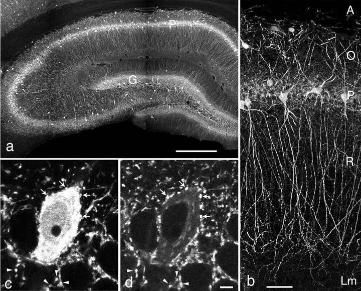
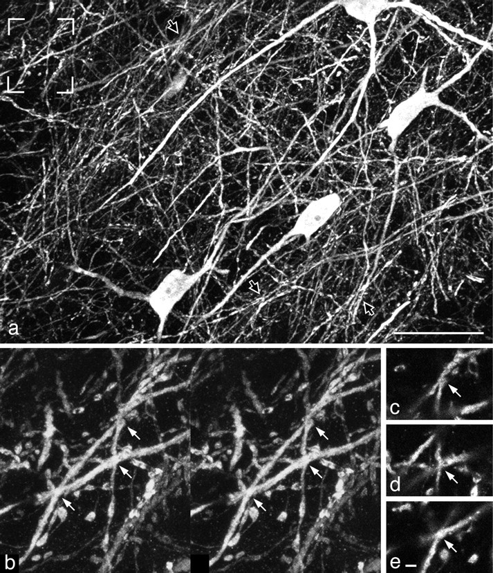
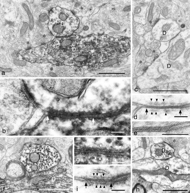
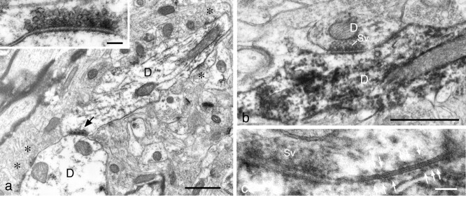
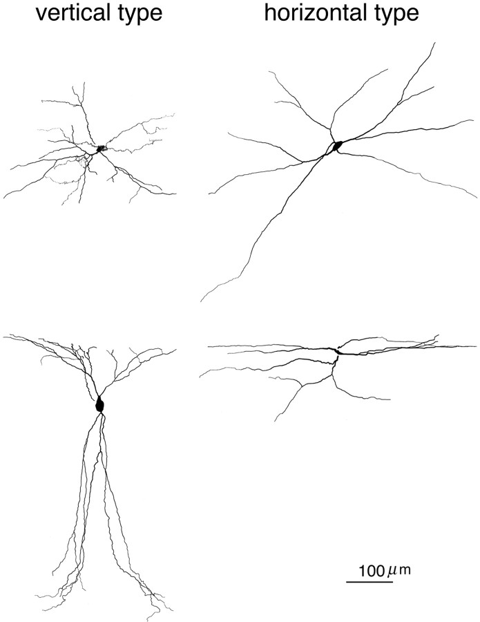
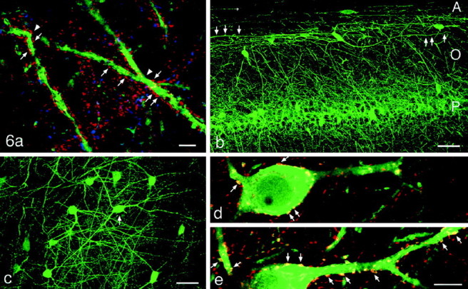
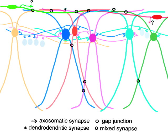
The network of GABAergic interneurons connected by chemical synapses is a candidate for the generator of synchronized oscillations in the hippocampus. We present evidence that parvalbumin (PV)-containing GABAergic neurons in the rat hippocampal CA1 region, known to form a network by mutual synaptic contacts, also form another network connected by dendrodendritic gap junctions. Distal dendrites of PV neurons run parallel to the alveus (hippocampal white matter) and establish multiple contacts with one another at the border between the stratum oriens and the alveus. In electron microscopic serial section analysis, gap junctions could be identified clearly at 24% of these contact sites. A dendrodendritic chemical synapse and a mixed synapse also were found between PV-immunoreactive dendrites. Three-dimensional reconstruction of the dendritic arborization revealed that both PV neurons of the well known vertical type (presumptive basket cells and axoaxonic cells) and those of another horizontal type constitute the dendritic network at the light microscopic level. The extent of dendritic fields of single PV neurons in the lateral direction was 538 +/- 201 micrometer (n = 5) in the vertical type and 838 +/- 159 micrometer (n = 6) in the horizontal type. Our previous and present observations indicate that PV-containing GABAergic neurons in the hippocampus form the dual networks connected by chemical and electrical synapses located at axosomatic and dendrodendritic contact sites, respectively. Gap junctions linking the dendritic network may mediate coherent synaptic inputs to distant interneurons and thereby facilitate the synchronization of oscillatory activities generated in the interneuron network.


Figures

Fig. 1.
Fig. 1.
Morphological characteristics of PV neurons viewed in conventionally oriented coronal sections. a, A montage of confocal laser-scanning microscopic images. Somata of PV neurons are located mainly in and around the pyramidal cell layer (P) and the granule cell layer (G). Their axon terminals also are distributed in the same layers, causing band-like staining in lower magnification.b, PV neurons in the pyramidal cell layer of the CA1 region give rise to dendrites spanning all hippocampal layers.A, Alveus; O, stratum oriens;R, stratum radiatum; Lm, stratum lacunosum-moleculare. c, d, Double immunostaining with antibodies against PV (c) and GAD (d). Note multiple PV-containing GAD-immunoreactive (ir) boutons (arrows) abutting on the PV-ir soma. Arrowheads indicate similarly double-labeled boutons, some of which may abut on unstained somata of pyramidal neurons. Scale bars: a, 0.5 mm; b, 50 μm; c, d, 5 μm.
Fig. 2.
Fig. 2.
CLSM images in a section cut tangentially along the border between the alveus and the stratum oriens of the CA1 region.a, PV-ir dendrites running two-dimensionally within a single 40-μm-thick section form an extremely dense network. Some dendrites appear to be bundled together (open arrows), whereas others cross one another with a fairly wide angle. Four PV-ir somata are of the horizontal type (see Results). b, Stereo pair of the enlargement of the framed area ina. PV-ir dendrites make multiple contacts with one another, only three of which are indicated by arrows.c–e, Single confocal optical images of the three contact sites designated in b, suggesting a direct contact between these dendrites. Scale bars: a, 50 μm;be, 1 μm.
Fig. 3.
Fig. 3.
Electron micrographs showing the dendrodendritic gap junctions. a, Two PV-ir dendrites (D) receiving multiple presynaptic terminals (asterisks) make direct contact with each other. b, Enlargement of the contact site ina. A gap junction is formed between the two PV-ir dendrites. Note the close apposition of the plasma membranes of the two contacting cells as demarcated by arrows. A synaptic bouton (asterisk) forming a synapse of asymmetrical type is located in close vicinity to the gap junction. c, Electron micrograph of a specimen without immunoreaction, demonstrating a profile similar to a. The contacting dendrites (D) receive presynaptic terminals (asterisks). d, Enlargement of the contact site in c with the same magnification as inb, g, and i to facilitate comparison. Plasma membranes of the two cells are closely apposed, as demarcated by arrows. Note a layer of cytoplasmic semidense material (arrowheads) undercoating either side of the junction, which is characteristic of neuronal gap junctions.e, Further enlargement of the contact site ind, showing a narrow central gap, 2.7 nm wide, between the outer leaflets of the apposed unit membranes. f–i, Other examples of gap junctions formed between PV-ir dendrites (D). The contact sites in f andh are enlarged in g and i, respectively, with the same magnification as in b andd. Scale bars: a, c,f, h, 1 μm; b,d, e, g, i, 0.1 μm.
Fig. 4.
Fig. 4.
Electron micrographs showing two novel forms of dendrodendritic contacts between PV neurons. a, Two PV-ir dendrites (D) receiving multiple presynaptic terminals (asterisks) make direct contact with each other (arrow). Inset, Enlargement of the contact site demonstrating synaptic vesicles, widening of the synaptic cleft, and less prominent thickening of the postsynaptic density; the last feature is consistent to conventional symmetrical synapses, including those formed by PV-ir axon terminals.b,c, Serial ultrathin sections with different magnifications demonstrating that two PV-ir dendrites (D), receiving multiple presynaptic terminals (data not shown in these sections), establish a mixed type of synapse between each other. Synaptic vesicles (Sv) accumulate in one side of the contact as in a, but in this case the gap junction can be identified in line with the chemical synapse inc. Note a layer of cytoplasmic semidense material (arrows) undercoating gap junction as in Fig.3d, g, i. Scale bars:a, b, 1 μm; Inset ina, c, 0.1 μm.
Fig. 5.
Fig. 5.
Comparison of the reconstructed dendritic arborization of PV neurons between the vertical type (left) and the horizontal type (right), viewed in two directions (top, top view;down, front view) with the use of a computer-assisted neuron tracing system. The top and front views are of the same cell in each type and are aligned.
Fig. 6.
Fig. 6.
Top. CLSM images showing several morphological aspects of PV-ir neurons viewed in tangential (a, c–e) and coronal (b) sections. a, Triple-immunostained section at the border between the stratum oriens and the alveus. PV-ir dendrites (green) receive numerous synaptophysin (red)-positive and GAD (blue)-negative boutons (arrows). The mutual contact sites in dendrites are shown byarrowheads. b, A PV neuron of the horizontal type (arrow) located at the border between the stratum oriens (O) and the alveus (A) gives rise to long dendrites (arrowheads) along the border. The stratum oriens contains many obliquely running dendrites originating from PV-ir somata located in and around the stratum pyramidale (P).c, Dendrites of horizontal PV neurons extend in all directions from their somata along the horizontal plane.d,e, Enlargement of the PV-ir neuron, indicated by an arrow in c, double-immunostained with antibodies against PV (green) and GAD (red). CLSM images in d and e were taken at different depths from the section surface. Note numerous PV-containing GABAergic boutons (arrows) abutting on the soma and proximal dendrites. Scale bars: a, 5 μm; b,c, 50 μm; d, e, 10 μm.
Fig. 7.
Fig. 7.
Bottom. A schematic drawing representing the dual networks of hippocampal PV neurons connected by chemical and electrical synapses. PV neurons are shown in color, whereas pyramidal cell somata are depicted as gray ovals. Frequent occurrence of gap junctions between horizontally oriented dendrites (top of the figure) was confirmed by quantitative electron microscopic analysis, but it remains unknown whether both or either of the two neuronal types (vertical and horizontal) actually forms gap junctions there. See Discussion for details.

References

    1. Ali AB, Thomson AM. Facilitating pyramid to horizontal oriens–alveus interneurone inputs: dual intracellular recordings in slices of rat hippocampus. J Physiol (Lond) 1998;507:185–199. - PMC - PubMed
    1. Bragin A, Jandó G, Nádasdy Z, Hetke J, Wise K, Buzsáki G. Gamma (40–100 Hz) oscillation in the hippocampus of the behaving rat. J Neurosci. 1995;15:47–60. - PMC - PubMed
    1. Brightman MW, Reese TS. Junctions between intimately apposed cell membranes in the vertebrate brain. J Cell Biol. 1969;40:648–677. - PMC - PubMed
    1. Buhl EH, Halasy K, Somogyi P. Diverse sources of hippocampal unitary inhibitory postsynaptic potentials and the number of synaptic release sites. Nature. 1994a;368:823–828. - PubMed
    1. Buhl EH, Han Z-S, Lörinczi Z, Stezhka VV, Karnup SV, Somogyi P. Physiological properties of anatomically identified axo-axonic cells in the rat hippocampus. J Neurophysiol. 1994b;71:1289–1307. - PubMed

Publication types

Substances

LinkOut - more resources