. 2000 Feb 15;20(4):1550-8.
doi: 10.1523/JNEUROSCI.20-04-01550.2000.

alpha-Melanocyte-stimulating hormone is contained in nerve terminals innervating thyrotropin-releasing hormone-synthesizing neurons in the hypothalamic paraventricular nucleus and prevents fasting-induced suppression of prothyrotropin-releasing hormone gene expression

Affiliations

alpha-Melanocyte-stimulating hormone is contained in nerve terminals innervating thyrotropin-releasing hormone-synthesizing neurons in the hypothalamic paraventricular nucleus and prevents fasting-induced suppression of prothyrotropin-releasing hormone gene expression

C Fekete et al. J Neurosci. .

Abstract

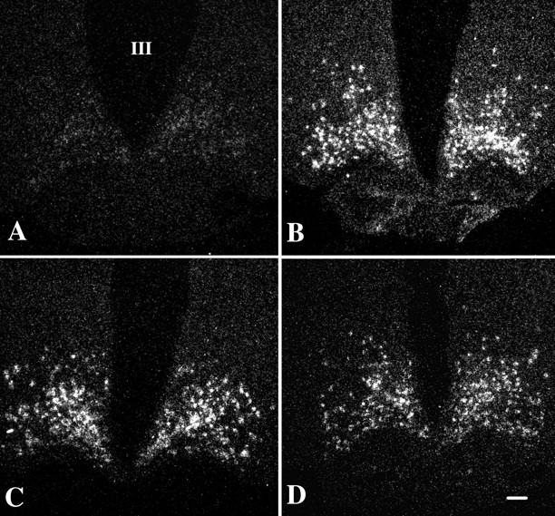
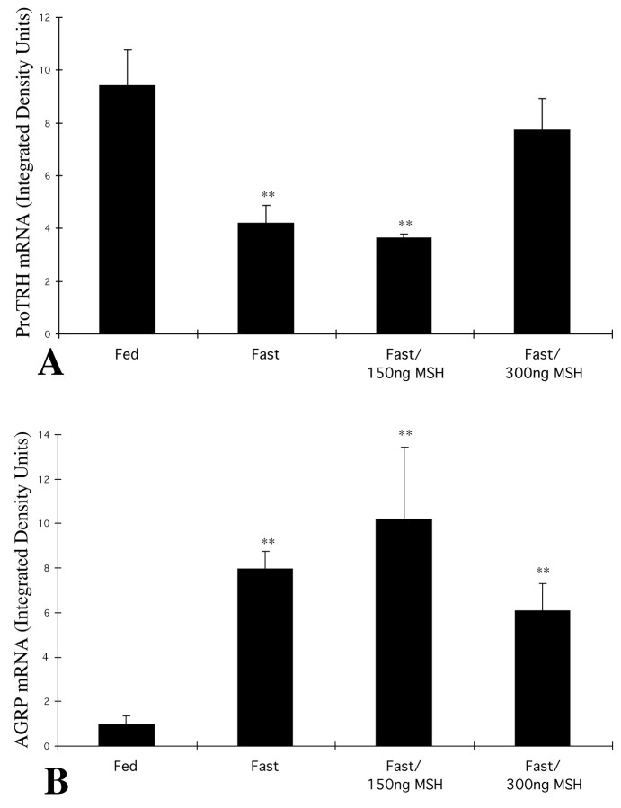
The hypothalamic arcuate nucleus has an essential role in mediating the homeostatic responses of the thyroid axis to fasting by altering the sensitivity of prothyrotropin-releasing hormone (pro-TRH) gene expression in the paraventricular nucleus (PVN) to feedback regulation by thyroid hormone. Because agouti-related protein (AGRP), a leptin-regulated, arcuate nucleus-derived peptide with alpha-MSH antagonist activity, is contained in axon terminals that terminate on TRH neurons in the PVN, we raised the possibility that alpha-MSH may also participate in the mechanism by which leptin influences pro-TRH gene expression. By double-labeling immunocytochemistry, alpha-MSH-IR axon varicosities were juxtaposed to approximately 70% of pro-TRH neurons in the anterior and periventricular parvocellular subdivisions of the PVN and to 34% of pro-TRH neurons in the medial parvocellular subdivision, establishing synaptic contacts both on the cell soma and dendrites. All pro-TRH neurons receiving contacts by alpha-MSH-containing fibers also were innervated by axons containing AGRP. The intracerebroventricular infusion of 300 ng of alpha-MSH every 6 hr for 3 d prevented fasting-induced suppression of pro-TRH in the PVN but had no effect on AGRP mRNA in the arcuate nucleus. alpha-MSH also increased circulating levels of free thyroxine (T4) 2.5-fold over the levels in fasted controls, but free T4 did not reach the levels in fed controls. These data suggest that alpha-MSH has an important role in the activation of pro-TRH gene expression in hypophysiotropic neurons via either a mono- and/or multisynaptic pathway to the PVN, but factors in addition to alpha-MSH also contribute to the mechanism by which leptin administration restores thyroid hormone levels to normal in fasted animals.


Figures

Fig. 1.
Fig. 1.
A–C, Low-power photomicrographs showing the distribution of α-MSH-IR axons (black) and pro-TRH-containing neurons (brown) in different levels of the PVN. A, Anterior level of the PVN.B, Mid level of the PVN. C, Caudal level of the PVN. D–G, High-power magnification of α-MSH-IR axon varicosities (arrows) contacting pro-TRH neurons in the anterior (D), periventricular (E), medial (F), and dorsal (G) parvocellular subdivisions of the PVN. Note that the majority of the pro-TRH-IR neurons are contacted by α-MSH-IR fibers in the anterior and periventricular subdivisions, whereas fewer pro-TRH-IR neurons in the medial and dorsal parvocellular subdivisions appear to be innervated by α-MSH-IR axons. H, I,Triple-labeling fluorescent immunocytochemistry showing dual innervation of periventricular (a) and medial (b) parvocellular proTRH neurons (blue) of the PVN by axon terminals containing α-MSH (red;arrowheads) and AGRP (green;arrows). Note that all pro-TRH-containing neurons establishing contacts with α-MSH-IR terminals are also contacted by several AGRP-IR varicosities, whereas other pro-TRH-IR neurons (asterisks) receive only an AGRP-IR innervation. AP, Anterior parvocellular subdivision; DP, dorsal parvocellular subdivision;MP, medial parvocellular subdivision; PV,periventricular parvocellular subdivision; VP, ventral parvocellular subdivision. Scale bars: A–C, 100 μm;D–I, 20 μm.
Fig. 2.
Fig. 2.
Electron micrographs showing synaptic associations (arrows) between pro-TRH-containing neurons in the PVN and α-MSH-containing axon terminals. The pro-TRH-IR dendrites and perikarya are labeled with highly electron-dense silver granules, whereas the α-MSH-IR terminals are recognized by the presence of the electron-dense DAB. A, Medium-power magnification view of an axodendritic synapse, shown in greater detail in theinset. B, High-power magnification of an axosomatic synapse. Scale bars: A, 1 μm; B, inset, 0.4 μm.
Fig. 3.
Fig. 3.
Dark-field illumination micrographs of pro-TRH mRNA in the medial and periventricular parvocellular subdivisions of the hypothalamic PVN in fed (A) and fasted (B) animals and fasted animals receiving an intracerebroventricular infusion of α-MSH at a dose of 150 ng (C) or 300 ng (D) every 6 hr for 64 hr. Note the reduction in the accumulation of silver grains over the PVN in fasted animals compared with the fed controls. Fasted animals receiving 150 ng of α-MSH every 6 hr show suppression of pro-TRH mRNA in the PVN similar to that of fasted animals receiving artificial CSF. Fasted animals receiving 300 ng of α-MSH every 6 hr, however, show a marked increase in pro-TRH mRNA that is similar to that of fed control animals. III, Third ventricle. Scale bar, 100 μm.
Fig. 4.
Fig. 4.
Dark-field illumination micrographs of AGRP mRNA in the arcuate nucleus of fed (A) and fasted (B) animals and fasted animals receiving an intracerebroventricular infusion of α-MSH at a dose of 150 ng (C) or 300 ng (D). Note the marked increase in AGRP mRNA in the fasted animals. No significant alteration in AGRP mRNA levels is apparent after α-MSH administration in any of the groups. III, Third ventricle. Scale bar, 100 μm.
Fig. 5.
Fig. 5.
Computerized image analysis of pro-TRH mRNA content in the PVN (A) and AGRP mRNA content in the arcuate nucleus (B) of fed and fasted animals and fasted animals receiving an intracerebroventricular infusion of α-MSH at a dose of 150 or 300 ng (**p < 0.01 compared with fed animals).

References

    1. Adan RA, Cone RD, Burbach JP, Gispen WH. Differential effects of melanocortin peptides on neural melanocortin receptors. Mol Pharmacol. 1994;46:1182–1190. - PubMed
    1. Beck B, Richy S. Hypothalamic hypocretin/orexin and neuropeptide Y: divergent interaction with energy depletion and leptin. Biochem Biophys Res Commun. 1999;258:119–122. - PubMed
    1. Blake NG, Eckland DJ, Foster OJ, Lightman SL. Inhibition of hypothalamic thyrotropin-releasing hormone messenger ribonucleic acid during food deprivation. Endocrinology. 1991;129:2714–2718. - PubMed
    1. Branchereau P, Van Bockstaele EJ, Chan J, Pickel VM. Ultrastructural characterization of neurons recorded intracellularly in vivo and injected with lucifer yellow: advantages of immunogold-silver vs. immunoperoxidase labeling. Microsc Res Tech. 1995;30:427–436. - PubMed
    1. Broberger C, Johansen J, Johansson C, Schalling M, Hokfelt T. The neuropeptide Y/agouti gene-related protein (AGRP) brain circuitry in normal, anorectic, and monosodium glutamate-treated mice. Proc Natl Acad Sci USA. 1998;95:15043–15048. - PMC - PubMed

Publication types

MeSH terms

LinkOut - more resources