Comparative Study
. 2000 Mar;74(5):2073-83.
doi: 10.1128/jvi.74.5.2073-2083.2000.

The rice tungro bacilliform virus gene II product interacts with the coat protein domain of the viral gene III polyprotein

Affiliations
Comparative Study

The rice tungro bacilliform virus gene II product interacts with the coat protein domain of the viral gene III polyprotein

E Herzog et al. J Virol. 2000 Mar.

Abstract

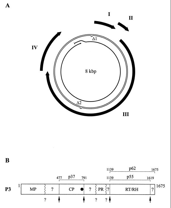
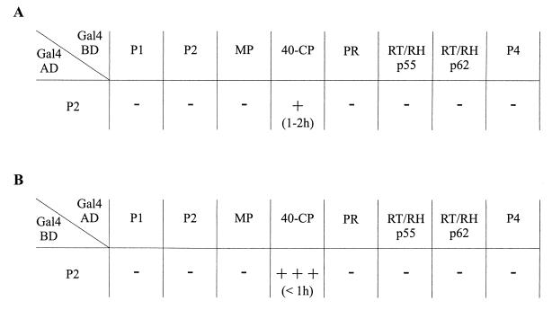
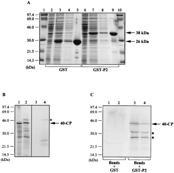
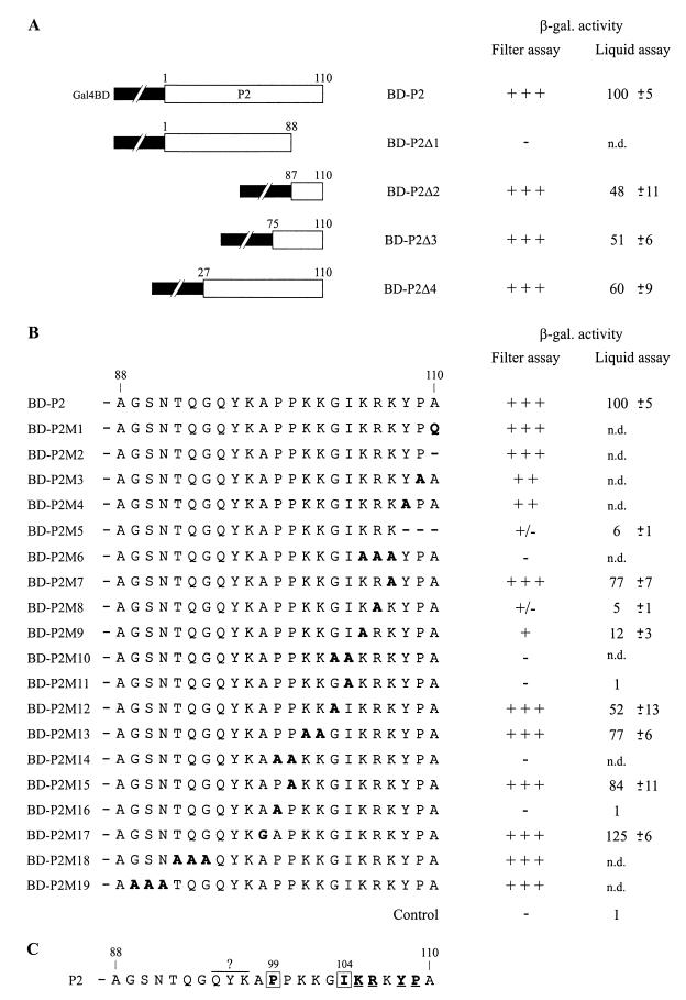
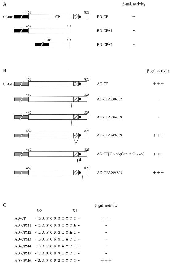
Rice tungro bacilliform virus (RTBV) is a plant pararetrovirus whose DNA genome contains four genes encoding three proteins and a large polyprotein. The function of most of the viral proteins is still unknown. To investigate the role of the gene II product (P2), we searched for interactions between this protein and other RTBV proteins. P2 was shown to interact with the coat protein (CP) domain of the viral gene III polyprotein (P3) both in the yeast two-hybrid system and in vitro. Domains involved in the P2-CP association have been identified and mapped on both proteins. To determine the importance of this interaction for viral multiplication, the infectivity of RTBV gene II mutants was investigated by agroinoculation of rice plants. The results showed that virus viability correlates with the ability of P2 to interact with the CP domain of P3. This study suggests that P2 could participate in RTBV capsid assembly.


Figures

FIG. 1
FIG. 1
Schematic representations of the RTBV genome and P3 polyprotein. (A) Genome organization. Viral DNA is represented by a thin double line with the sites of the two discontinuities (Δ1 and Δ2) indicated. The thick arrows outside the DNA represent the four viral genes (I, II, III, and IV). The pregenomic RNA is shown as a thin arrow inside the DNA. (B) P3 polyprotein. The locations of the domains corresponding to MP, CP, PR, RT, RH within P3 are shown. Domains with unknown functions are indicated by question marks. Positions of the cleavage sites characterized by Laco et al. (27) and Marmey et al. (30) are indicated by vertical lines and arrows. Other presumed cleavage sites are symbolized by zigzag lines and question marks. Positions of the amino and carboxy termini of CP (p37) and RT (p55 and p62) are indicated. The black circle indicates the position of the zinc finger motif in CP.
FIG. 2
FIG. 2
Interaction between P2 and other RTBV proteins in the yeast two-hybrid system. Yeast strain HF7c was cotransformed with a plasmid encoding the Gal4AD-P2 fusion protein and a plasmid expressing one of the other RTBV proteins fused to the Gal4BD (A) or with plasmid pairs encoding the various Gal4 domain-RTBV protein fusions in the opposite combination (B). The β-galactosidase activity produced by the resulting transformants was assessed by filter assay, with the appearance of a blue color indicating interaction between the proteins tested. The strength of the interactions was estimated by the intensity of the blue coloration after 6 h: +++, dark blue; +, light blue; −, no color. The time taken for the blue color to appear is indicated in parentheses.
FIG. 3
FIG. 3
In vitro binding of P2 to 40-CP. (A) Expression and purification of the GST-P2 fusion protein. P2 fused to GST was expressed in E. coli and isolated by binding to glutathione-Sepharose 4B beads. GST alone was also expressed and purified. The proteins present in the total bacterial lysates before (lanes 2 and 6) and after (lanes 3 and 7) induction, in the soluble fractions (lanes 4 and 8), and in the fractions bound to the beads (lanes 5 and 9) were subjected to SDS-PAGE and visualized by Coomassie blue staining. The positions of GST (26 kDa) and GST-P2 (38 kDa) are indicated on the right; the sizes of the protein markers (lanes 1 and 10) are indicated on the left. (B) Synthesis of 40-CP by in vitro translation. 40-CP was translated from a transcript corresponding to nucleotides 2370 to 3462 of the RTBV plus-strand sequence in the presence of [35S]methionine in either wheat germ extract (lane 1) or reticulocyte lysate (lane 2). Labelled translation products were analyzed by SDS-PAGE and autoradiography. Lanes 3 and 4 correspond to the translation controls (without addition of RNA) for the wheat germ extract and reticulocyte lysate, respectively. The positions of 40-CP and the size markers are indicated on the right and left, respectively. ⧫, position of the 46-kDa protein (lanes 2 and 4) mentioned in the text. (C) Interaction of 40-CP with GST-P2. 40-CP synthesized in vitro was incubated with GST-P2 or GST bound to the glutathione-Sepharose 4B beads. The labelled proteins produced in either wheat germ extract (lanes 1 and 3) or in reticulocyte lysate (lanes 2 and 4) and coprecipitated with the GST-P2- or GST-bead complexes were separated by SDS-PAGE and visualized by autoradiography. The positions of 40-CP and the size markers are indicated on the right and left, respectively. Asterisks, positions of the truncated forms of CP (lanes 3 and 4), which are discussed in the text.
FIG. 4
FIG. 4
Mapping of the CP-interacting domain of P2 with the yeast two-hybrid system. (A and B) Yeast strain HF7c was cotransformed with the plasmids encoding CP fused to Gal4AD (pGAD-CP) and P2 or modified P2 fused to the Gal4BD (pGBT-P2, pGBT-P2Δ1 to pGBT-P2Δ4, pGBT-P2M1 to pGBT-P2M19), and the transformants obtained were tested for β-galactosidase (β-gal.) activity. Mutations introduced in the P2 coding sequence of pGBT-P2 correspond to large deletions (A) and small deletions or amino acid substitutions (B). (A) P2 and truncated P2 forms are depicted schematically by open boxes, and Gal4BD is depicted by the interrupted black boxes. (B) Amino acid substitutions and small deletions in the C-terminal region of P2 fusion proteins are indicated by bold letters and bold hyphens, respectively. Numbers above the open boxes and the amino acid sequences refer to positions in P2. β-Galactosidase activity was determined by filter and liquid assay procedures. For the filter assay quantifications, β-galactosidase activity was assessed by the intensity of the blue color of yeast clones after 6 h: +++, dark blue; ++, intermediate color; +, light blue; +/− pale blue; −, no color. For the liquid-assay quantifications, the values indicate percent β-galactosidase activities relative to those obtained with the wild-type P2 construct, measured in extracts from yeast cells expressing AD-CP and one of the BD-P2 versions. Values given are the mean of the relative β-galactosidase activities (and their standard deviations) of five independent clones. The β-galactosidase activity of yeast clones harboring the P2 wild-type construct was, on average, 2.7 β-galactosidase units, as defined in Materials and Methods. The control corresponds to a yeast clone transformed with pGBT9 and pGAD424; n.d., not determined. (C) Role of the C-terminal residues of P2 in establishing the interaction with CP. Residues which are involved are shown in bold type. Boxed residues are crucial and underlined residues are important for the association with CP. The importance of residues QYK97 was not assessed.
FIG. 5
FIG. 5
Modifications of the CP affecting its interaction with P2 in the yeast two-hybrid system. Yeast strain HF7c was cotransformed with plasmids encoding P2 in fusion with the Gal4AD (pGAD-P2) and the CP or CP derivatives in fusion with the Gal4BD (pGBT-CP, pGBT-CPΔ1, and pGBT-CPΔ2) (A) or with plasmids encoding P2 in fusion with the Gal4BD (pGBT-P2) and the CP versions in fusion with Gal4AD (pGAD-CP, pGAD-CPΔ730–732, pGAD-CPΔ736–739, pGAD-CPΔ749–769, pGAD-CP[C772A;C774A;C777A], and pGAD-CPΔ799–803; pGAD-CPM1 to pGAD-CPM6) (B and C). The β-galactosidase (β-gal.) activity was estimated by filter assay. Open boxes represent CP or CP derivatives in the CP fusion proteins, and interrupted black and gray boxes represent Gal4BD and Gal4AD, respectively. The basic domain of CP is indicated by a shaded box, and the zinc finger motif is indicated by a black circle. (B) Small deletions and replacements by alanines are indicated by V and arrows, respectively. (C) Alanine substitutions introduced in Gal4AD-CP fusion protein are shown in bold. Numbers indicated above open boxes and amino acid sequences refer to positions in the P3 polyprotein.

Similar articles

Cited by

References

    1. Bao Y, Hull R. Characterization of the discontinuities in rice tungro bacilliform virus DNA. J Gen Virol. 1992;73:1297–1301. - PubMed
    1. Beaudet-Miller M, Zhang R, Durkin J, Gibson W, Kwong A D, Hong Z. Virus-specific interaction between the human cytomegalovirus major capsid protein and the C terminus of the assembly protein precursor. J Virol. 1996;70:8081–8088. - PMC - PubMed
    1. Cheng C P, Lockhart B E, Olszewski N E. The ORF I and II proteins of Commelina yellow mottle virus are virion-associated. Virology. 1996;223:263–271. - PubMed
    1. Cheng R H, Olson N H, Baker T S. Cauliflower mosaic virus: a 420 subunit (T=7), multilayer structure. Virology. 1992;186:655–668. - PMC - PubMed
    1. Dahal G, Hibino H, Saxena R C. Association of leafhopper feeding behavior with transmission of rice tungro to susceptible and resistant rice cultivars. Phytopathology. 1990;80:371–377.

Publication types

LinkOut - more resources